3D imaging video gallery
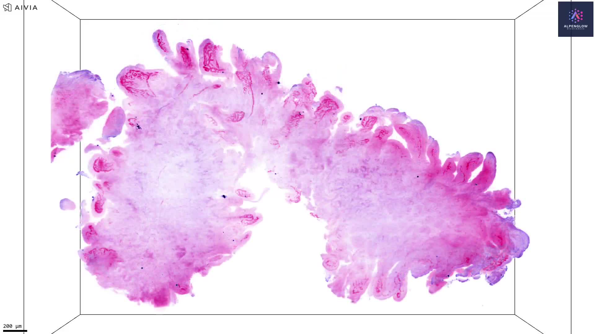
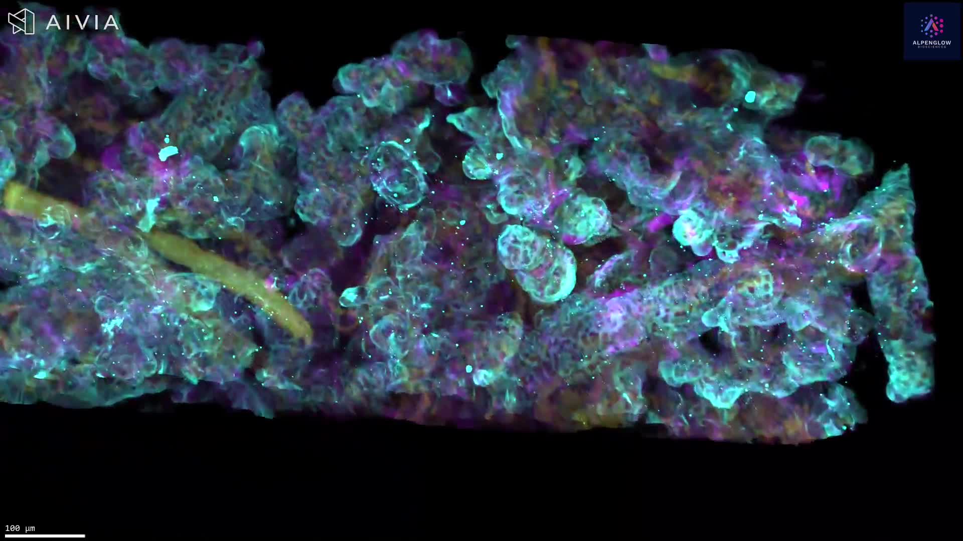
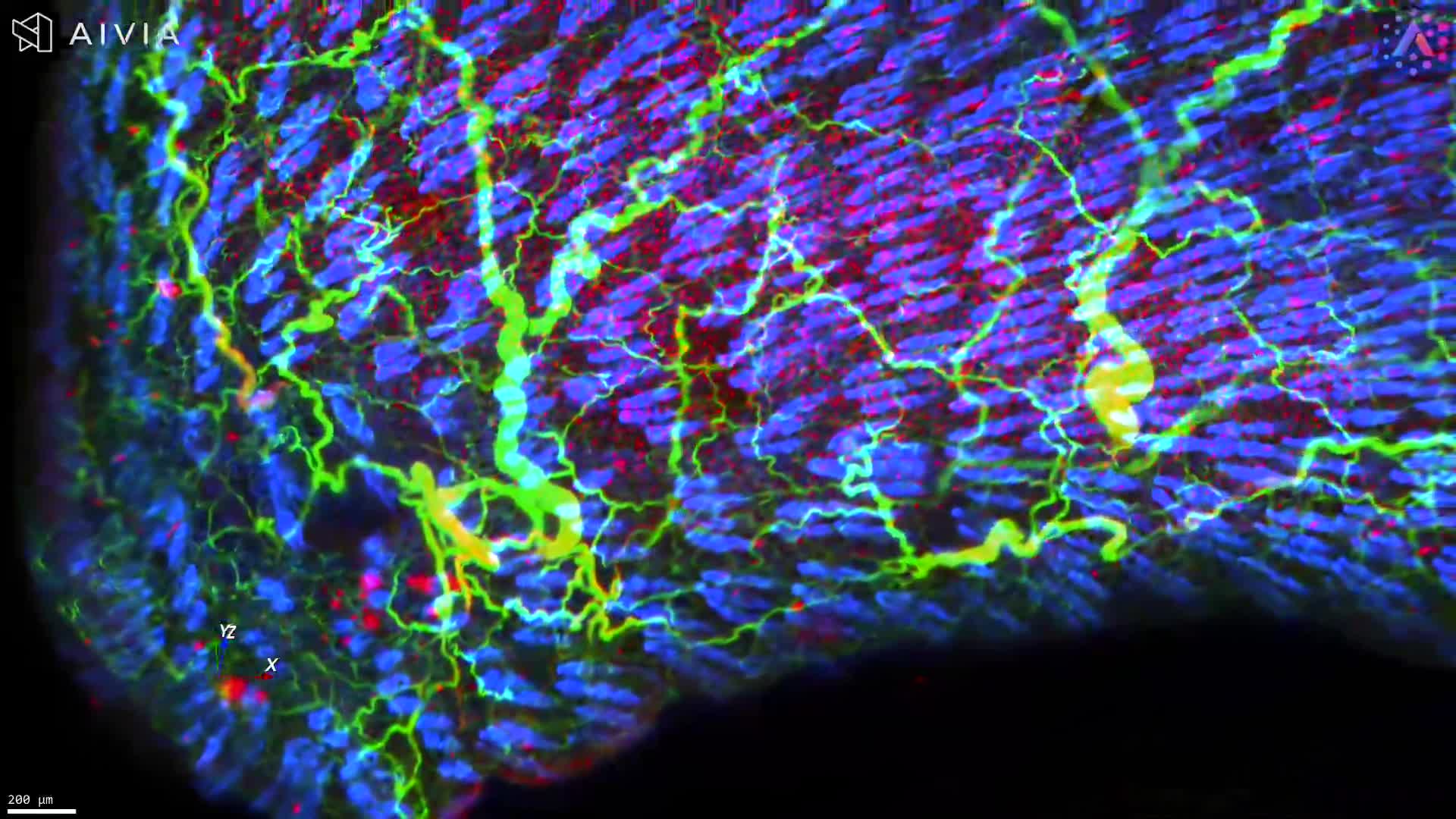
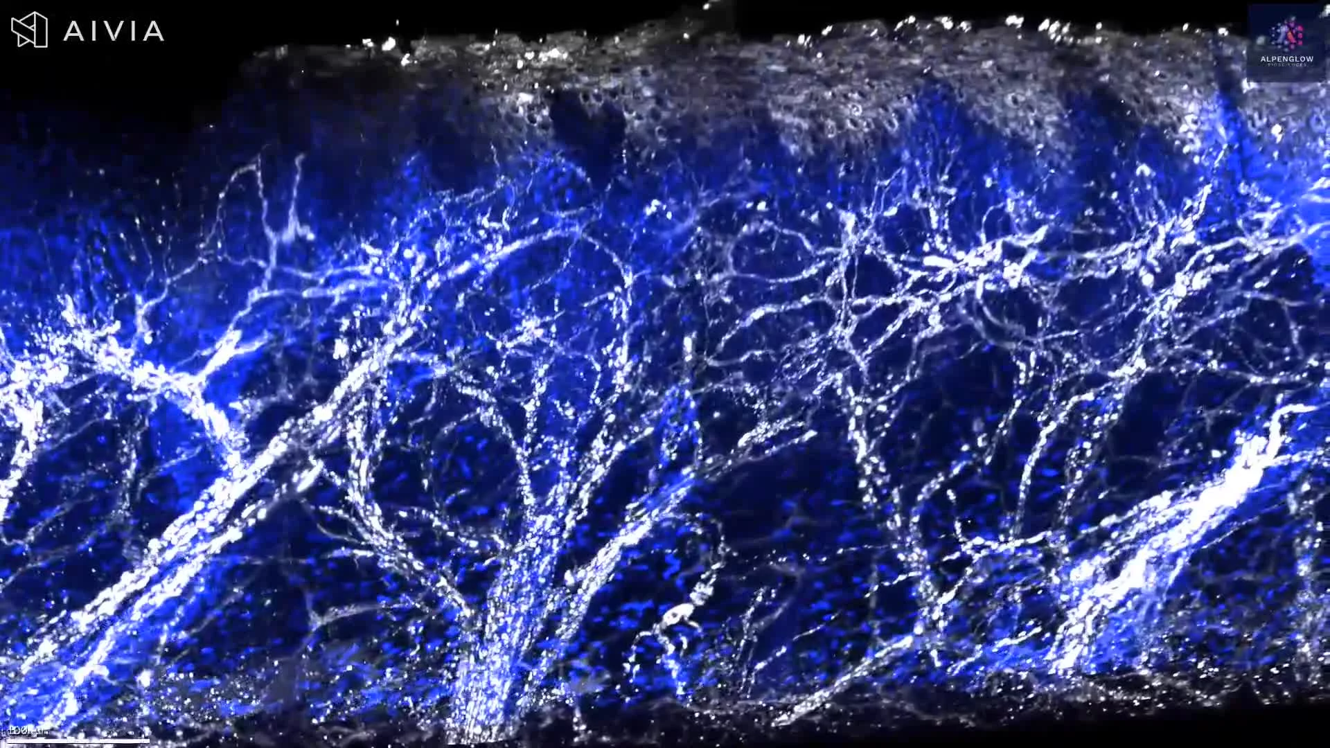
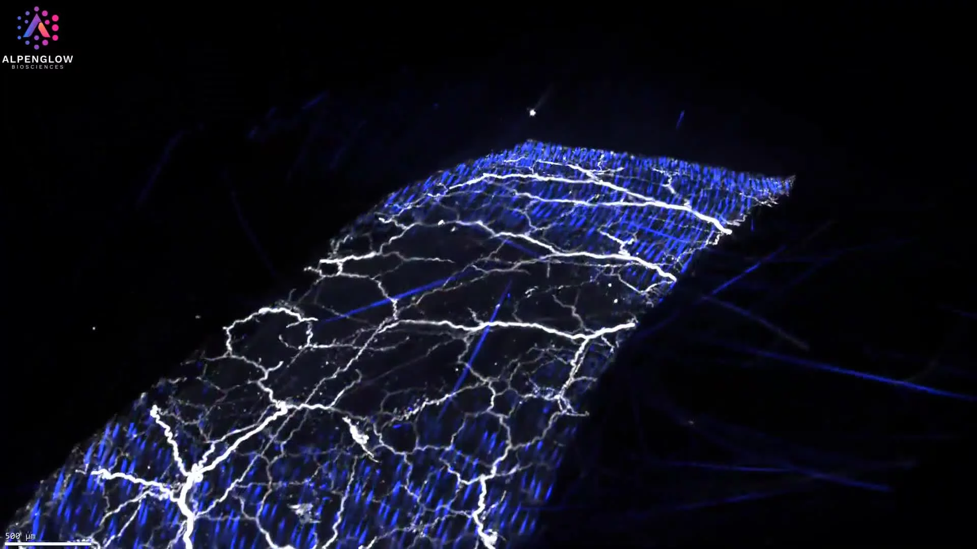
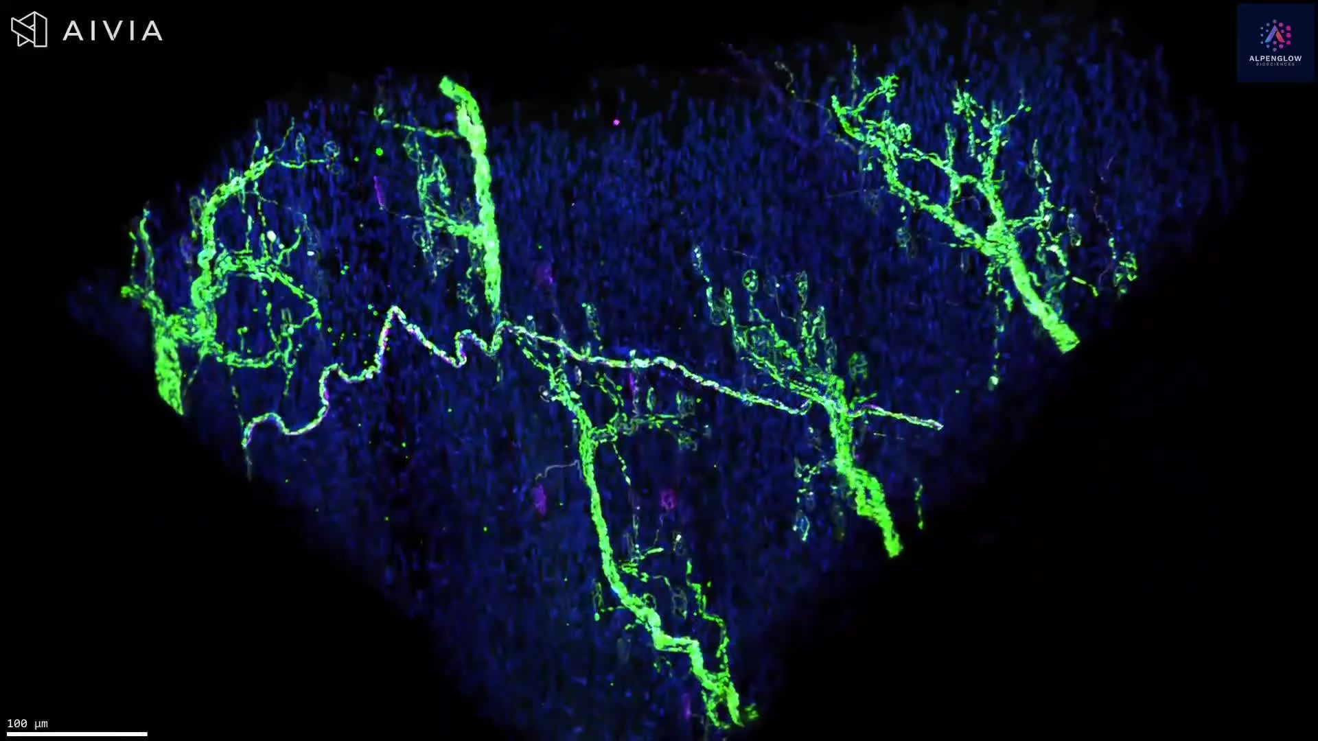
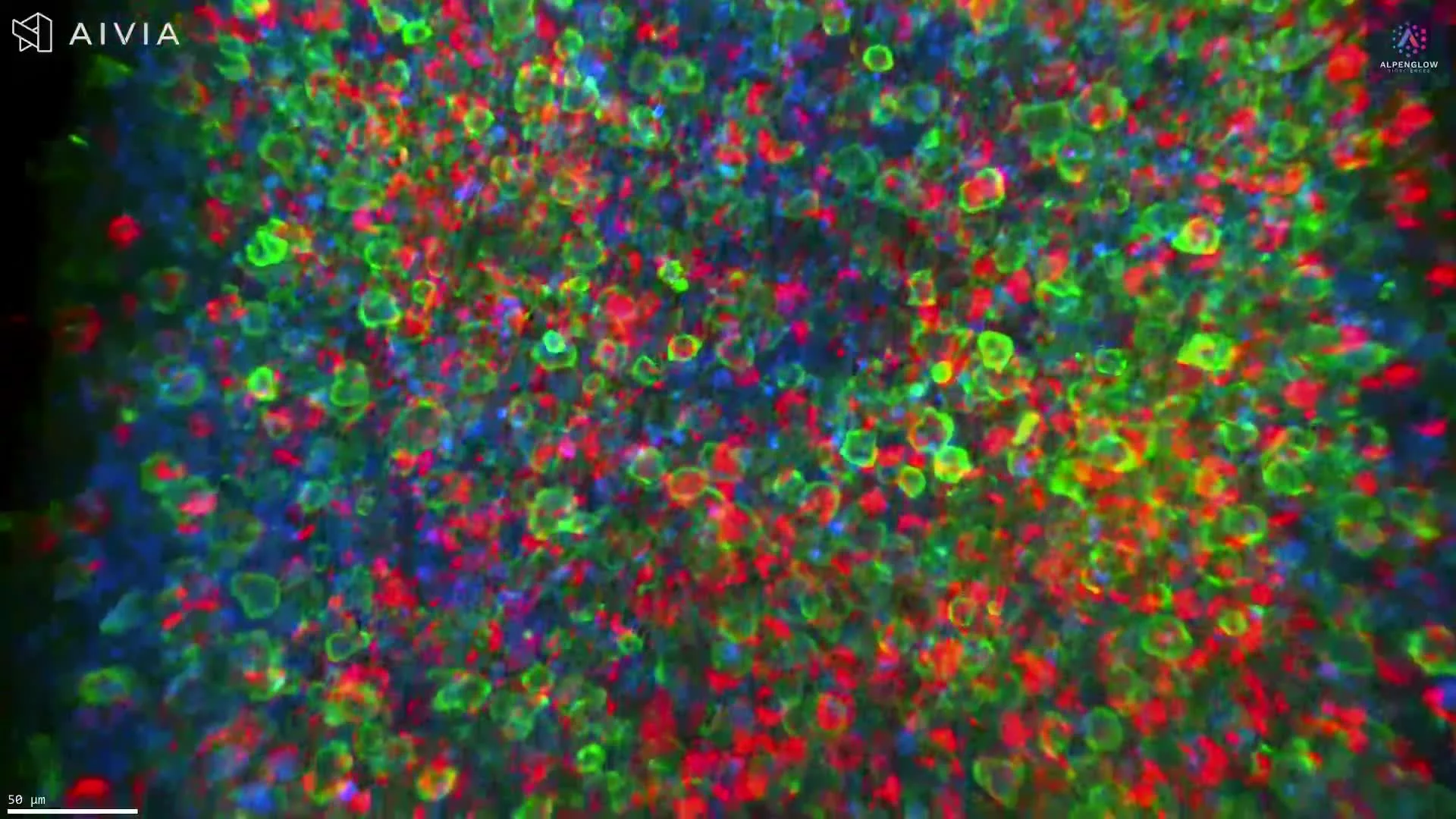
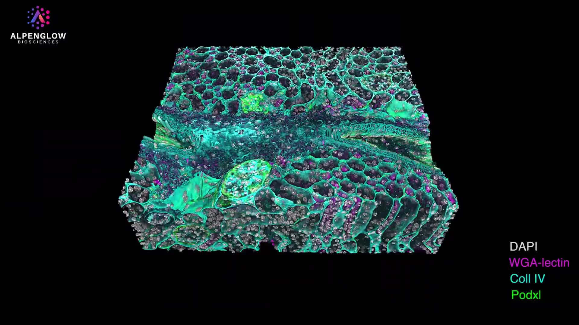
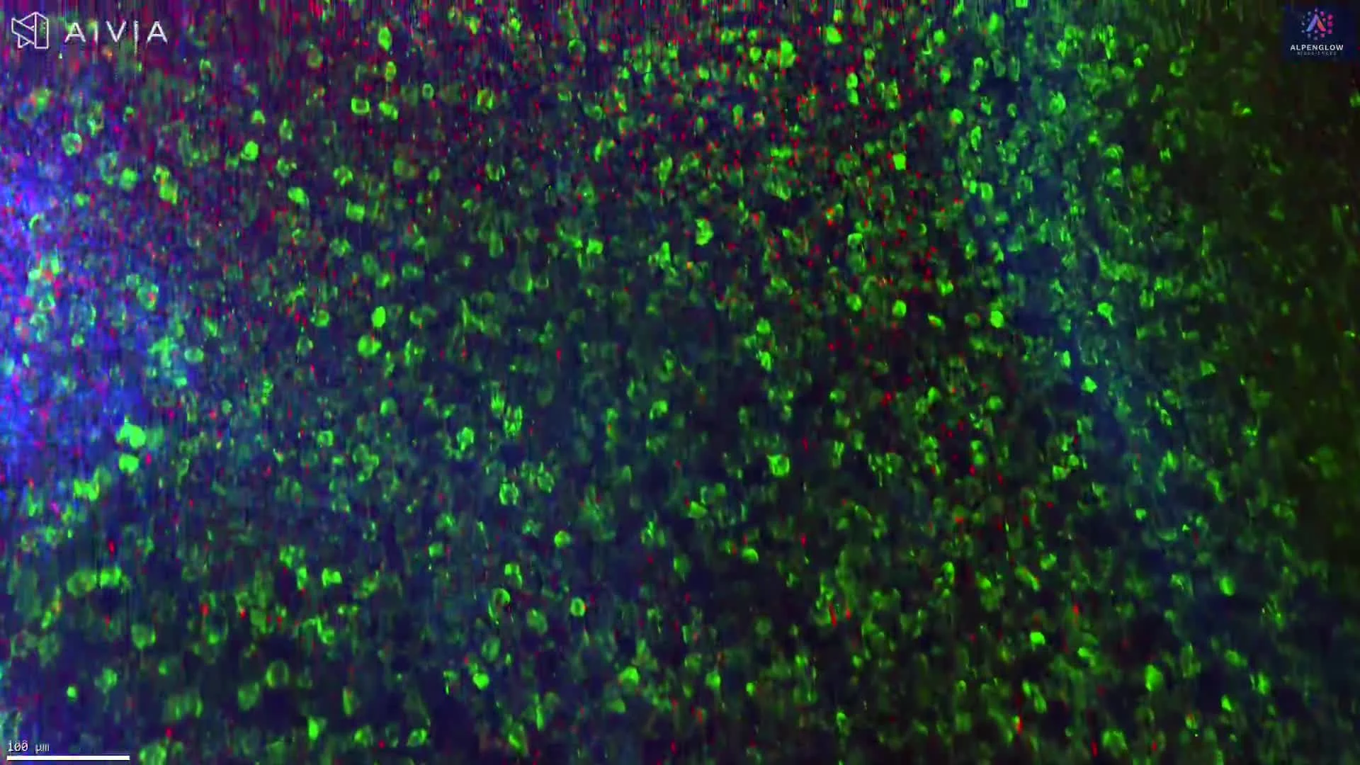
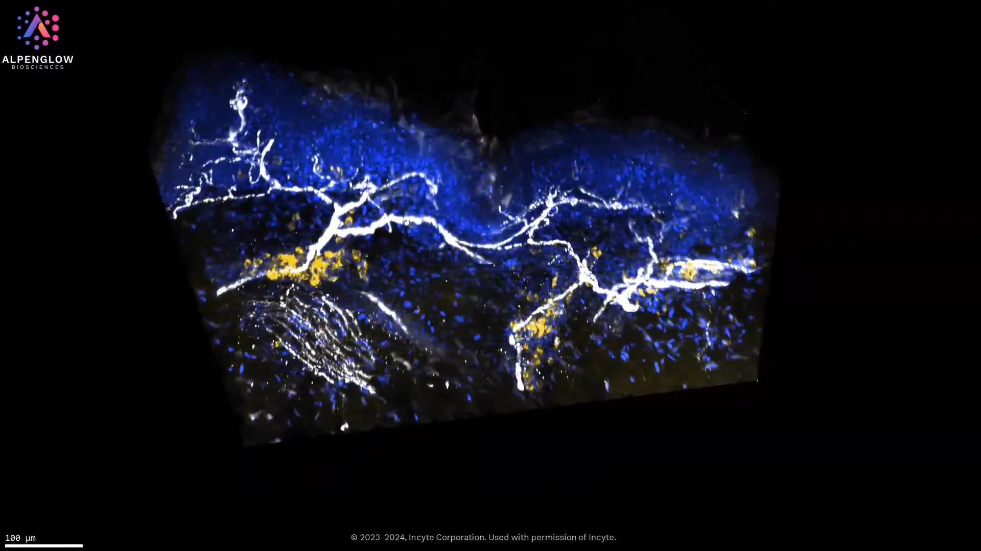
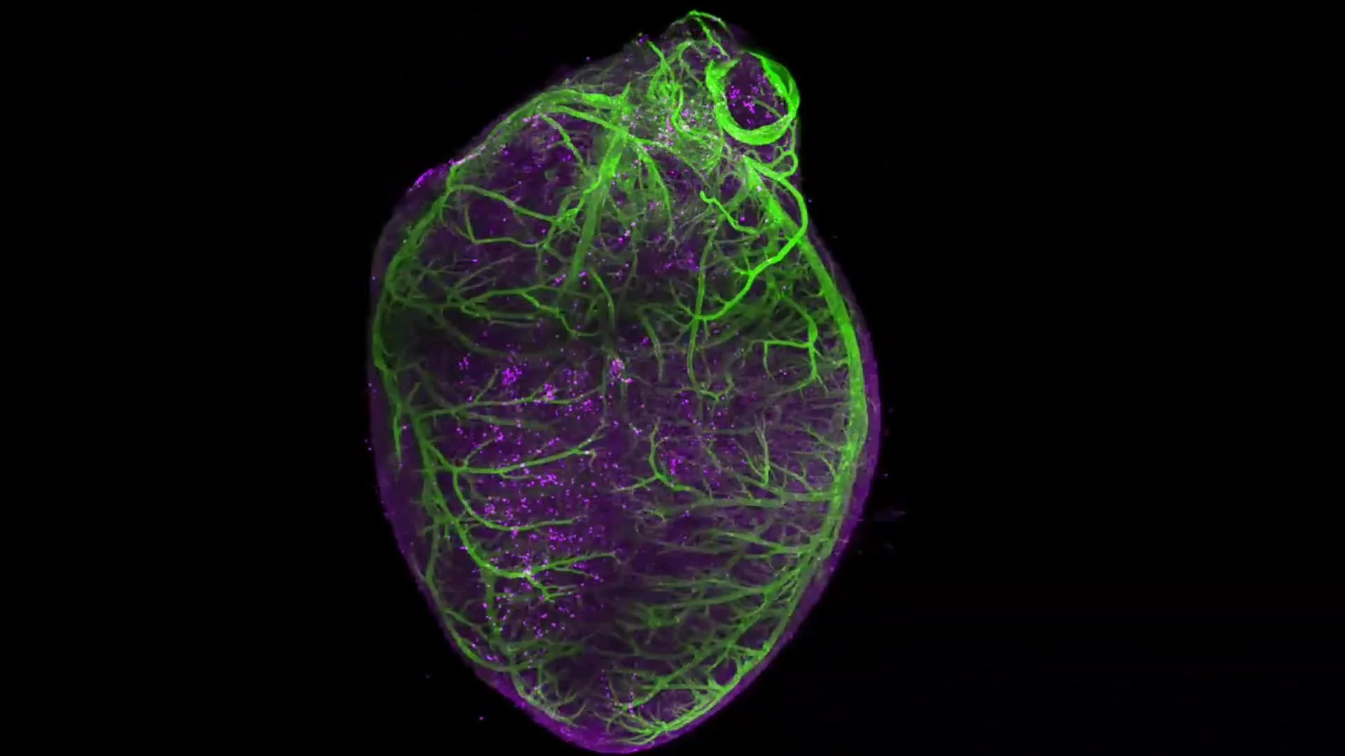
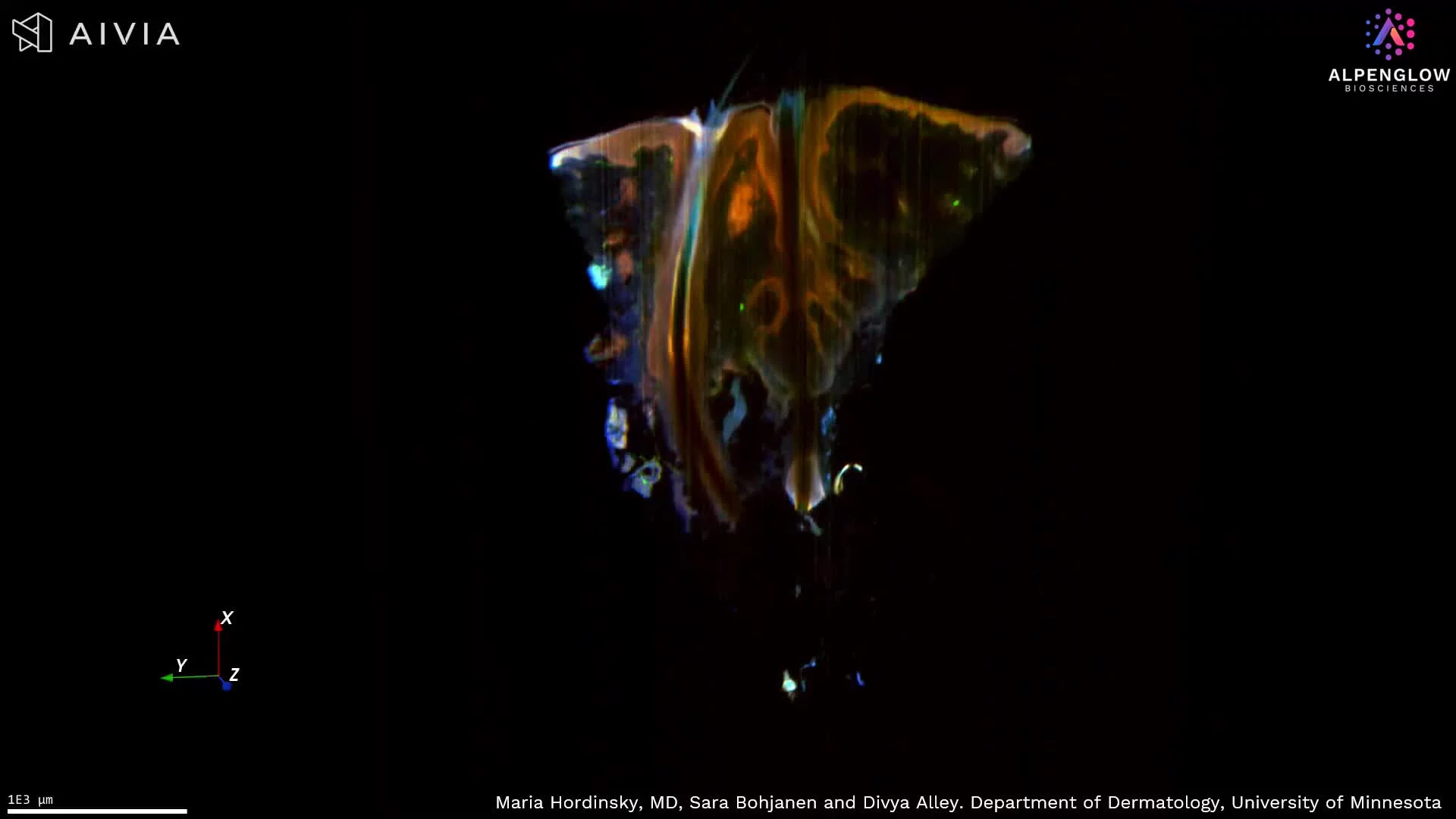
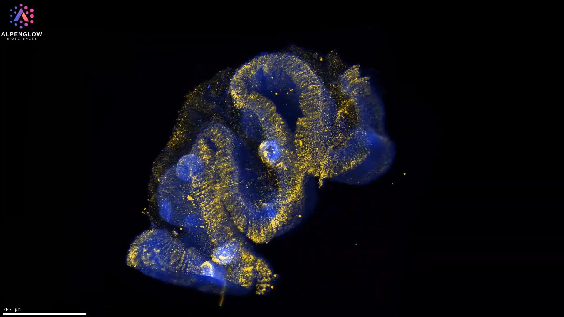
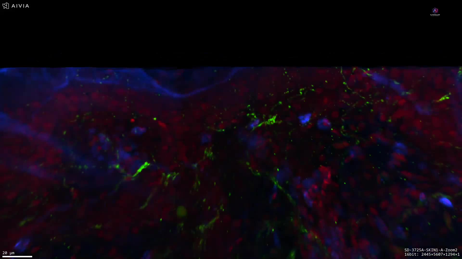
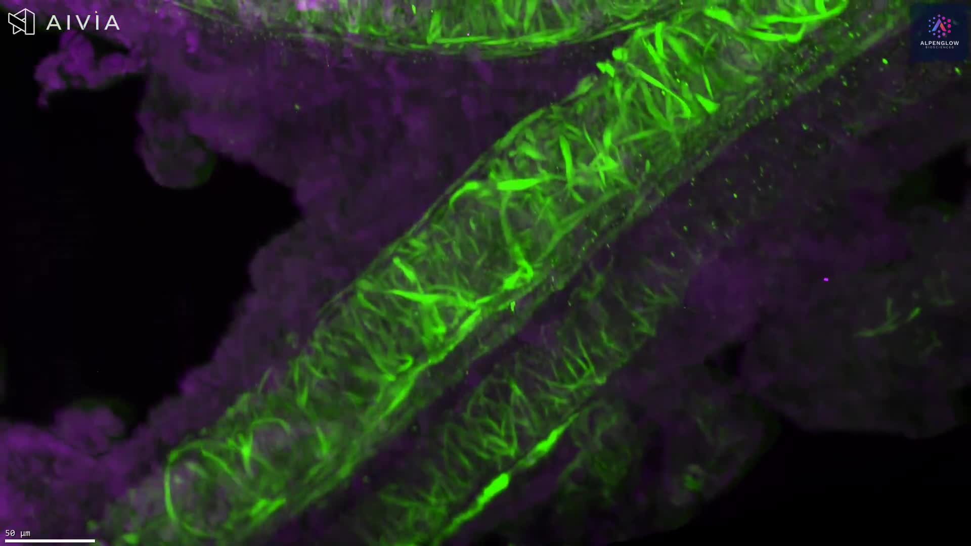
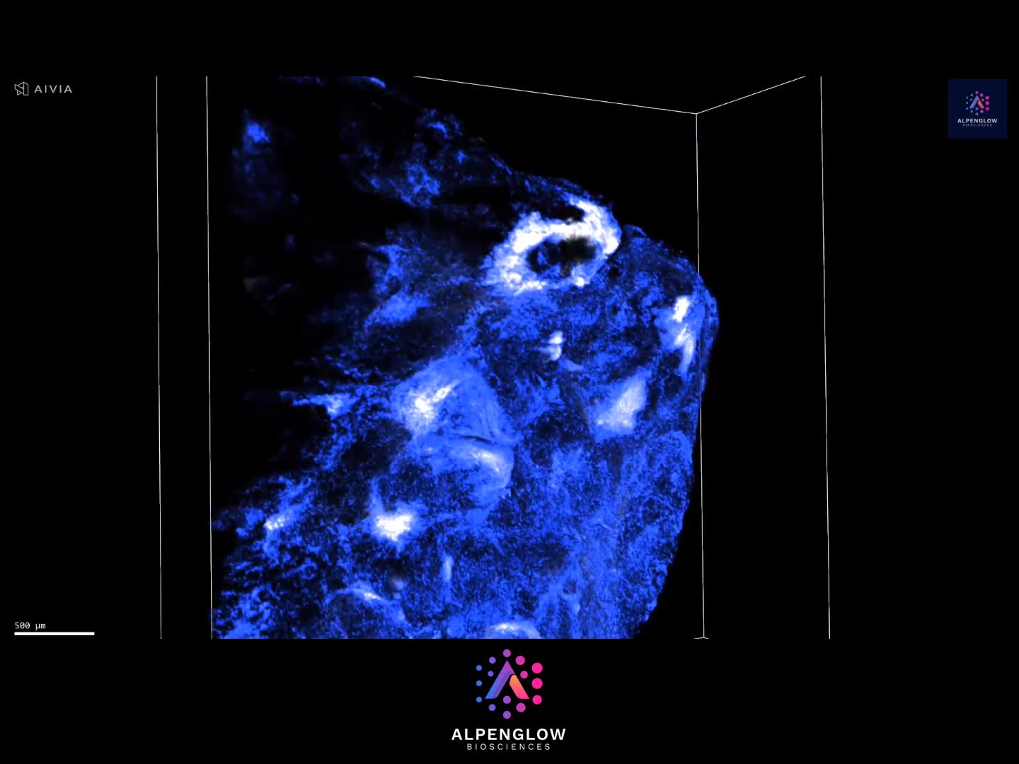
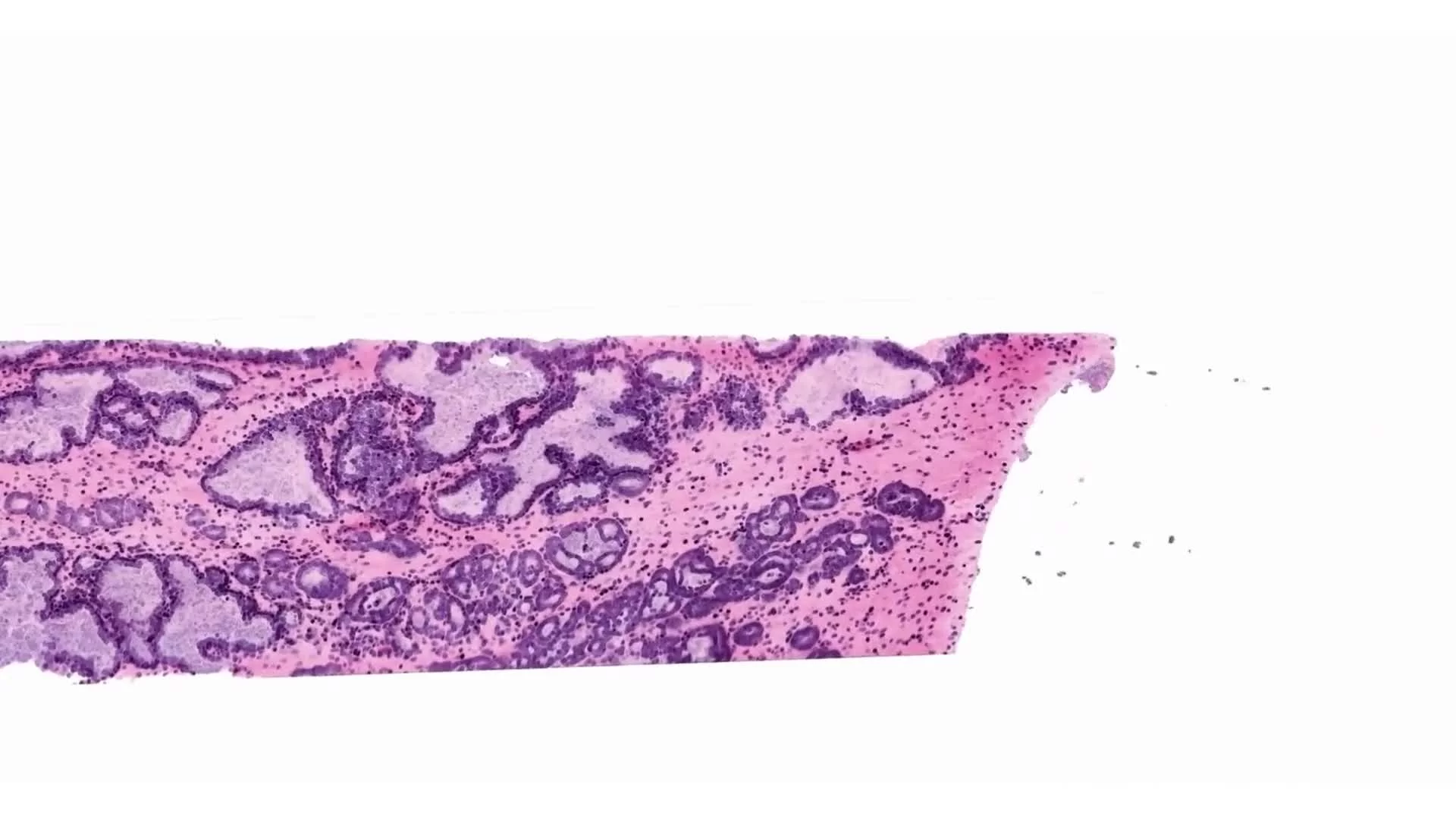
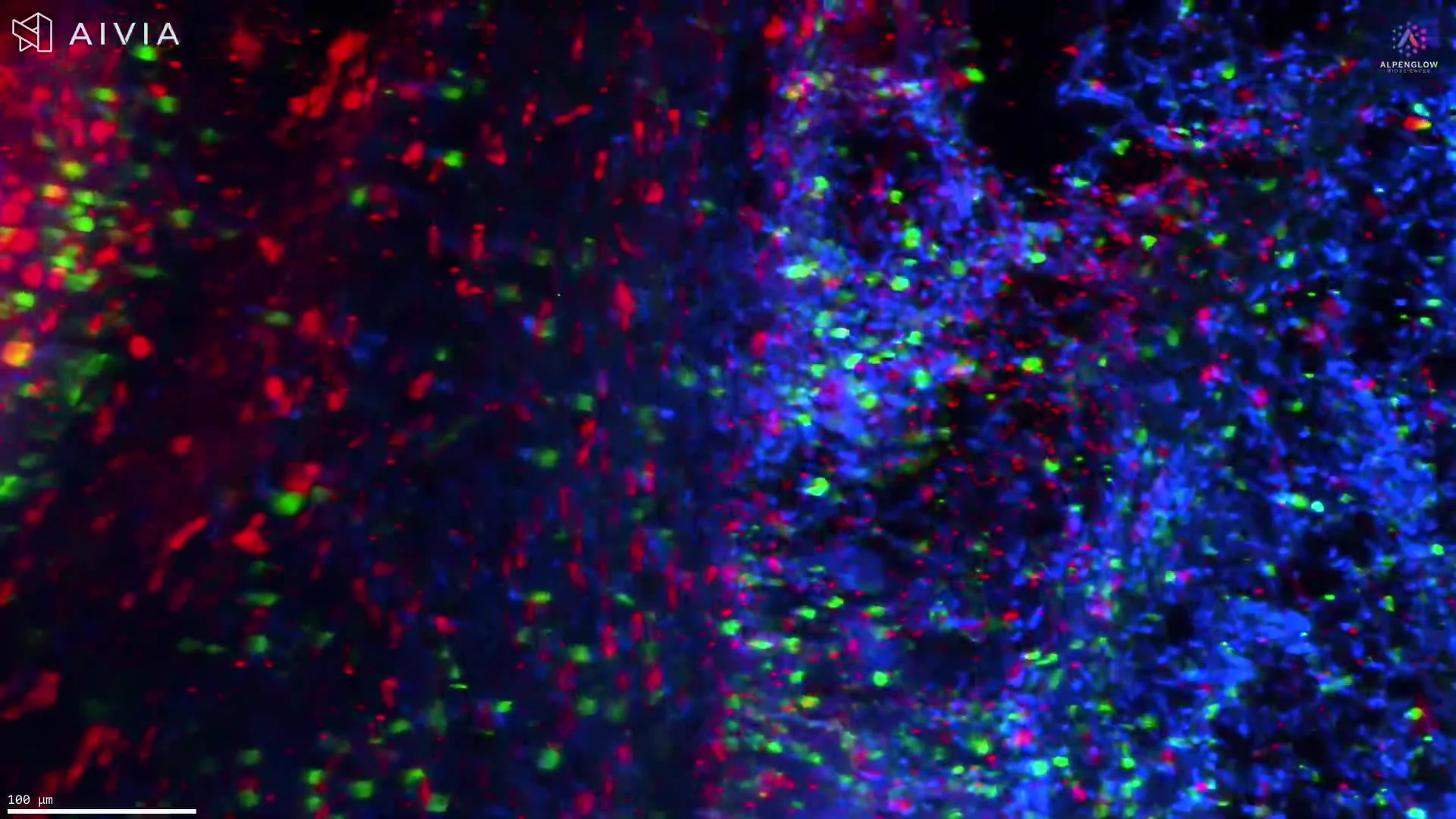
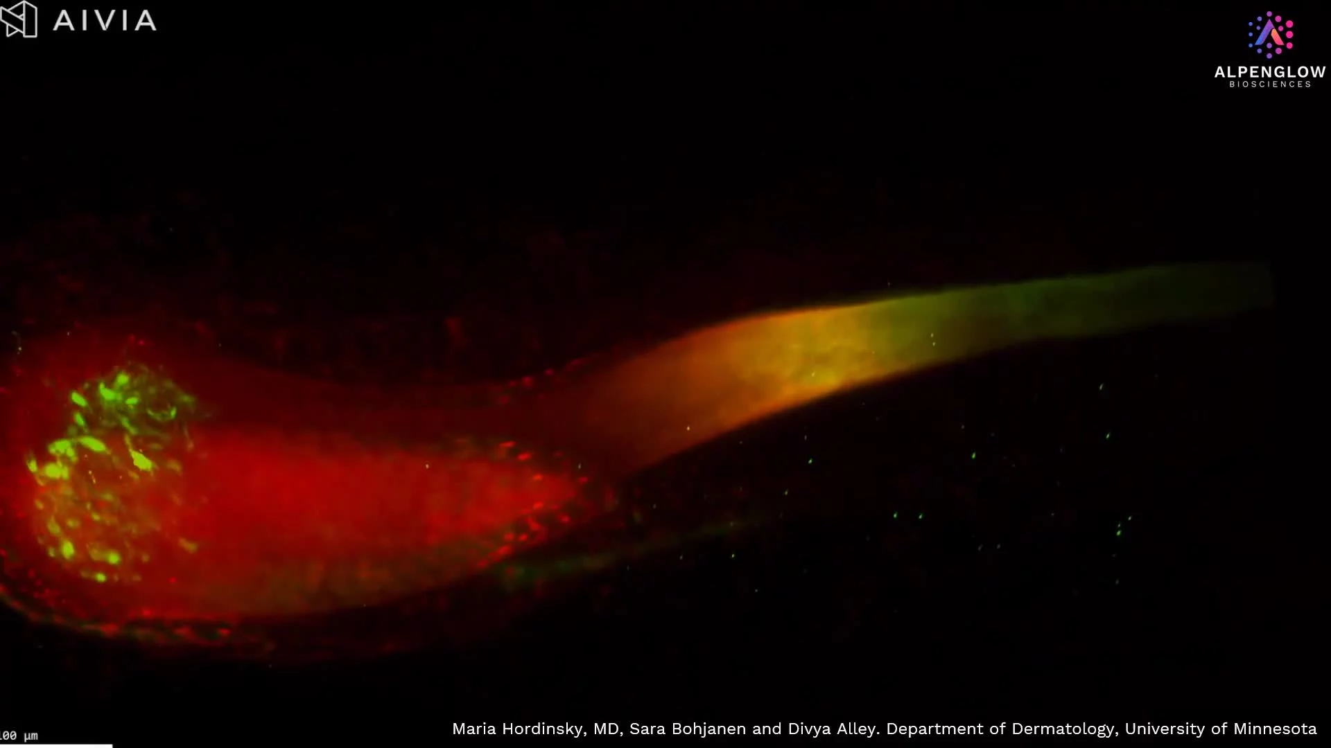
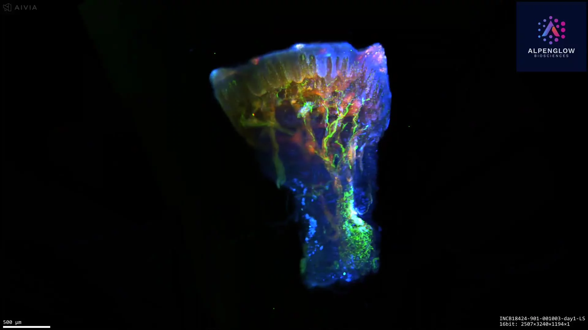
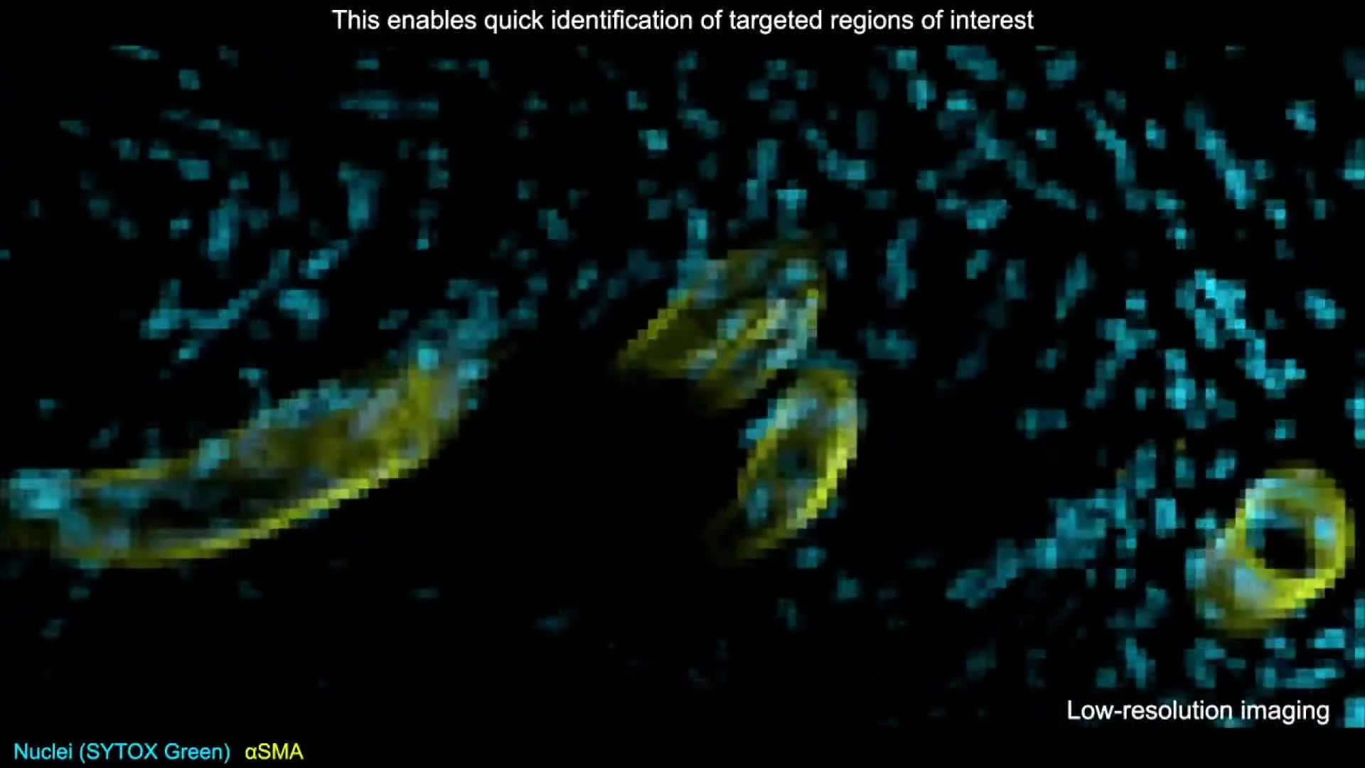
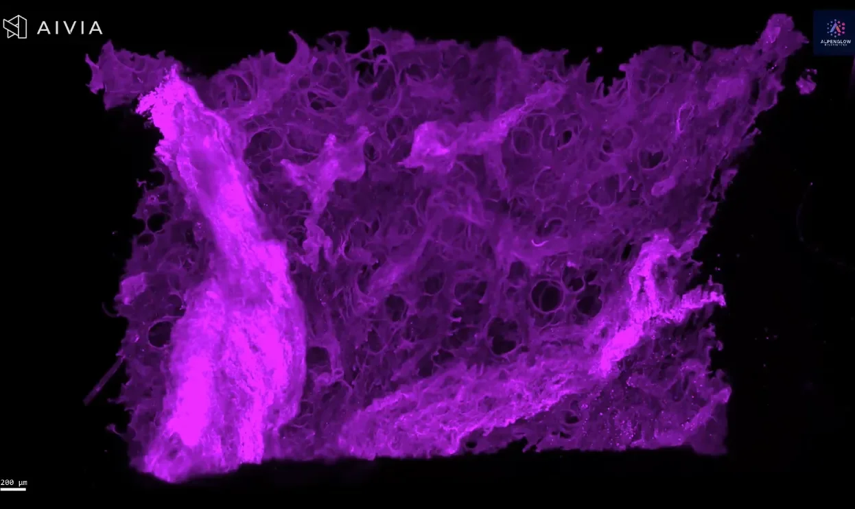
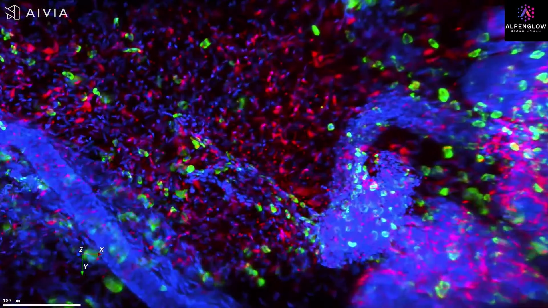
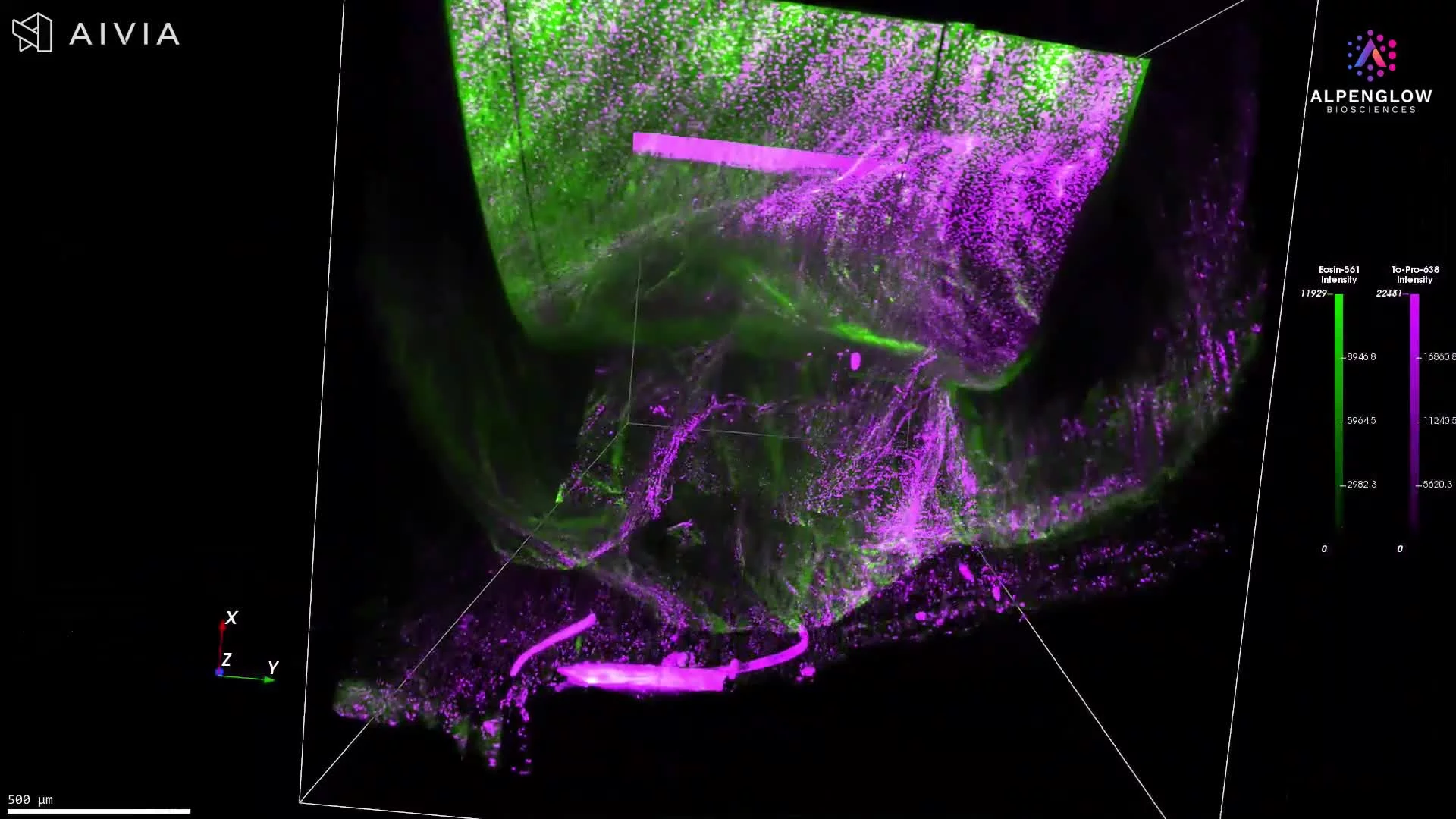
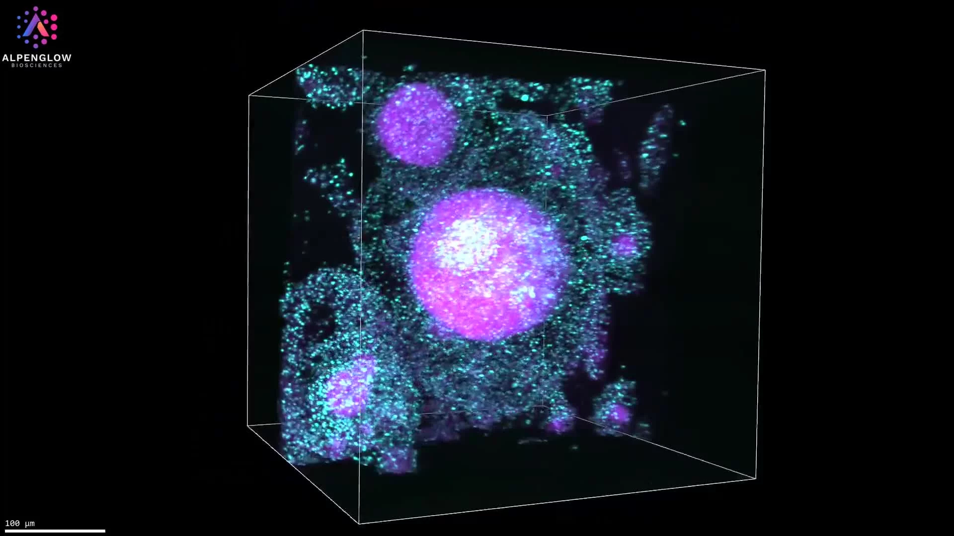
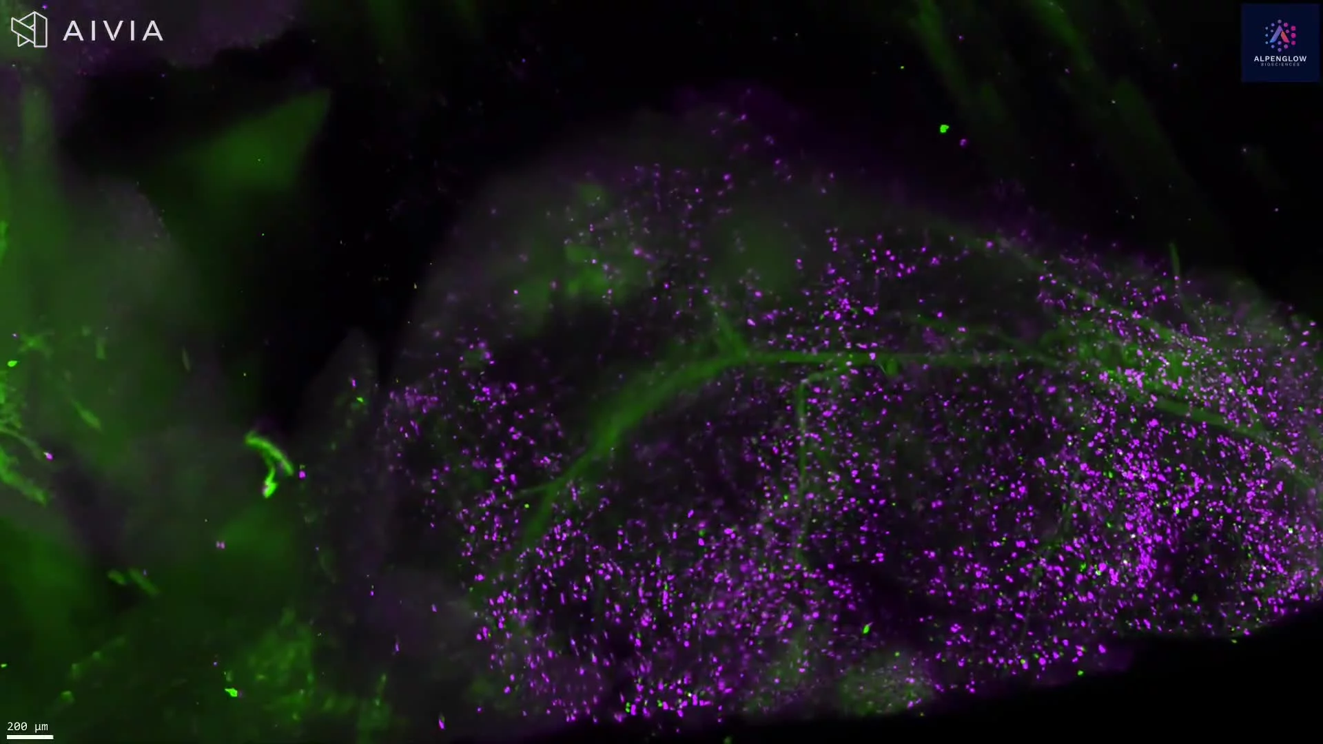
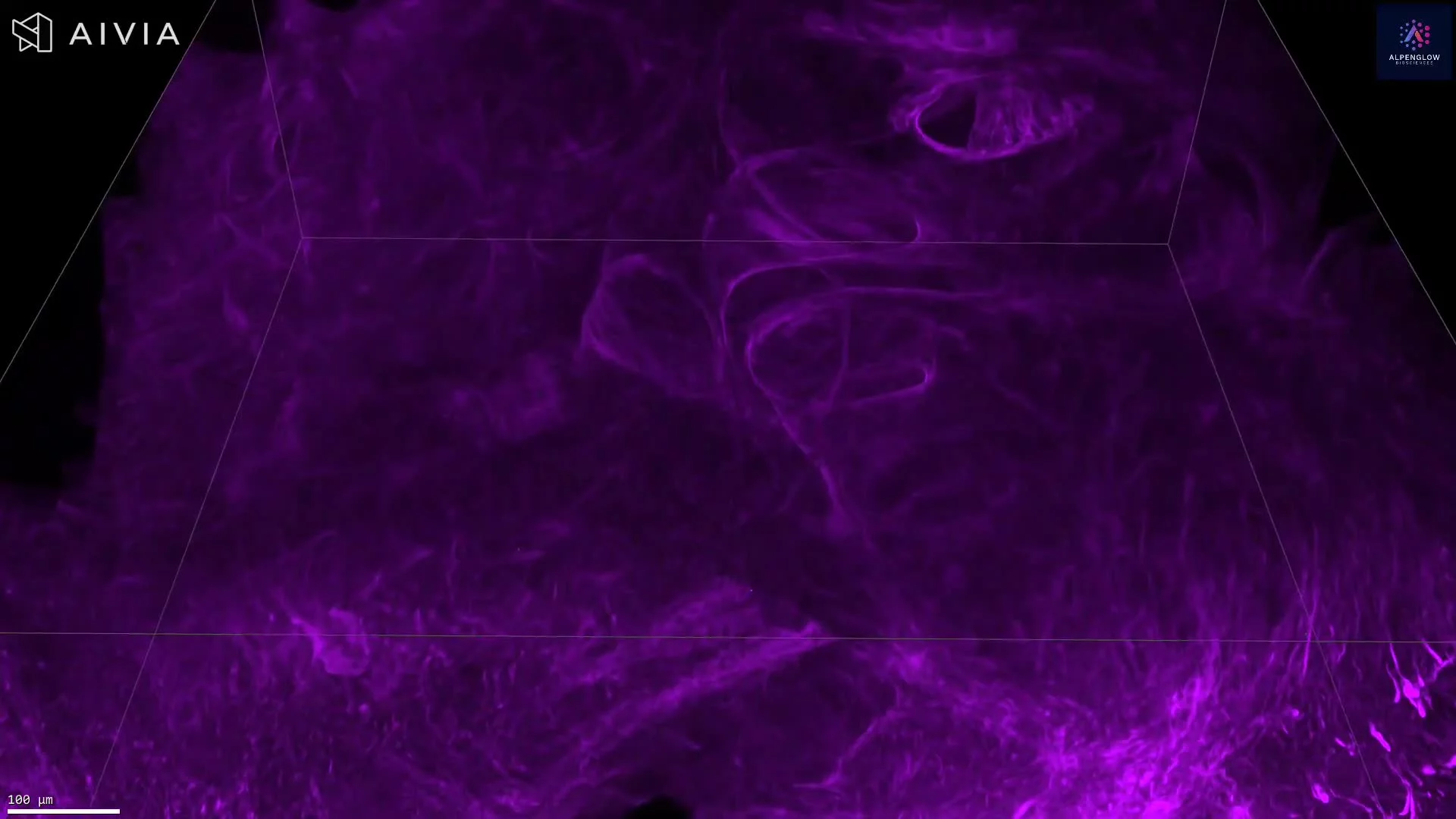
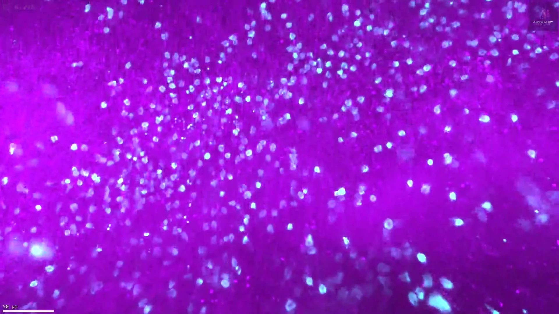
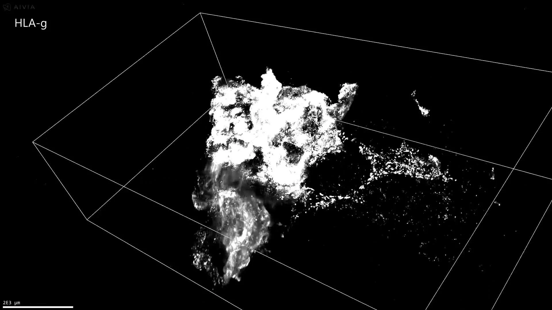
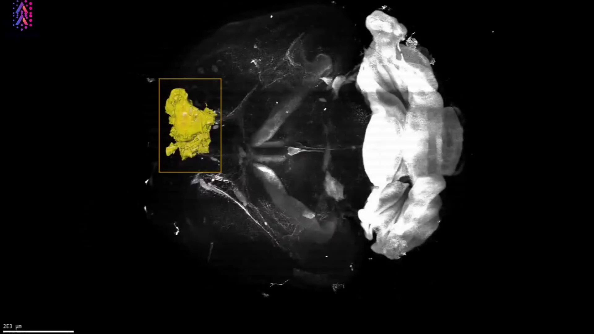
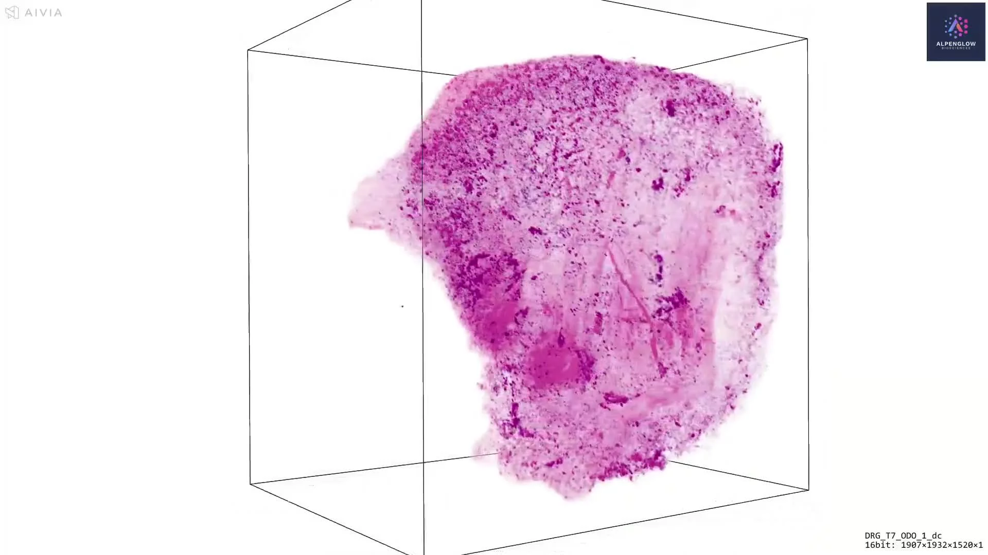
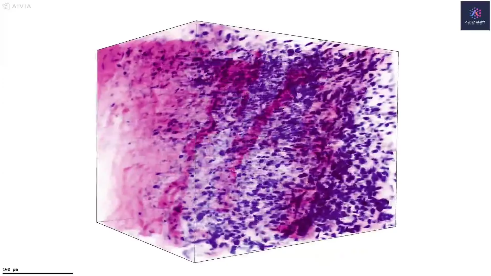
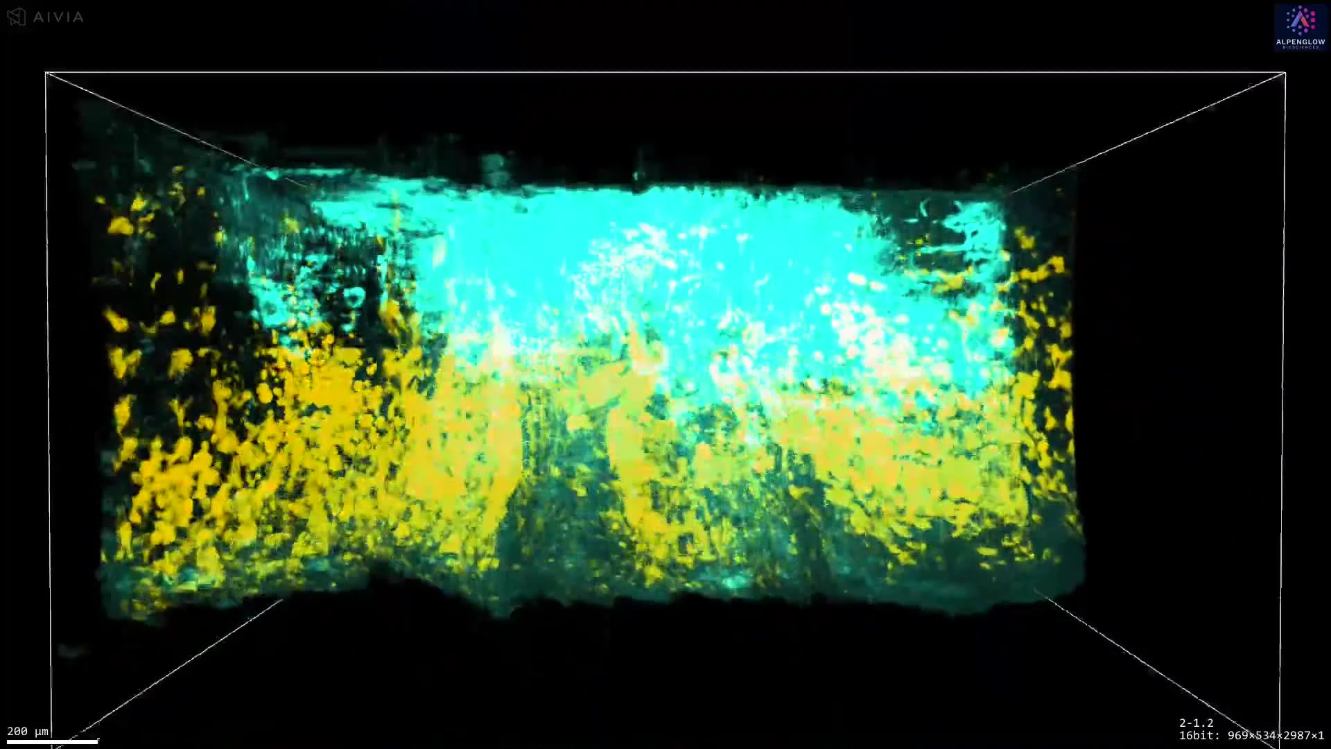
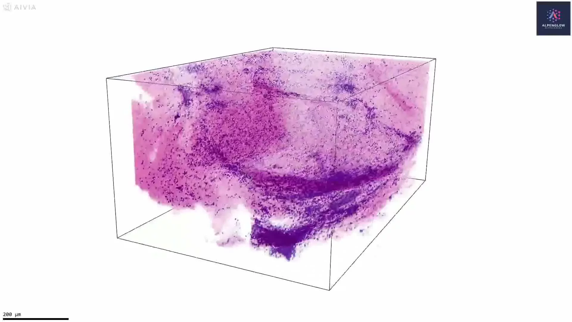
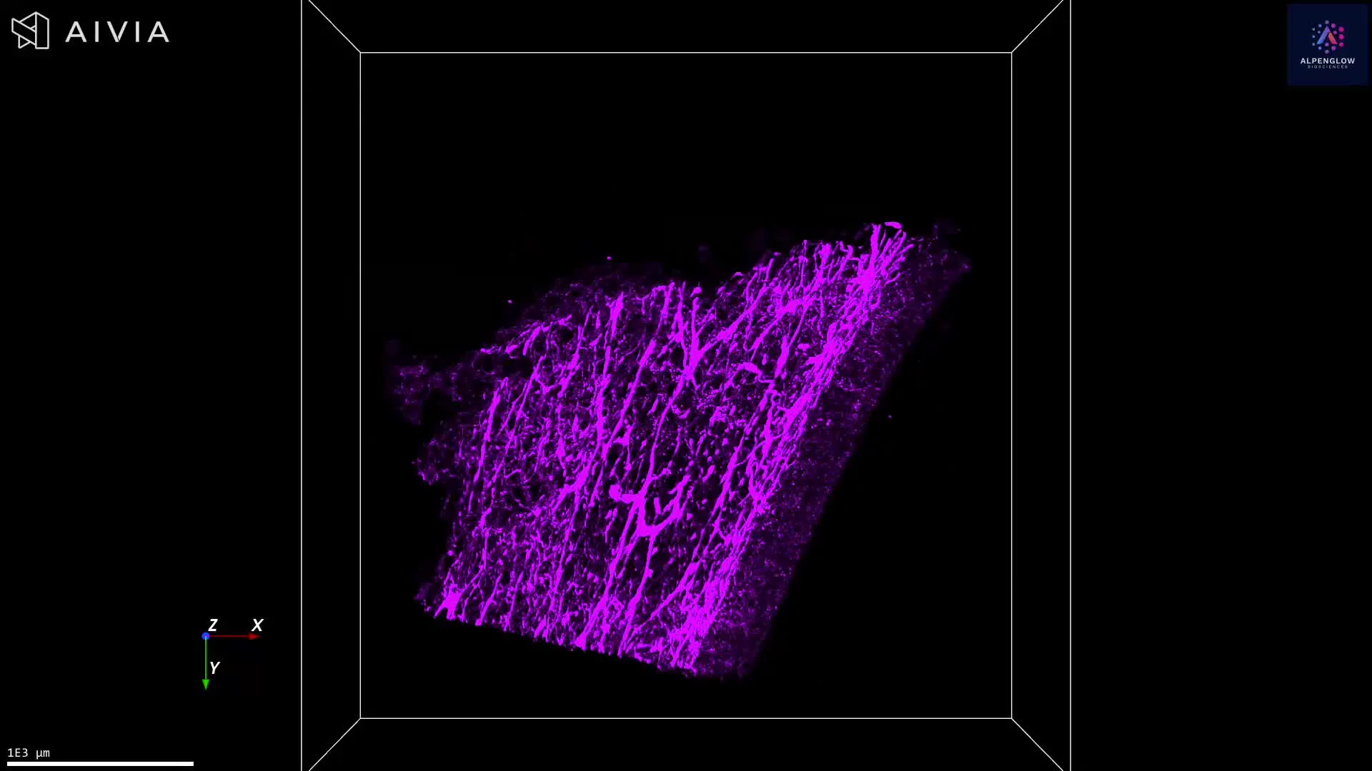
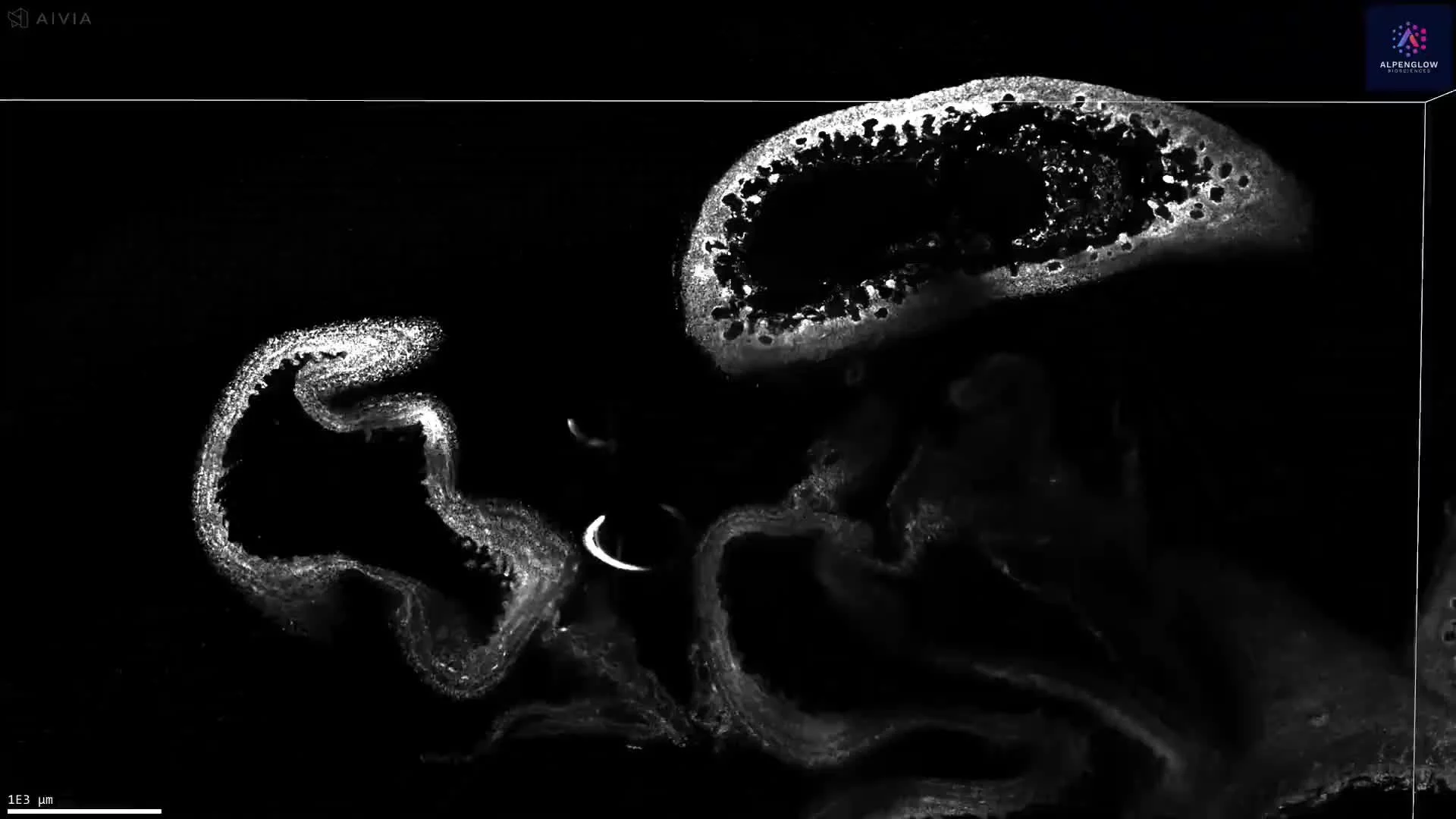
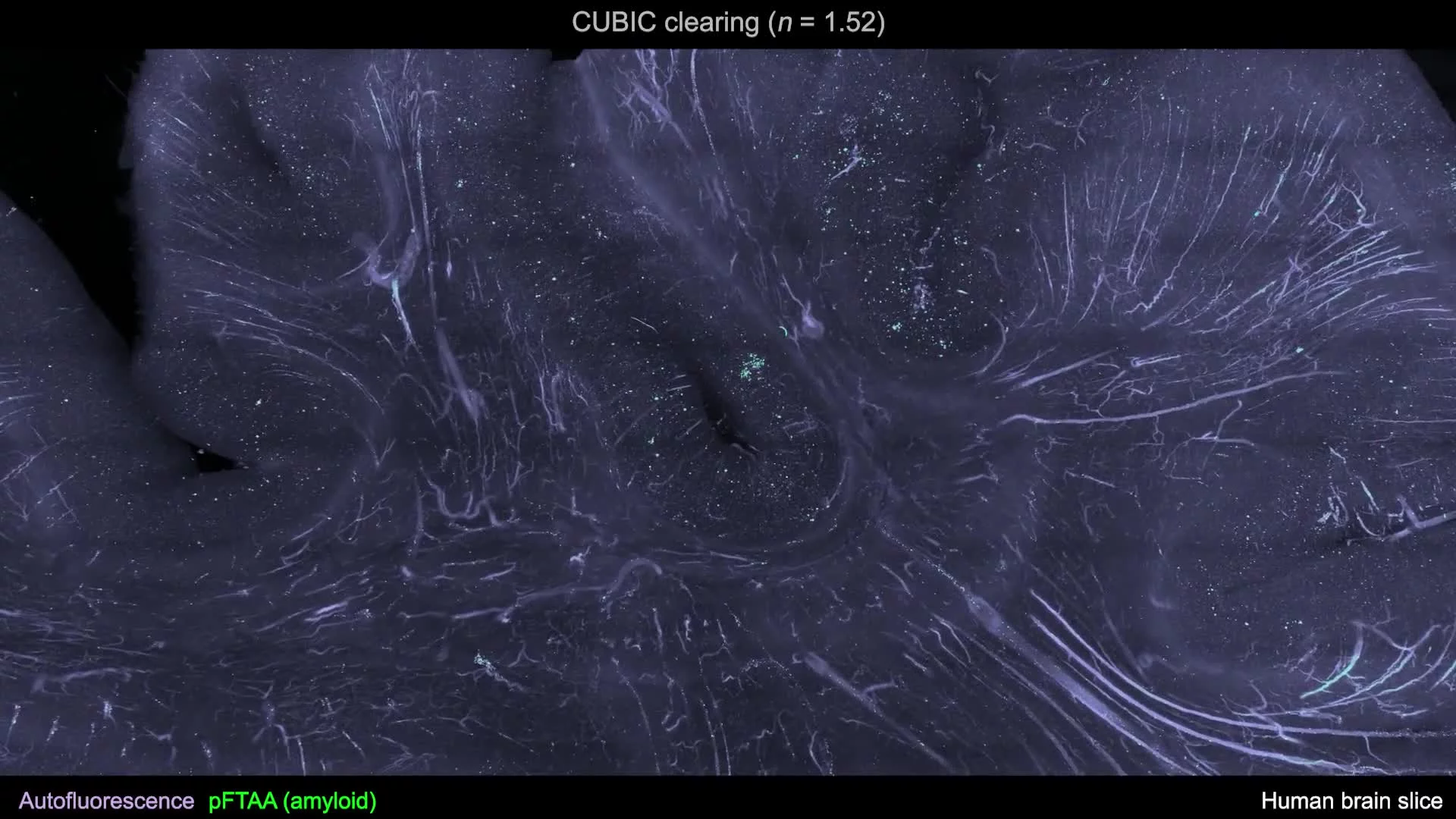
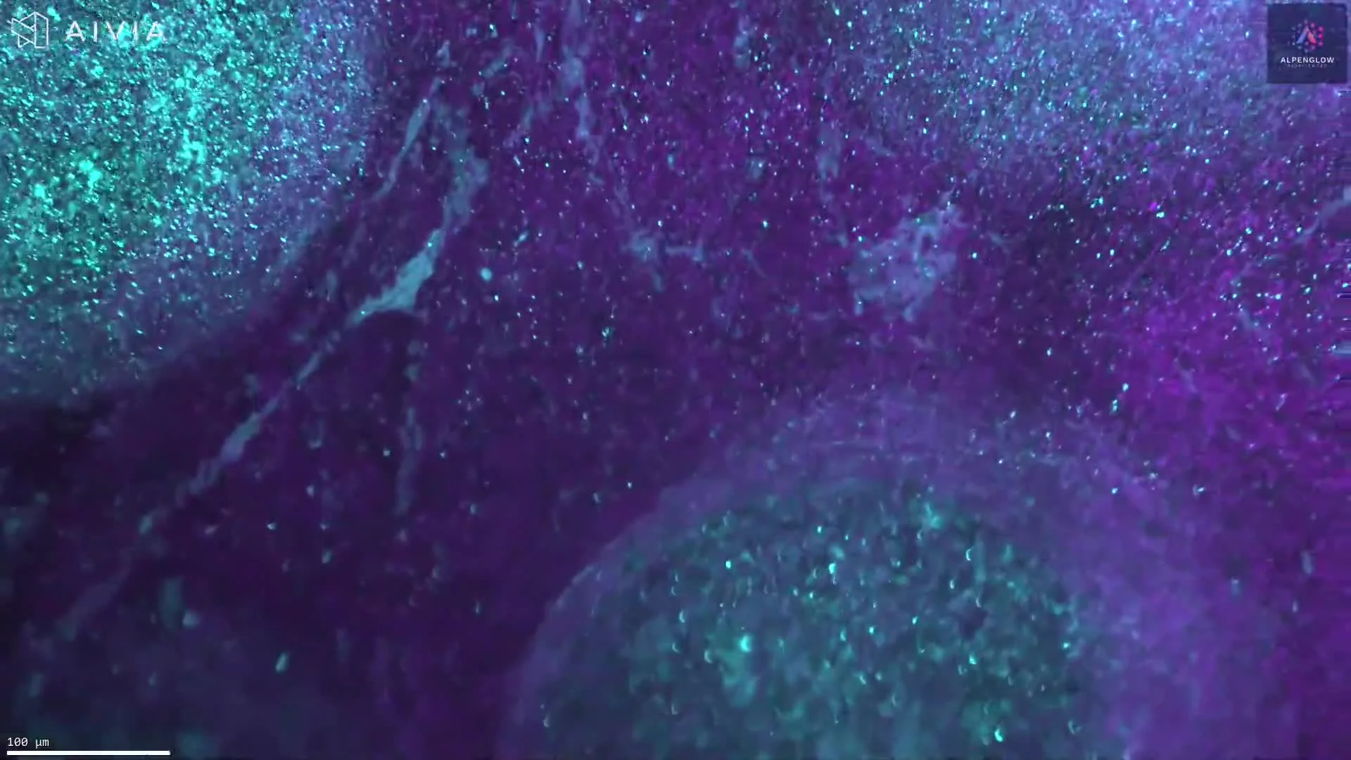
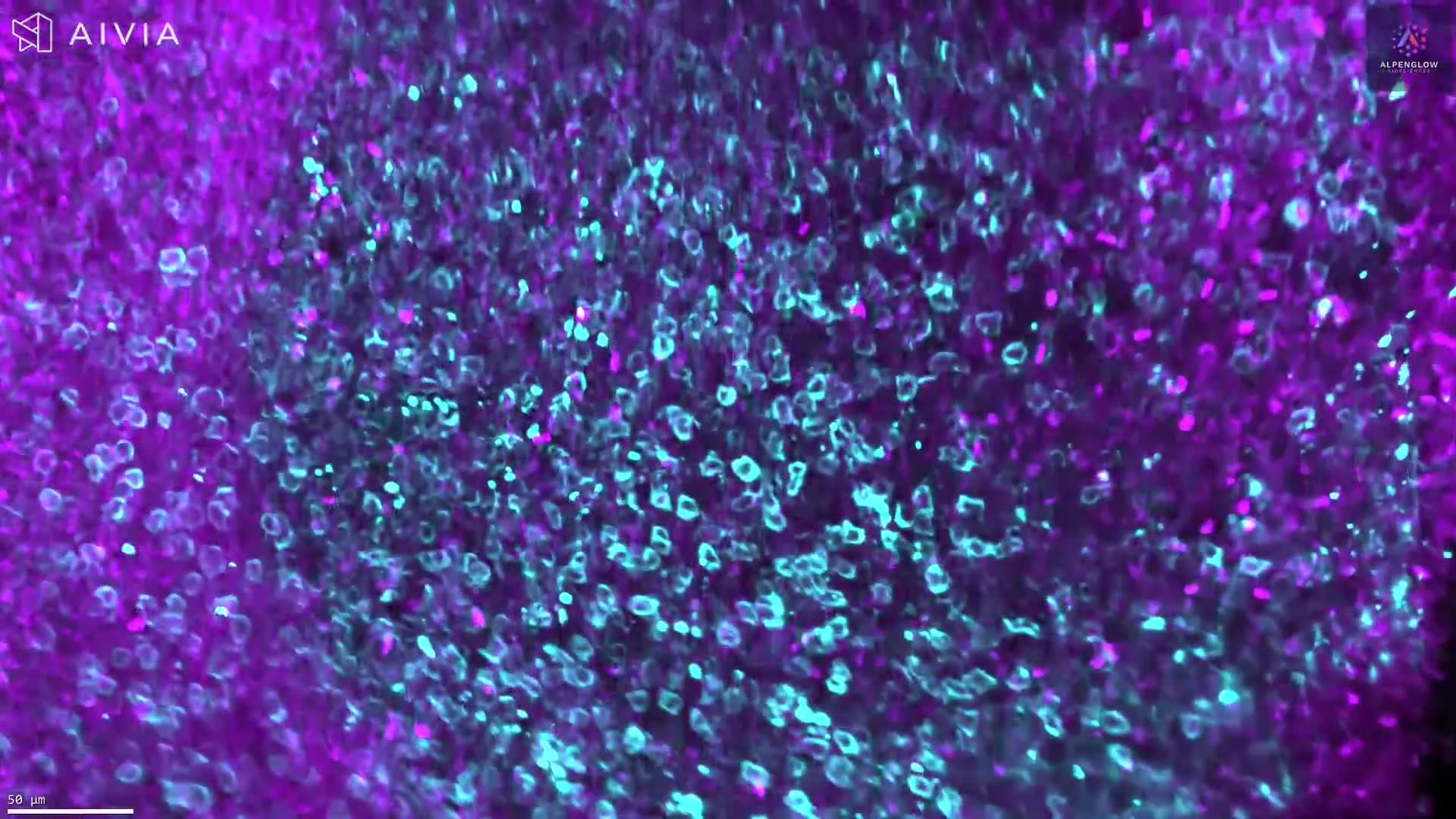
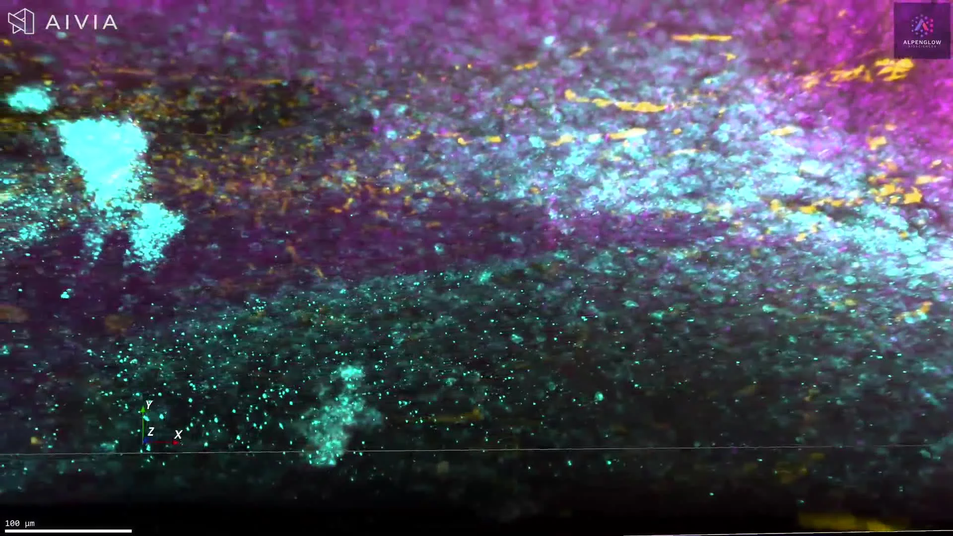
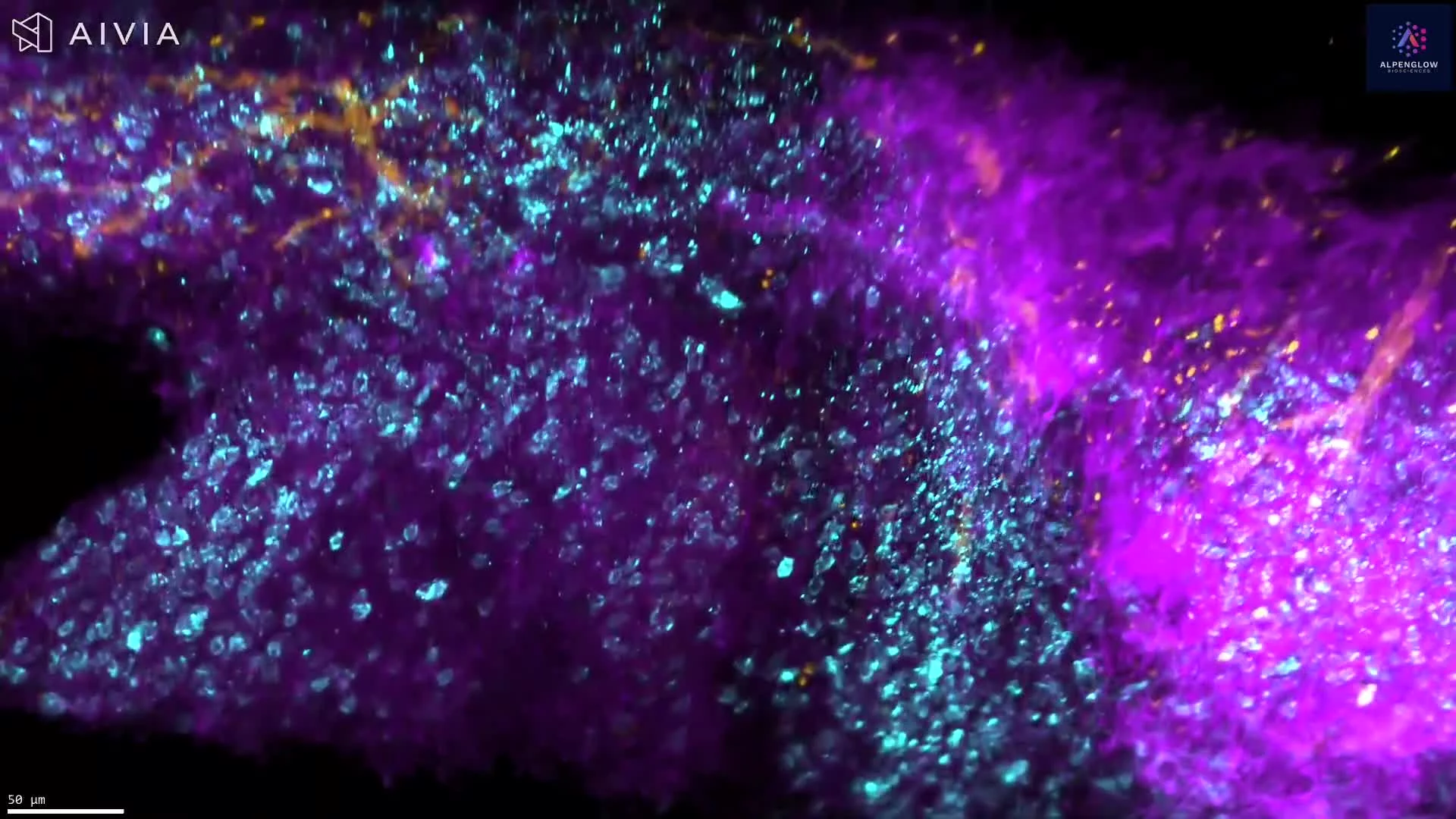
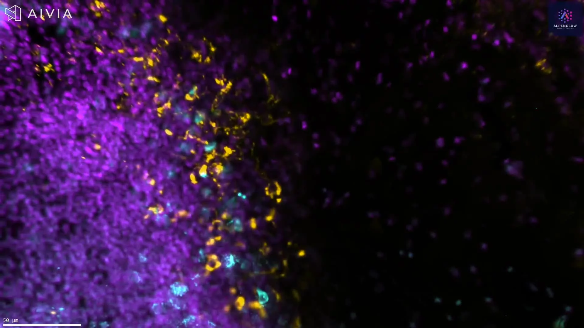
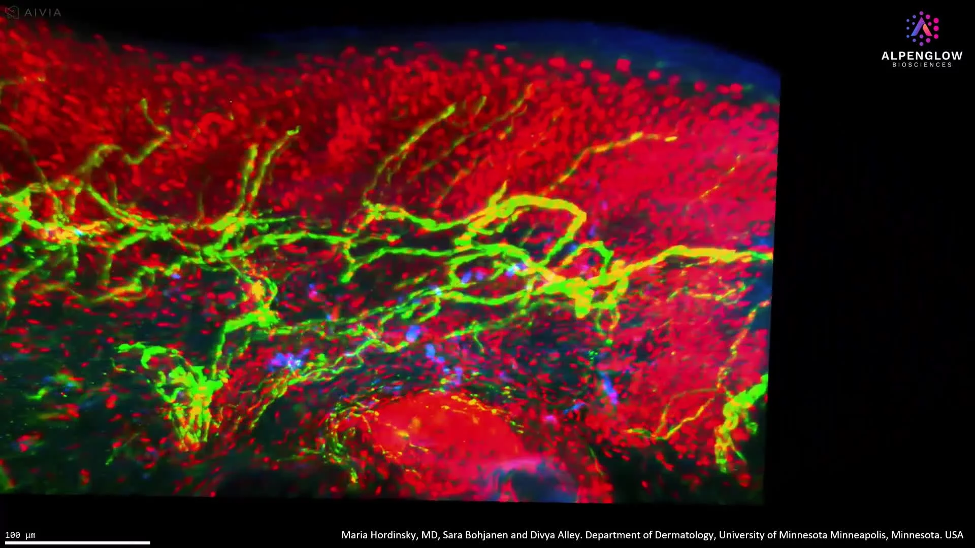
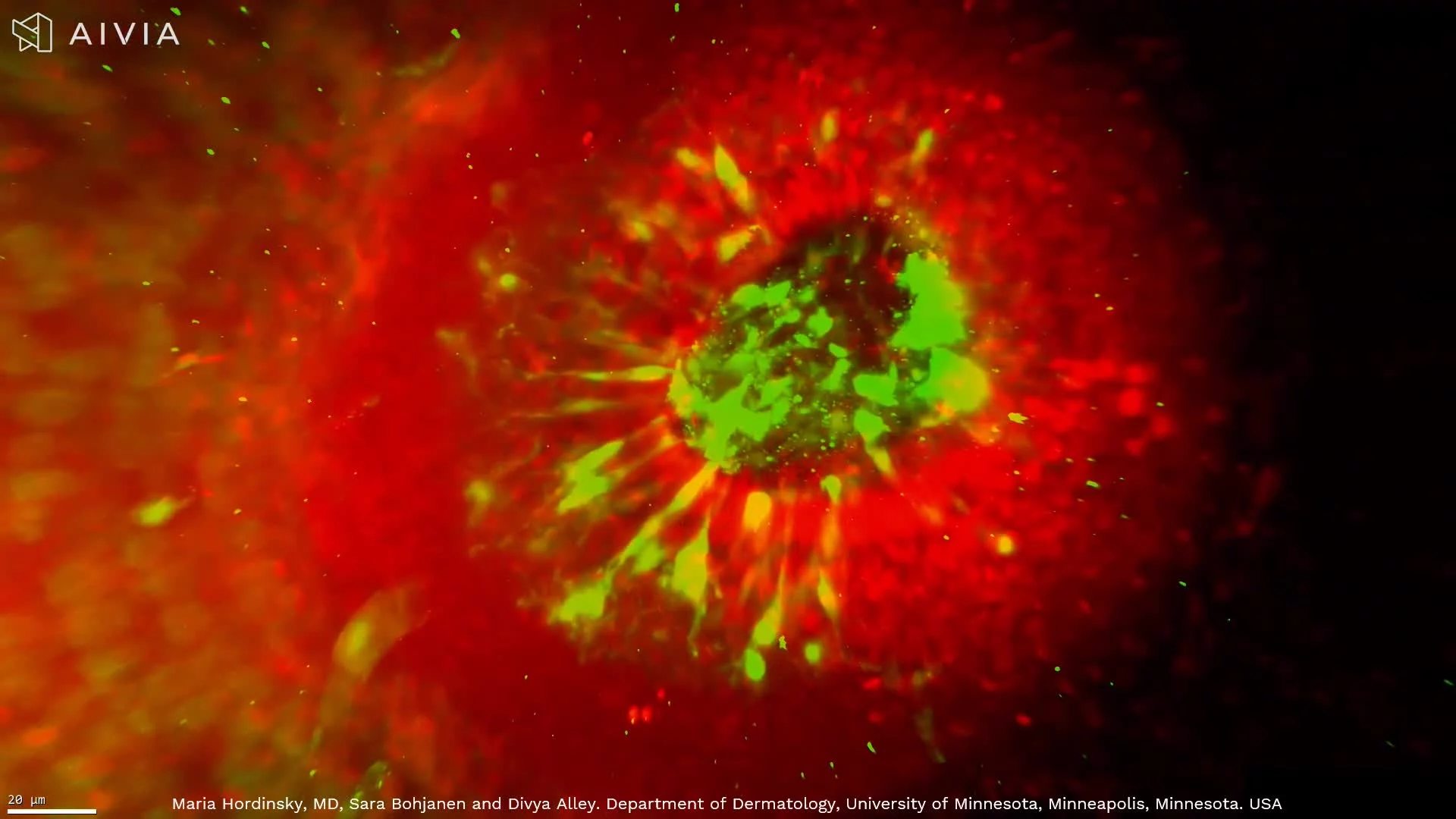
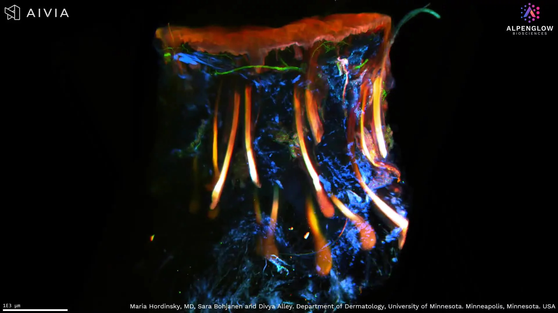
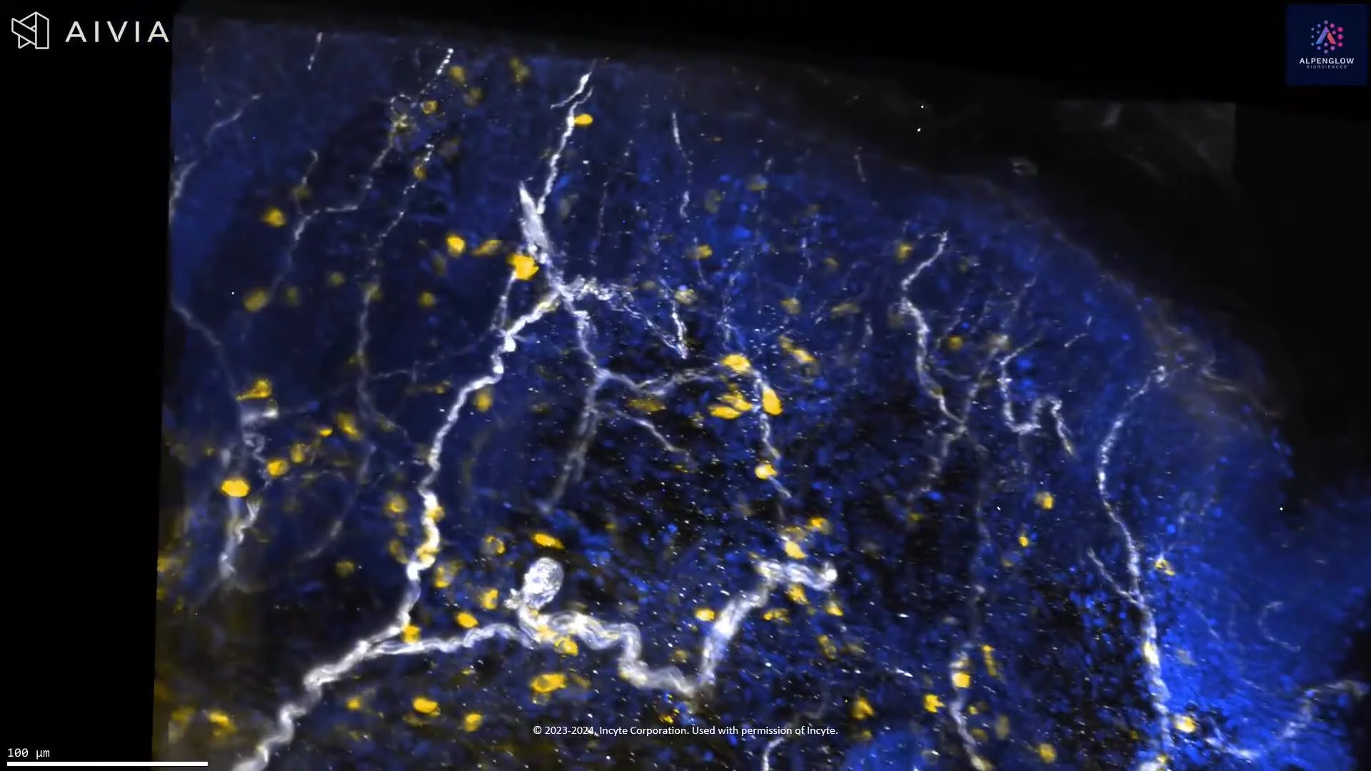
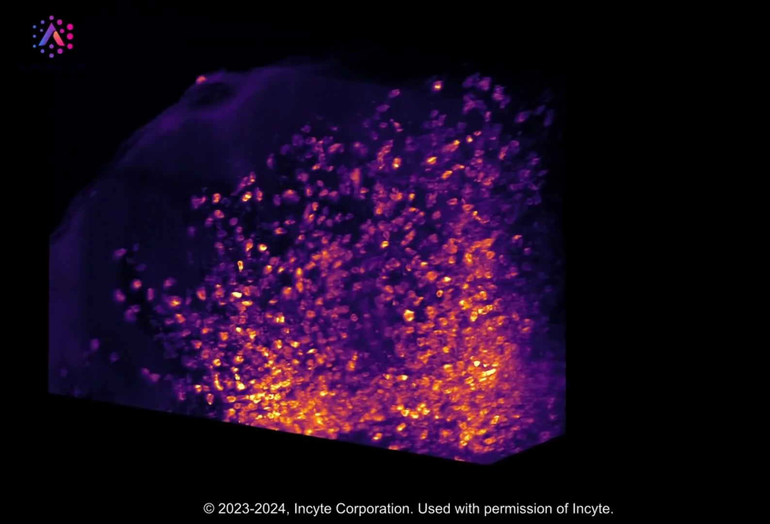
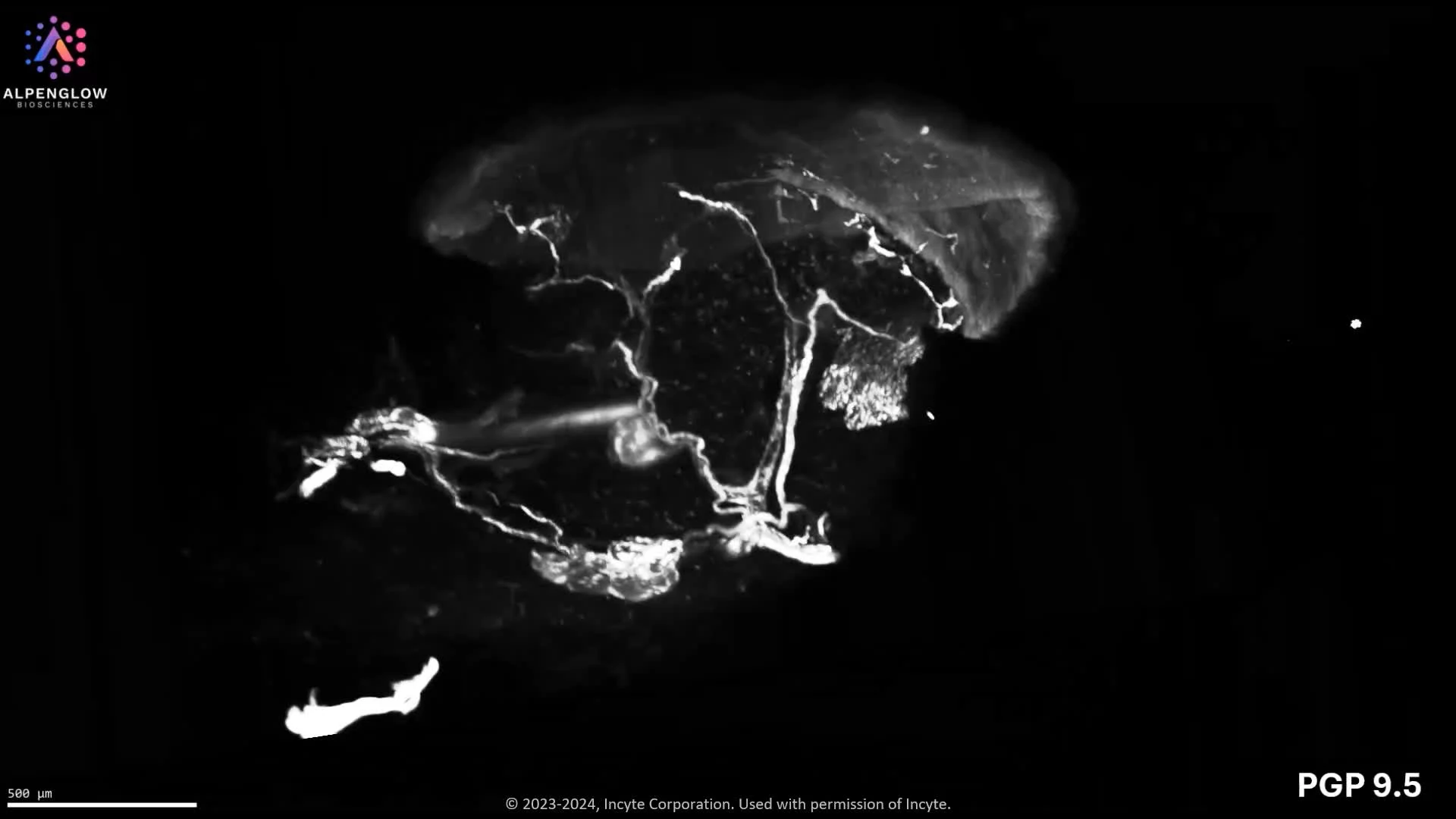
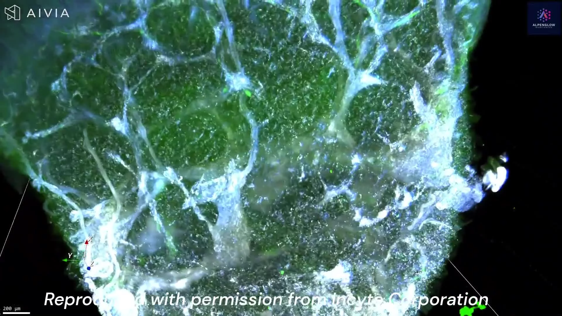
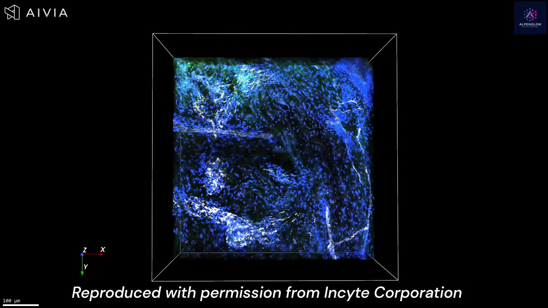
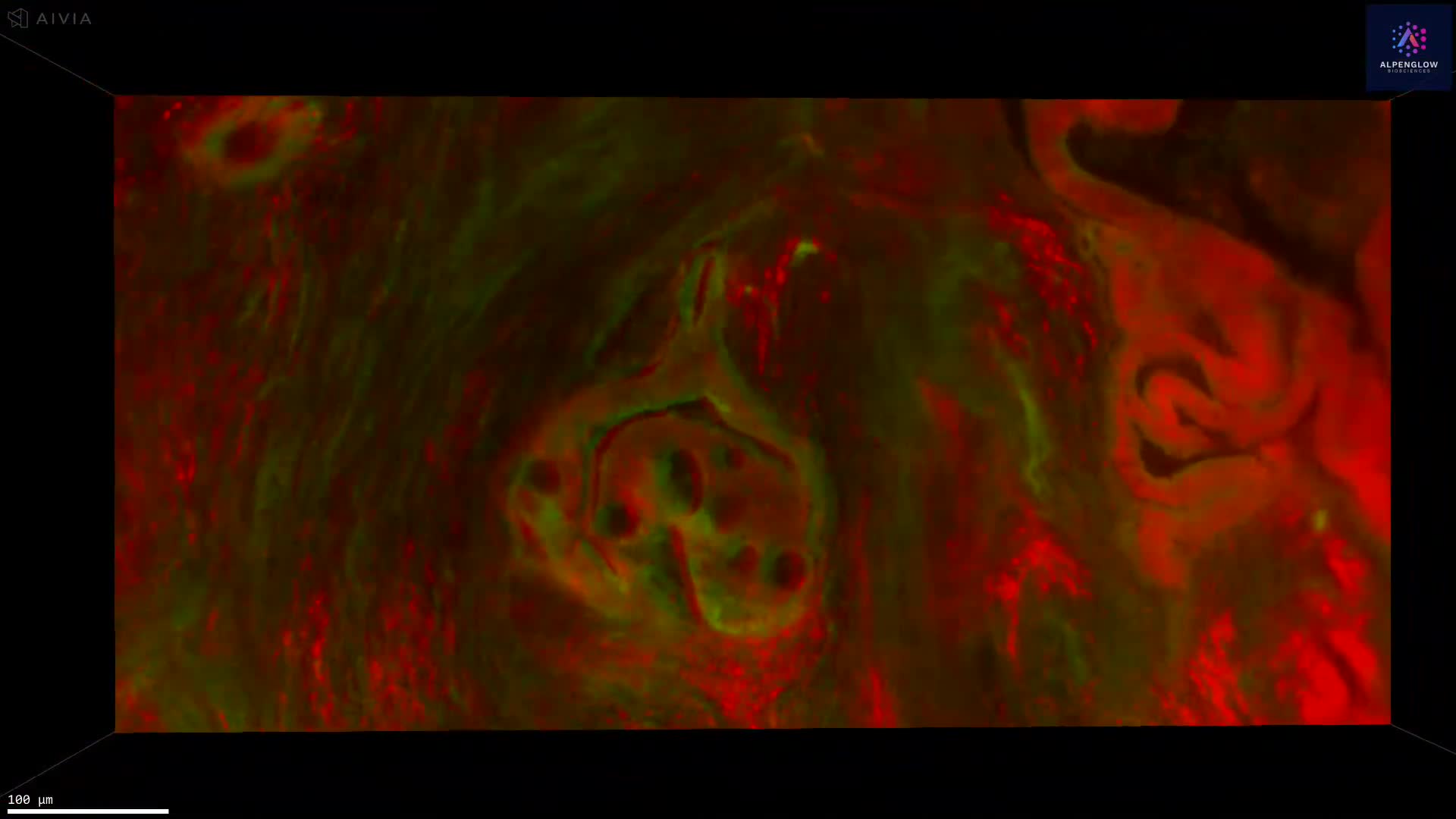
See 3D tissue imaging in motion.
Explore videos from Alpenglow Biosciences showing whole tissue imaging, volumetric datasets, digital pathology views, spatial profiling, and AI-powered analysis across biological applications.