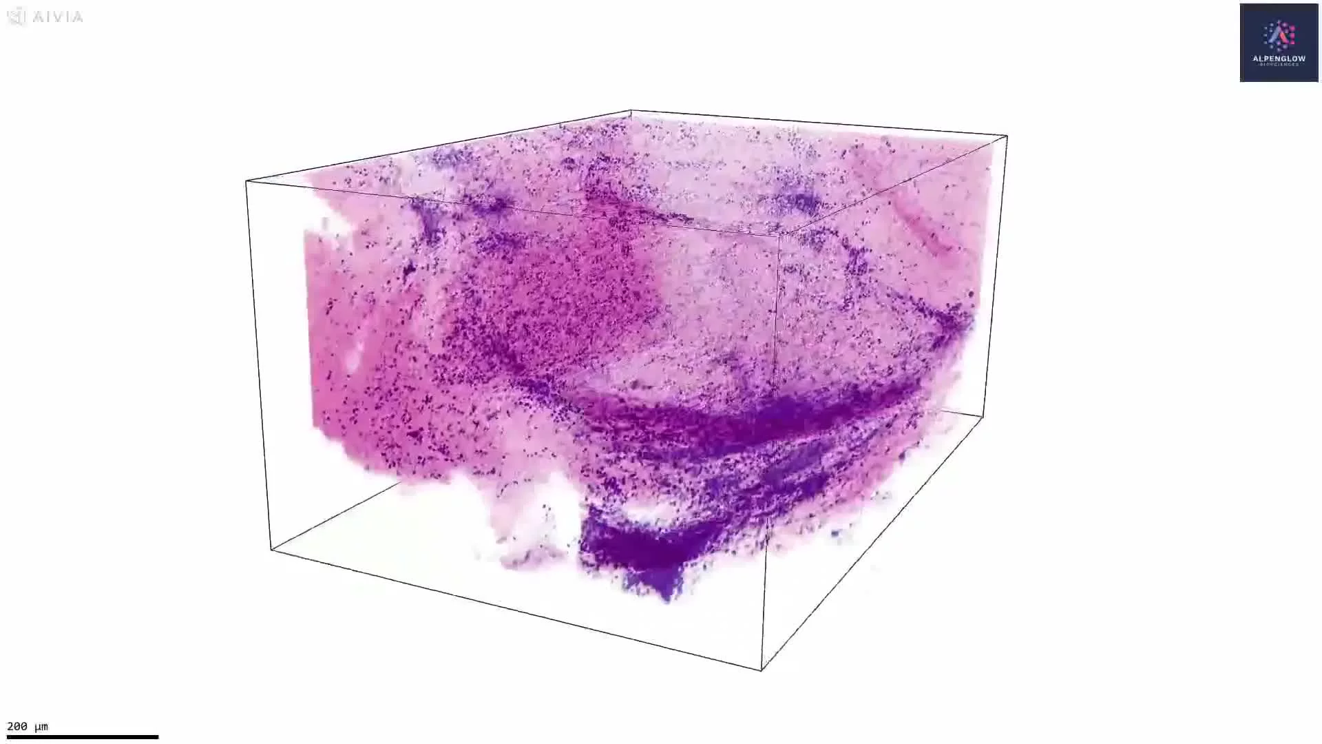
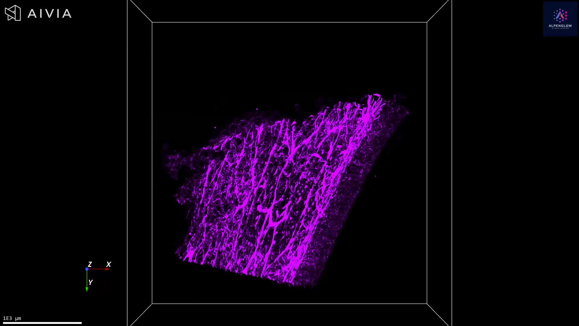
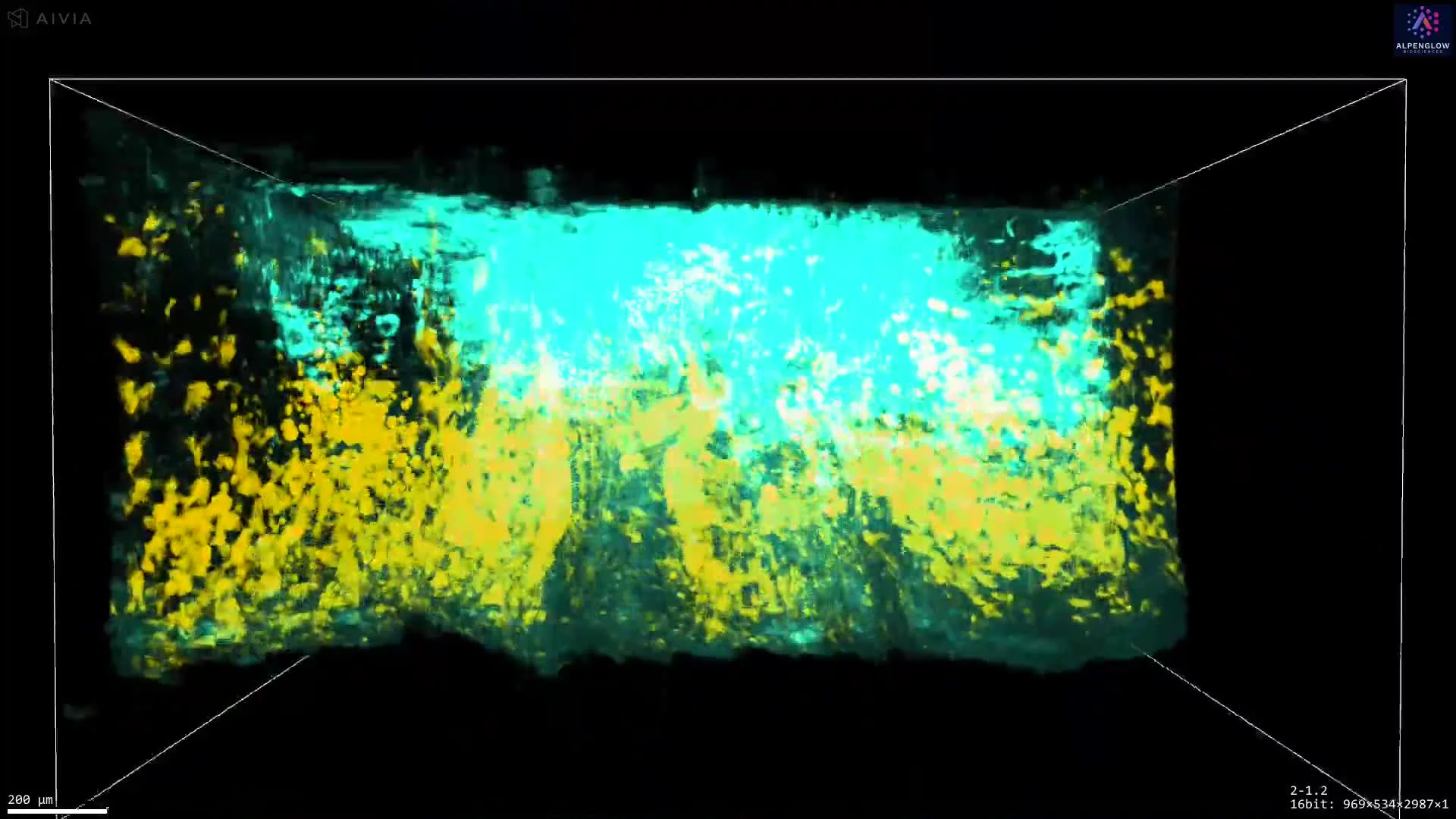
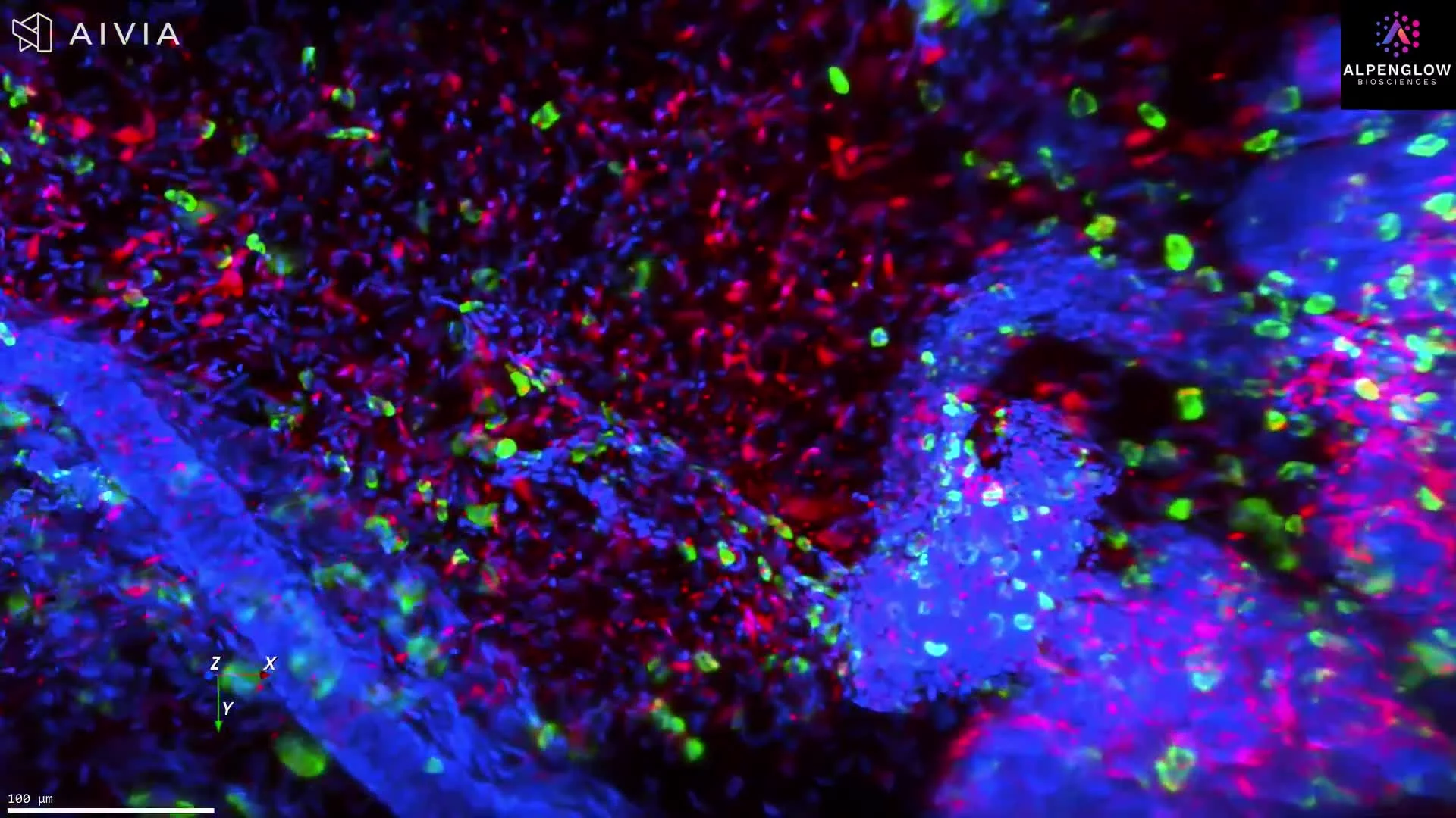
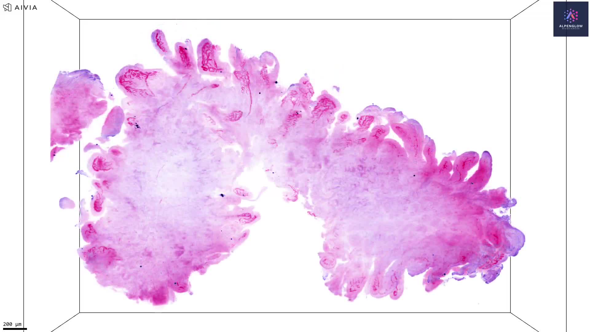
3D Imaging of Human Tonsil Tissue Highlighting Mast Cells with Tryptase
This visualization demonstrates the power of 3D fluorescence imaging at Zoom resolution using the Aurora™ 3Di Hybrid Open Top Light Sheet (HOTLS) microscope. A human tonsil sample (~800 µm in length) is imaged in full depth, capturing structural and cellular relationships that are lost in traditional 2D slices.
Stains used:
YO-PRO-1 (Purple): Labels nuclei
Tryptase (Cyan): Highlights mast cells — critical players in allergic responses, inflammation, and tumor microenvironments
By combining whole-tissue integrity with high-resolution 3D histology, this dataset reveals the spatial distribution of mast cells in their native context. Integration with 3Dm data management and 3Dai AI-powered segmentation enables quantification of mast-cell density and proximity to other immune or stromal structures.
Understanding mast cell biology in situ is crucial for advancing immunology research, dermatology, and immuno-oncology, where mast cells play a significant role in both inflammation and cancer progression.
Curious about the antibody used? This is from Abcam, Cat: ab2378, Clone: [AA1], https://www.abcam.com/en-us/products/primary-antibodies/mast-cell-tryptase-antibody[…]sltid=AfmBOoqottEqHLRMk43Xza7e0WYltzxI64-3fpUIGDxi0DXcz7tJhEA_