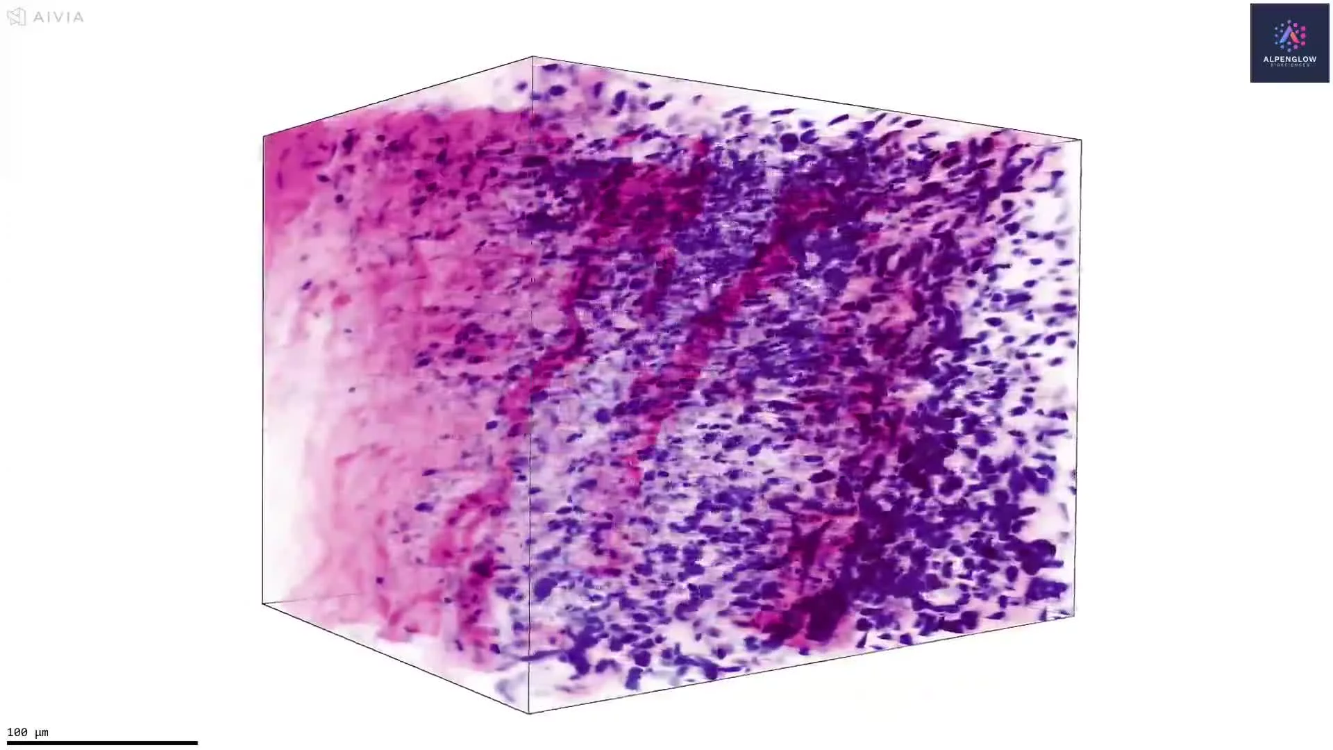
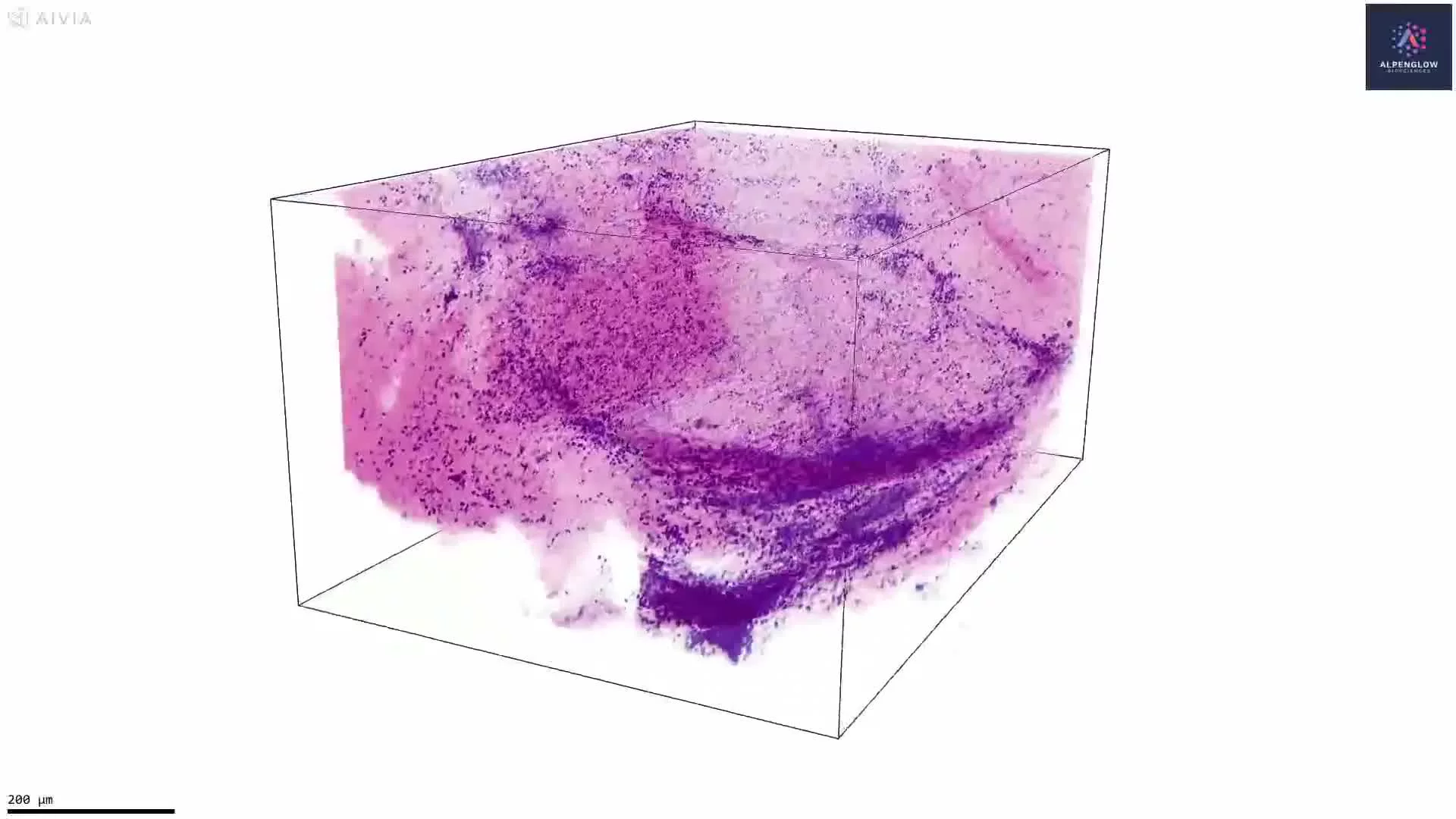
3D Imaging of Human Placental Vascular Architecture
This 3D dataset presents human placental tissue, preserving vessel-associated structures and cellular organization across the imaged volume.
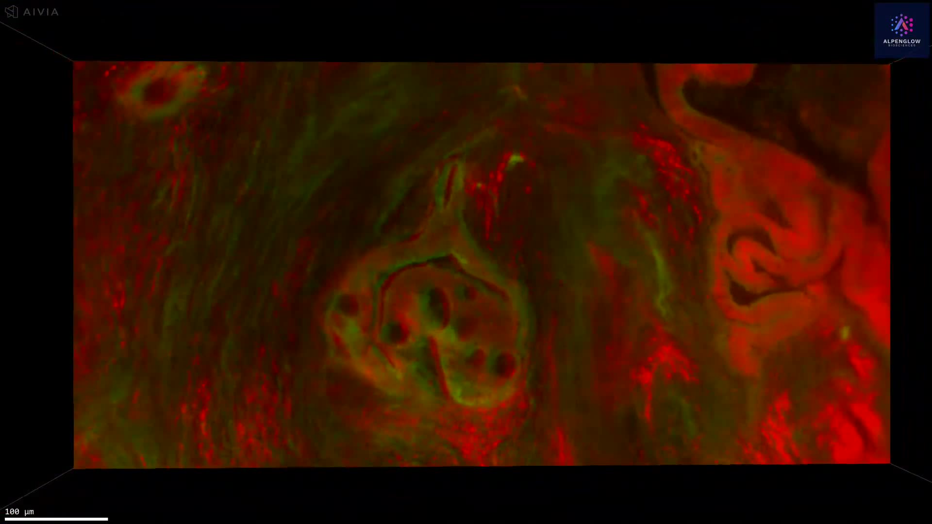
Alpha-smooth muscle actin, shown in green, highlights smooth muscle actin-positive cells associated with placental vessels. TO-PRO-3, shown in magenta, labels nuclei and provides cellular context throughout the tissue.
The volumetric view reveals vessel trajectories, branching, diameter variation, and the organization of smooth muscle actin-positive cells around larger vascular structures. Preserving these features across depth provides spatial context that can be fragmented across individual tissue sections.
The dataset supports quantitative analysis of vessel-associated structure density, branching, diameter, tortuosity, and regional variation across the placental tissue volume. These measurements are relevant to research on placental vascular development, remodeling, and maternal-fetal biology.
The tissue was imaged using the Aurora 3D™ Spatial Biology Solution, including the 3Di™ Hybrid Open-Top Light-Sheet microscope.