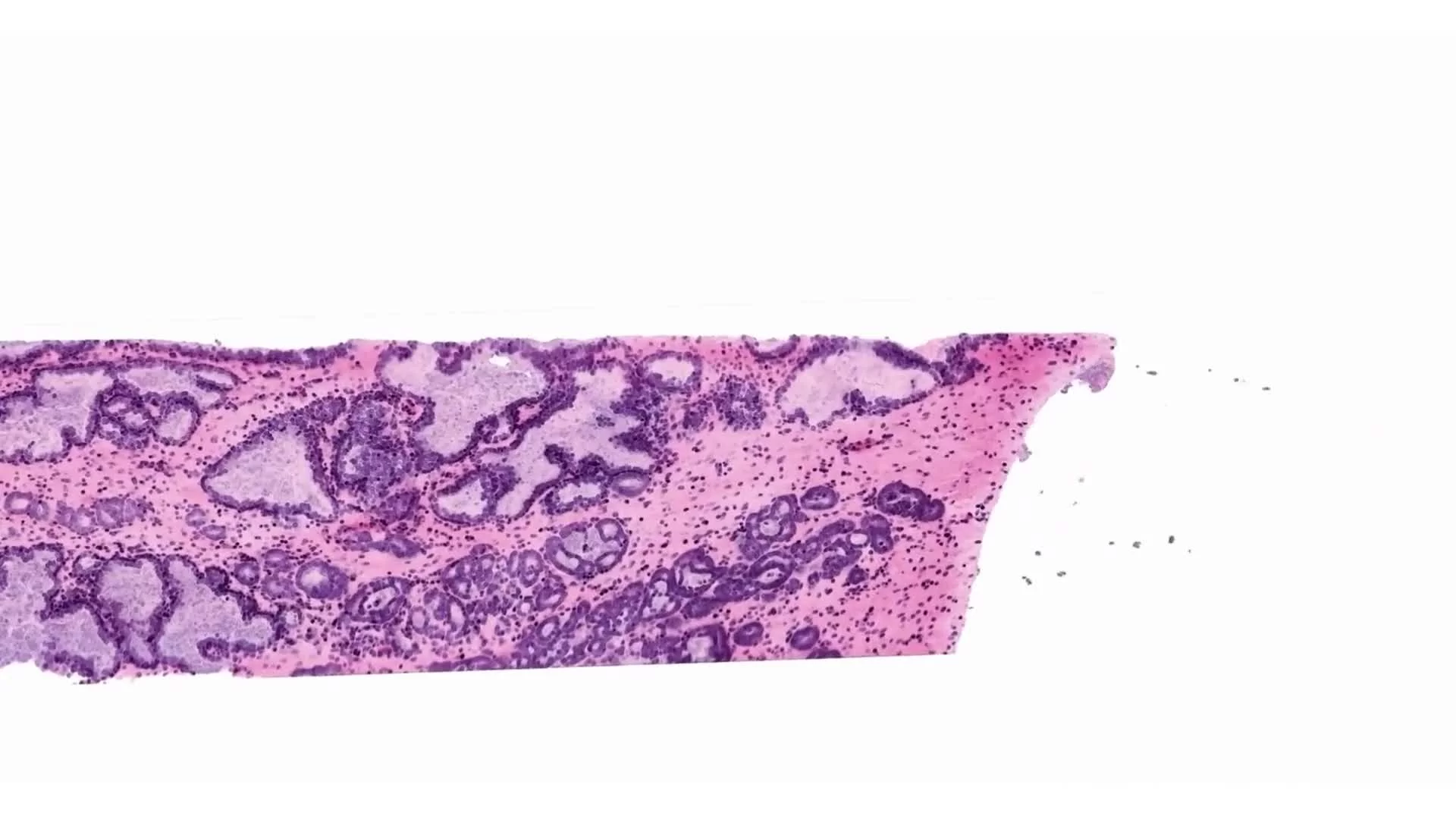
Human Placenta in 3D with SMA, HLA-G, and CD31 Staining
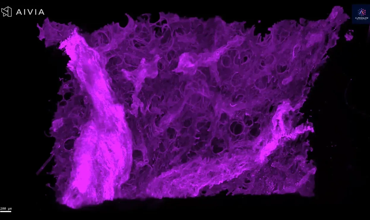
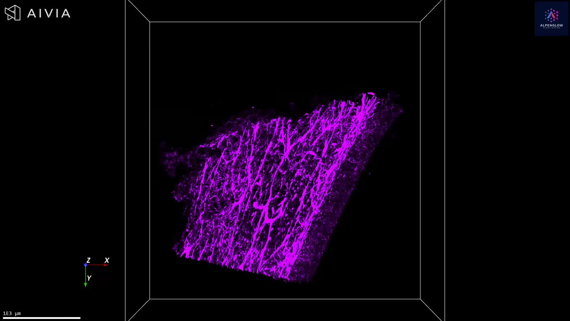
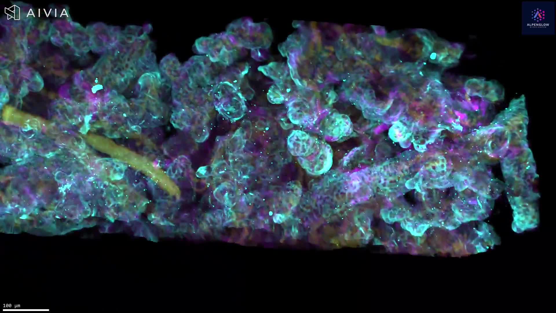
This 3D dataset showcases the intricate architecture of the human placenta, imaged on the Aurora™ 3Di Hybrid Open Top Light Sheet (HOTLS) microscope. The video begins with individual fluorescence channels and builds to a composite view, capturing the tissue’s complexity in vivid detail.
Stains used:
SMA (Red): Highlights smooth muscle structures
HLA-G (Yellow): Marks placental trophoblast cells
CD31 (Cyan): Traces vascular endothelium
The workflow demonstrates seamless transitions from low-resolution Scout scans to high-resolution Zoom imaging of regions of interest (ROIs), reaching single-cell resolution. At the 00:47 mark, the video zooms in to reveal detailed cellular features within the placenta’s structural framework.
By integrating3Dm data management with 3Dai AI-powered segmentation, this dataset supports quantitative analysis of trophoblast organization, vascular architecture, and immune interactions. Such multi-scale insights exemplify the power of 3D histology to capture intact tissue biology.
Applications extend to reproductive biology, placental health, and translational research in maternal–fetal medicine, where understanding cellular and vascular networks is essential.