3D Imaging of Enteric Nerve Architecture in Hirschsprung Disease
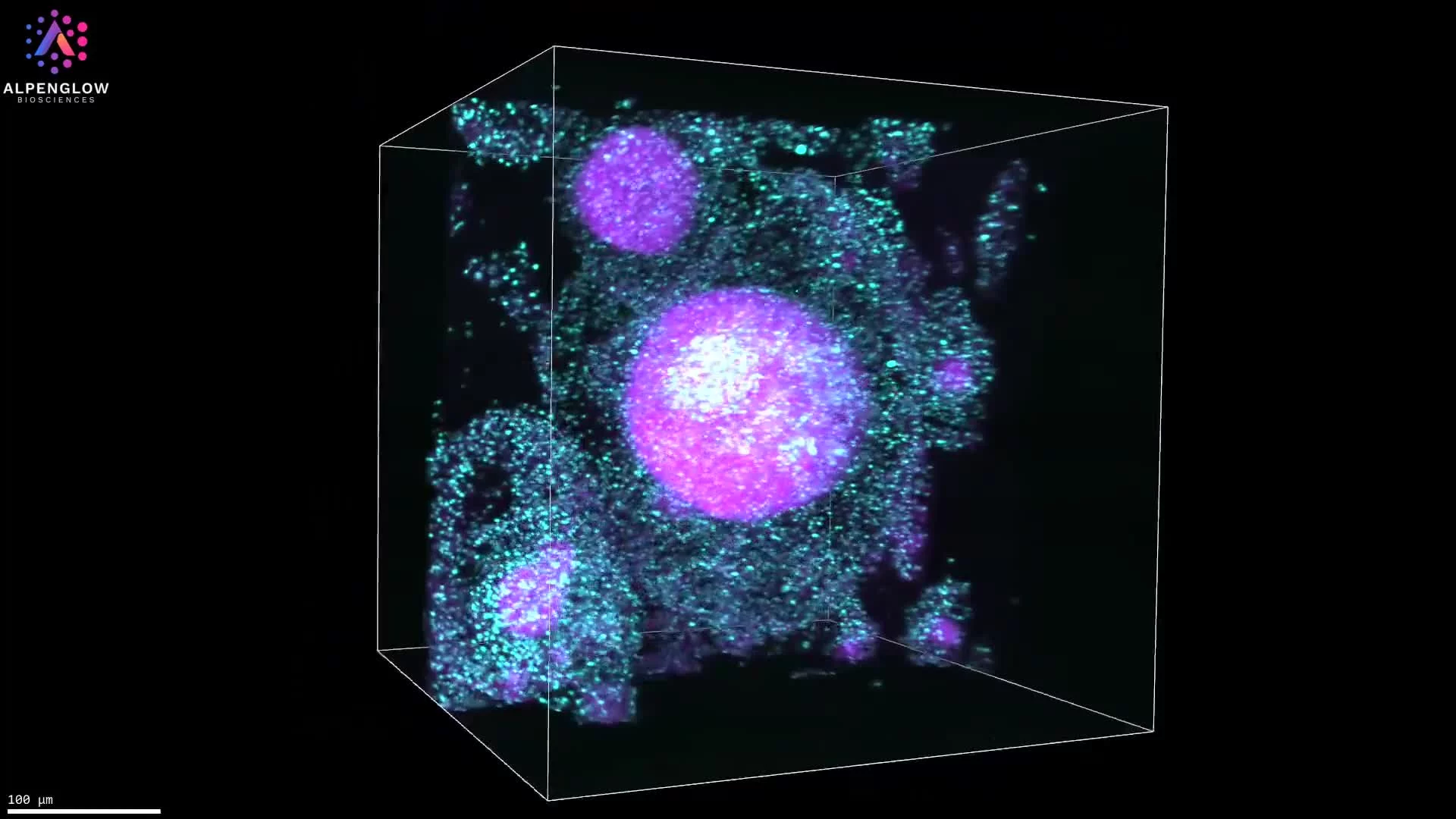
This dataset presents 3D imaging of human colon tissue affected by Hirschsprung disease, a congenital disorder characterized by the absence of enteric ganglion cells along a variable length of the bowel.
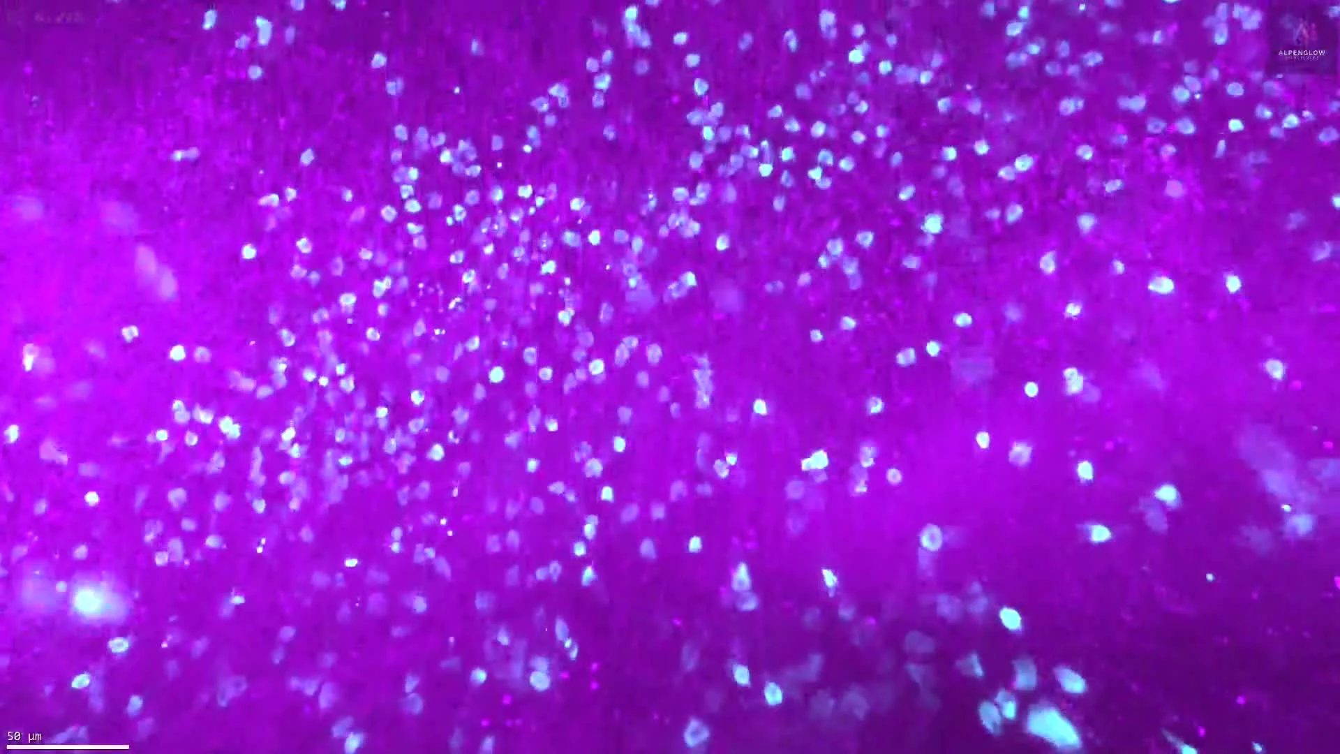
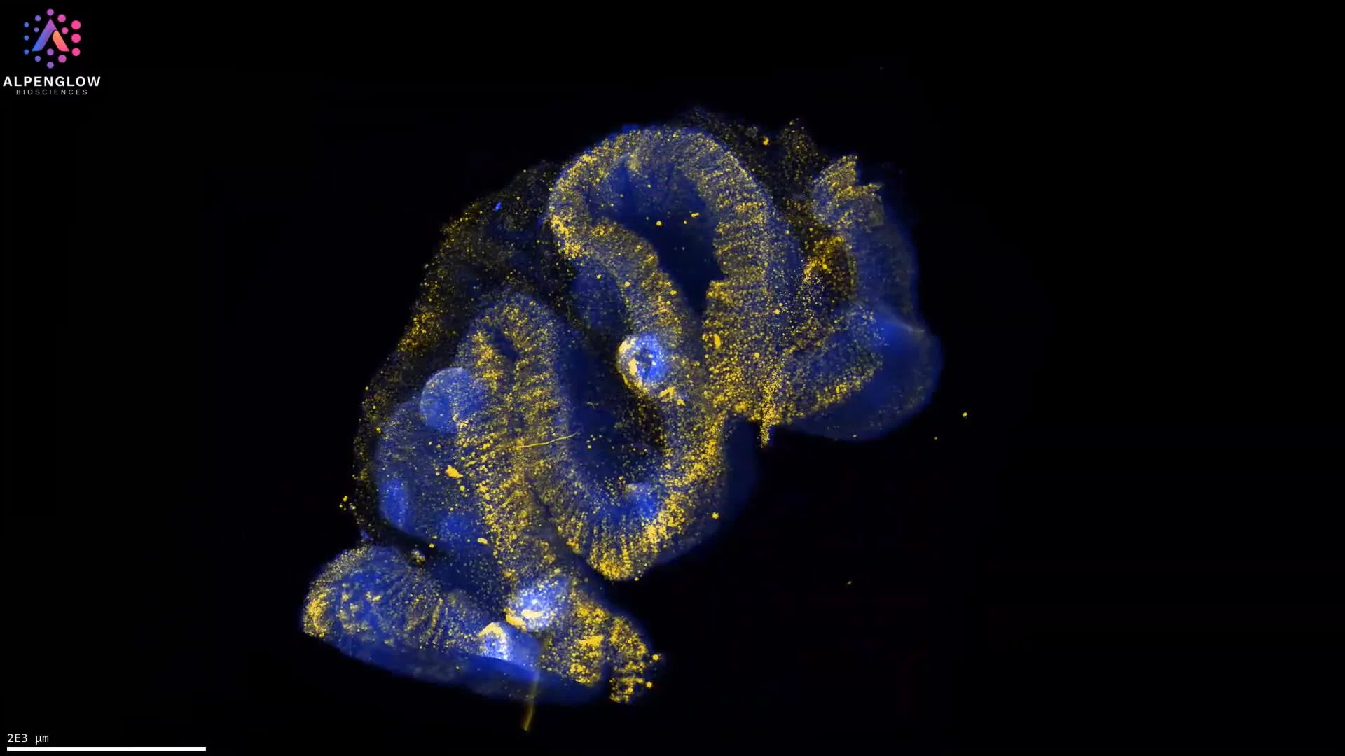
TO-PRO-3, shown in green, labels nuclei, while PGP9.5, shown in red, highlights enteric neural structures throughout the tissue volume.
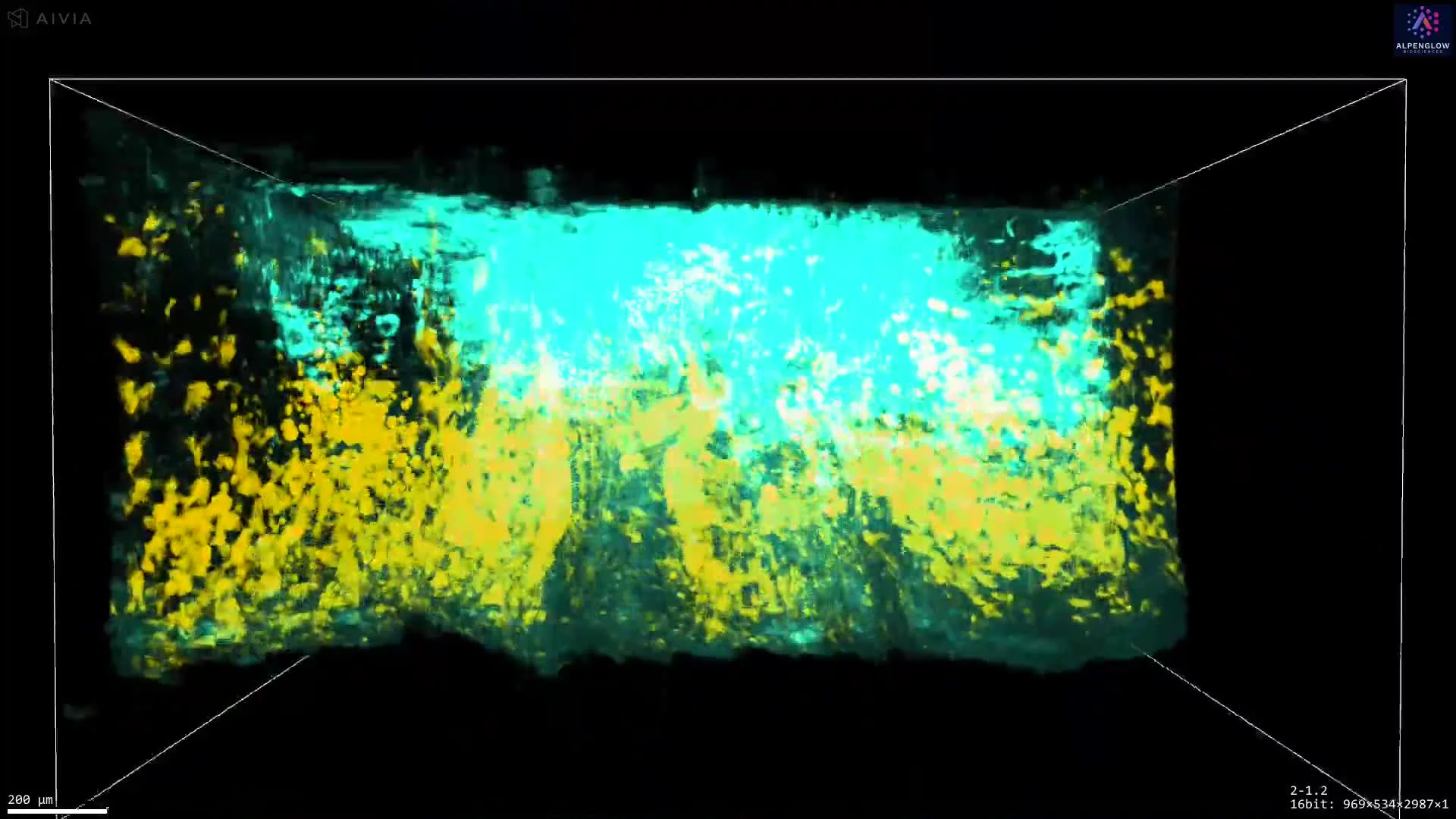
The dataset reveals hypertrophic nerve fibers extending through the submucosa, with no ganglion cells visible within the imaged region. Preserving the tissue in 3D enables these neural structures to be followed across depth and provides continuous spatial context for their distribution and organization.
The dataset supports analysis of nerve fiber thickness, density, branching, orientation, and regional distribution, as well as assessment of enteric neural architecture across the imaged tissue volume. These measurements can support research into congenital gastrointestinal disorders and disease-associated changes in the enteric nervous system.
The tissue was imaged using the Aurora 3D™ Spatial Biology Solution, including the 3Di™ Hybrid Open-Top Light-Sheet microscope.