
Use cases & brochures
Explore our applications and resources
Discover how Alpenglow's technologies are applied across different tissues, research questions, and scientific applications. Explore use cases and brochures featuring research examples, imaging workflows, and platform capabilities.
Use case library
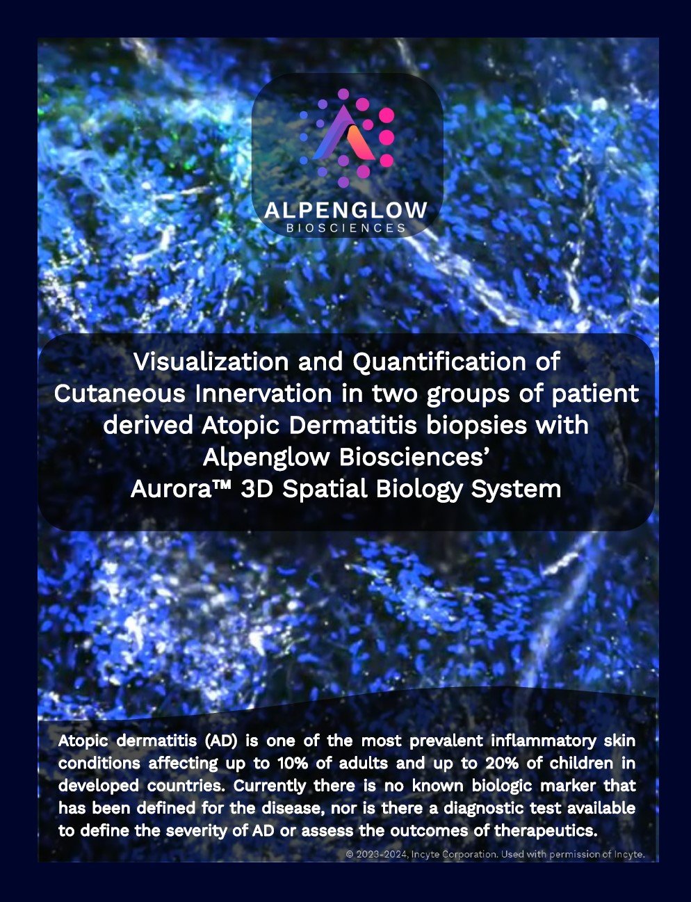
3D imaging of atopic dermatitis
See how full-volume 3D imaging of skin biopsies reveals immune–epithelial interactions, nerve fibers, and microvascular changes. The use case demonstrates how intact-tissue analysis can quantify inflammatory patterns and spatial changes associated with treatment.
Access use case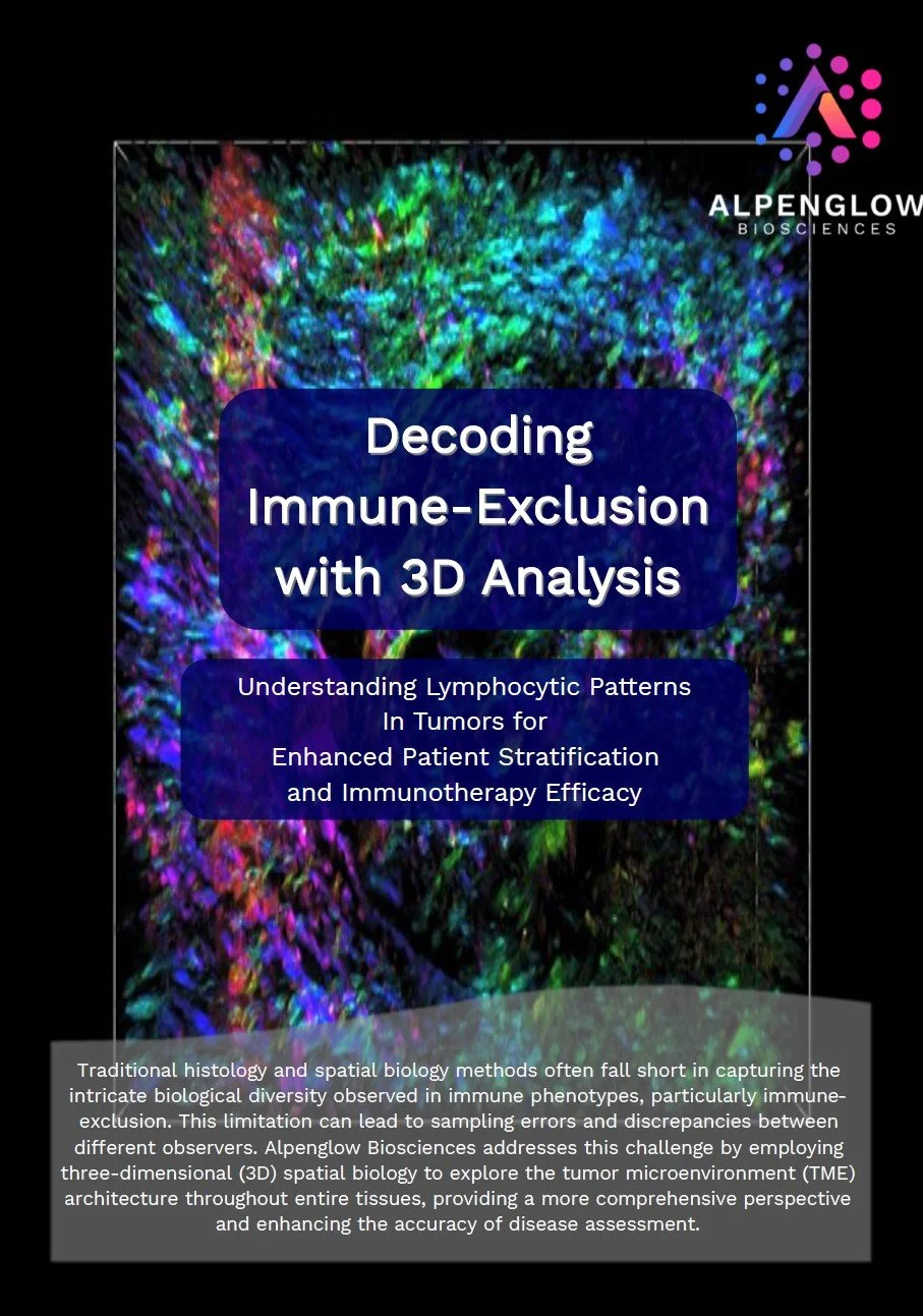
Decoding immune exclusion in 3D
Explore how 3D tissue imaging and AI-powered analysis map lymphocyte distribution, exclusion zones, and tumor microenvironment architecture across intact tumor samples. The study supports investigation of spatial patterns associated with differential treatment response.
Access use case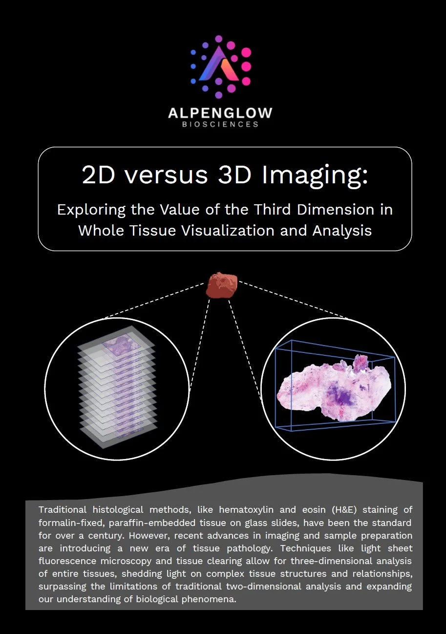
2D versus 3D tissue analysis
Compare conventional 2D histology with 3D imaging of the same specimen. The use case shows how isolated sections can undercount structures, miss rare features, or incompletely represent tissue architecture, while whole-tissue datasets provide a more complete spatial reference.
Access use case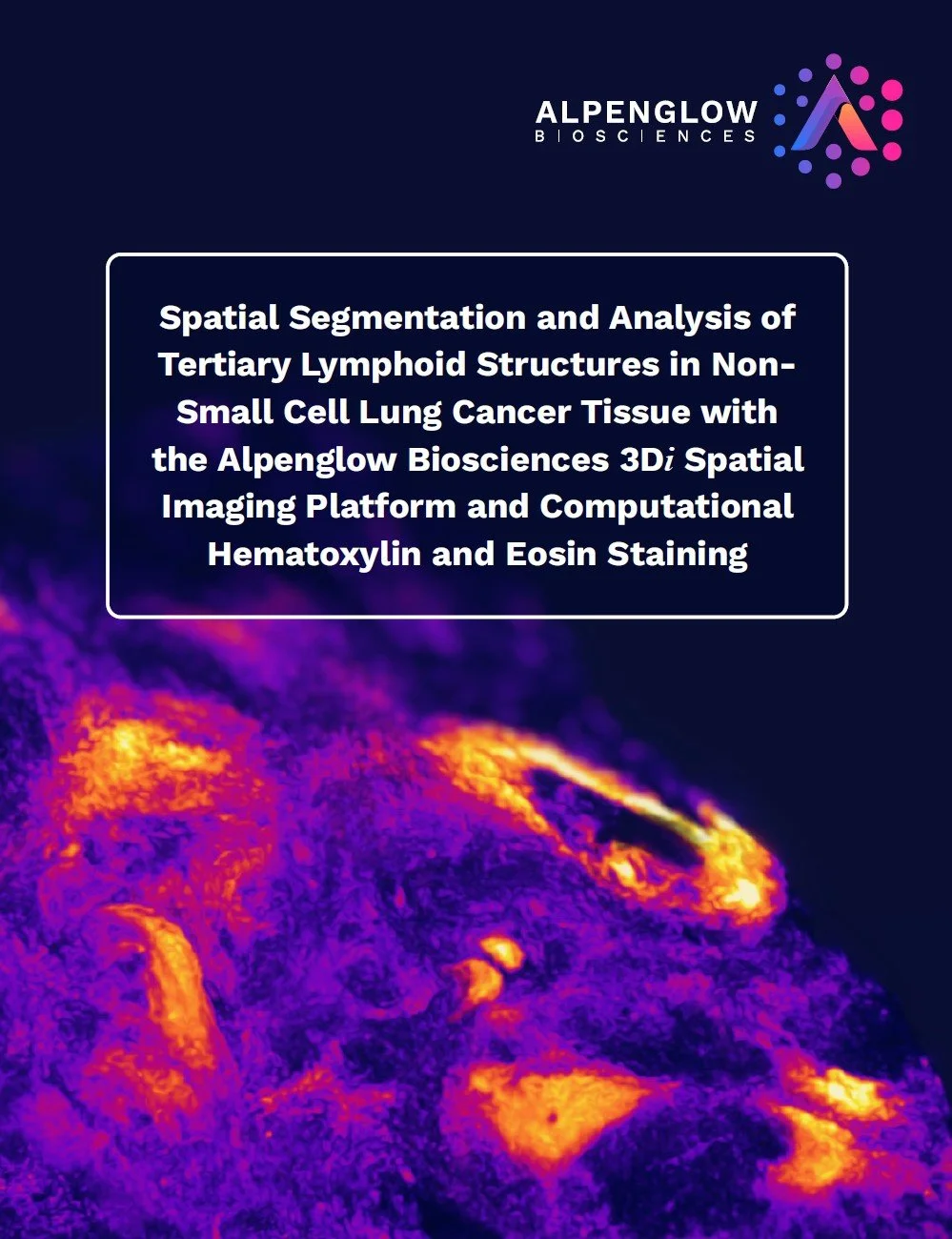
3D segmentation of tertiary lymphoid structures
See how tertiary lymphoid structures in non-small cell lung cancer can be segmented and quantified in 3D, including their size, location, volume, and spatial organization. The workflow supports TLS biomarker research in immuno-oncology.
Access use case
Ground-truth 3D data in immuno-oncology
Explore how whole-tissue 3D imaging, Aurora workflows, and quantitative analysis can support immuno-oncology research through full-volume tissue context and image-linked spatial measurements.
View PDFBrochure library
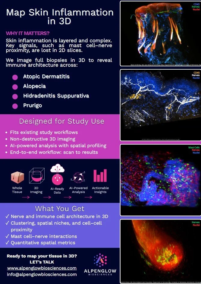
Map skin inflammation in 3D
Explore how intact-tissue 3D imaging can visualize inflammatory patterns, immune cells, vasculature, nerves, and structural changes across full-volume skin samples.
View brochure
3D I/O Pro Assay
Review an intact-tissue workflow for mapping immune-cell distribution, tumor architecture, spatial relationships, and quantitative features across the tumor microenvironment.
View brochure
3D Derm Score Assay
Explore quantitative 3D analysis of skin biopsies, including immune infiltration, nerves, vasculature, follicles, epidermal structure, and tissue-scale spatial relationships.
View brochure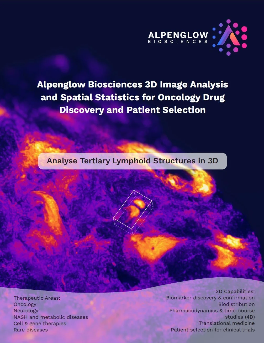
Quantifying tertiary lymphoid structures in 3D
See how tertiary lymphoid structures can be detected, segmented, and measured across intact tissue, including their volume, location, morphology, and spatial organization.
View brochure
3Di Hybrid Open-Top Light-Sheet Microscope
Review the open-top microscope architecture, Scout and Zoom imaging paths, sample versatility, imaging volume, and workflow for high-resolution 3D tissue imaging.
View brochure
3D immunomarker staining in intact tissue
Explore immunomarkers used across intact-tissue 3D imaging studies, supporting visualization of immune cells, nerves, vasculature, epithelial structures, and additional tissue features.
View brochureNext step
Want to discuss a 3D imaging use case for your tissue samples?
Connect with Alpenglow Biosciences to discuss sample type, imaging goals, spatial profiling, AI-powered analysis, and quantitative tissue analysis for your research program.