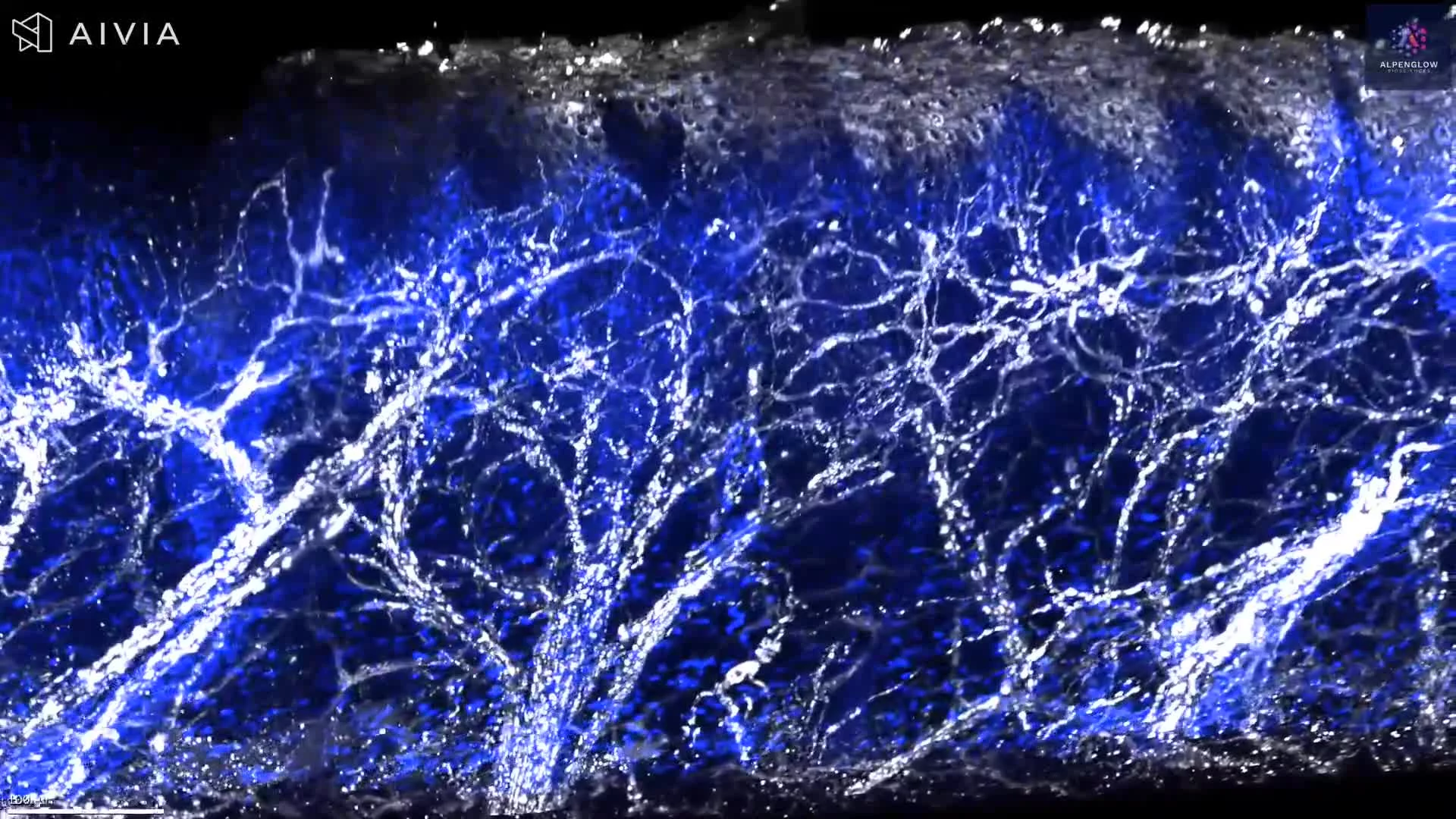
3D Visualization of TLS in Colorectal Tumor Tissue
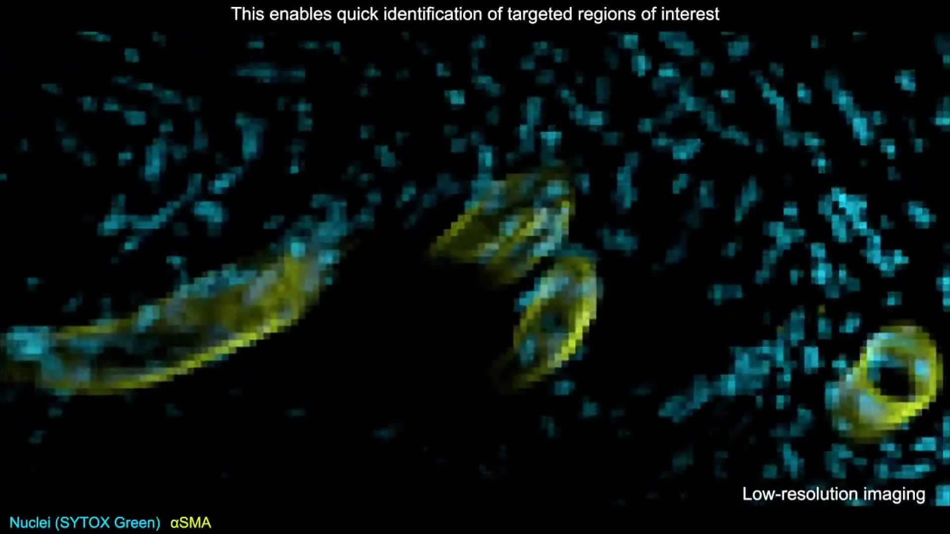
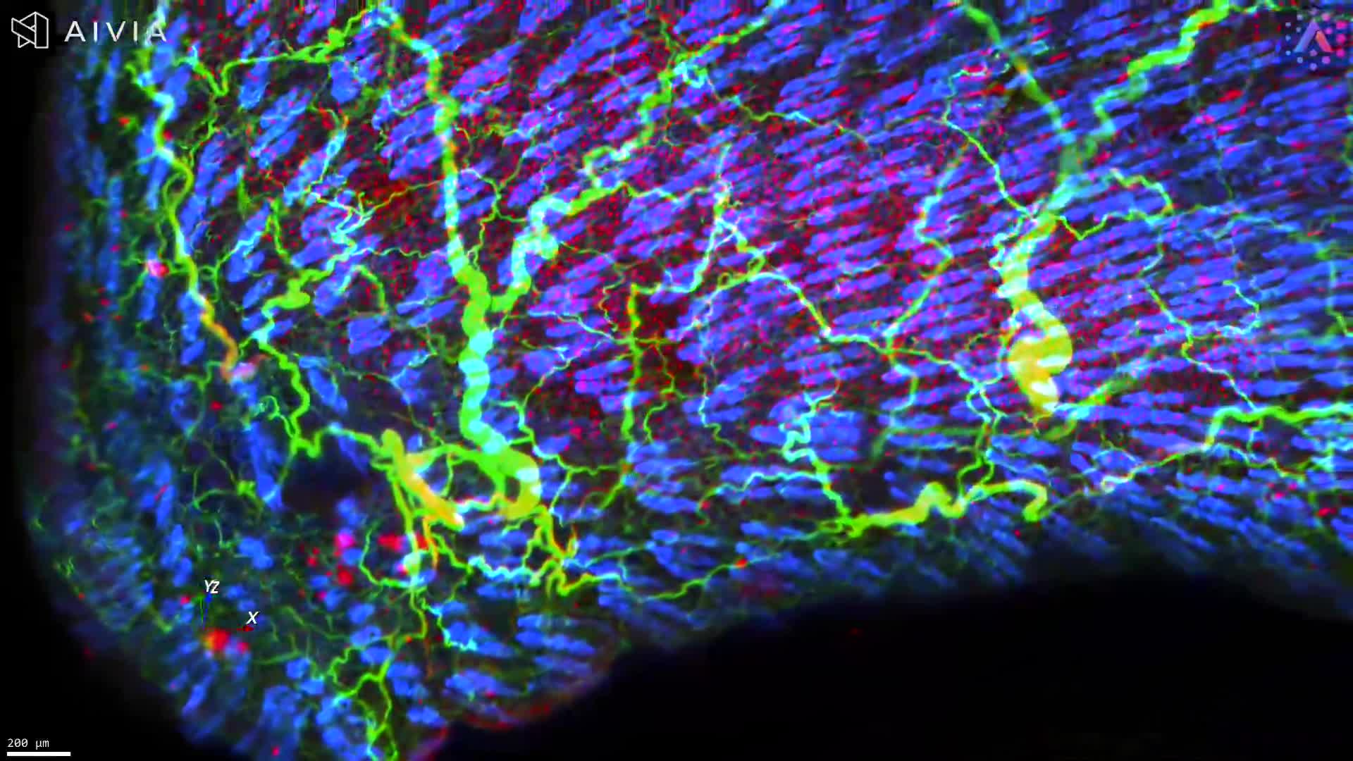
This dataset showcases a 3D visualization of a mouse colorectal tumor, imaged on the Aurora™ 3Di Hybrid Open Top Light Sheet (HOTLS) microscope to reveal key immune populations in their native spatial context.
Stains used:
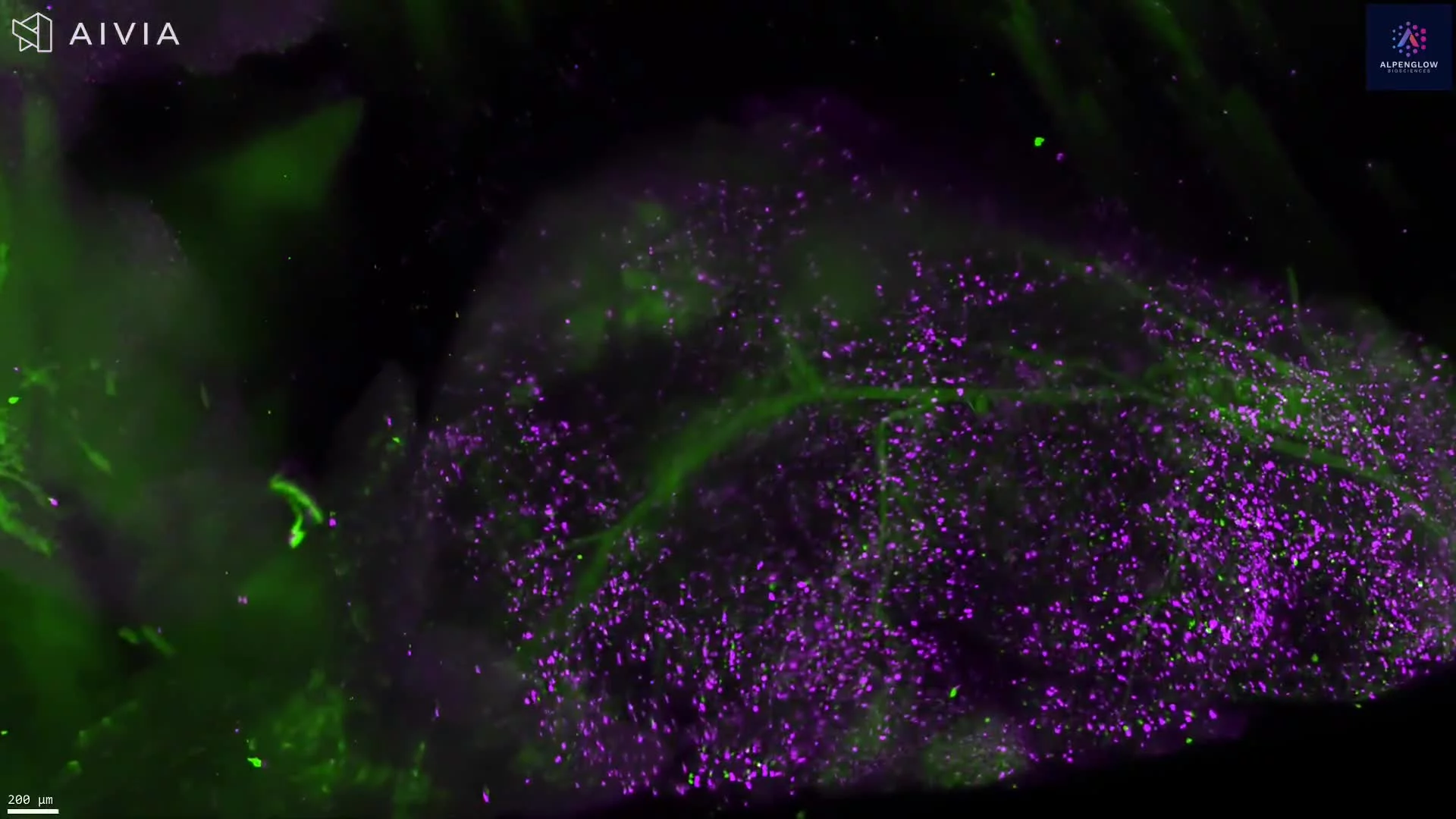
CD3 (Purple): Labels T cells
B220 (Green): Marks B cells
YoPro1 (Blue): Highlights nuclei
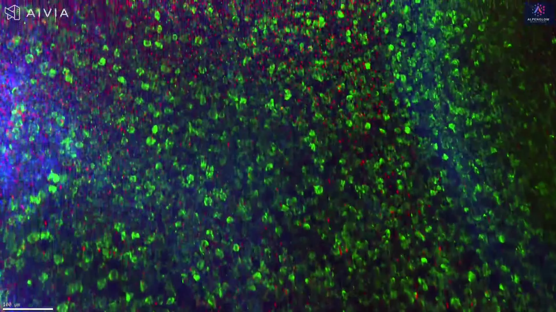
Within the tumor microenvironment, two tertiary lymphoid structures (TLS) are clearly visible, their complex organization preserved in full volumetric detail. TLS are dynamic, three-dimensional immune aggregates that are often overlooked in thin-section histology.
By combining light-sheet imaging with 3Dm data management and 3Dai AI-powered segmentation, the full tumor volume was imaged without slicing, enabling TLS segmentation, quantification, and spatial profiling. Researchers can measure TLS size, density, immune cell composition, and their relationships to tumor structures with unprecedented accuracy.
This dataset highlights how 3D histology, digital pathology, and spatial profiling uncover critical immune features, delivering insights essential for immuno-oncology research and advancing biomarker discovery.
Discover more in our TLS White Paper.