Aurora Workflow: 3D Imaging of Murine Heart Vasculature
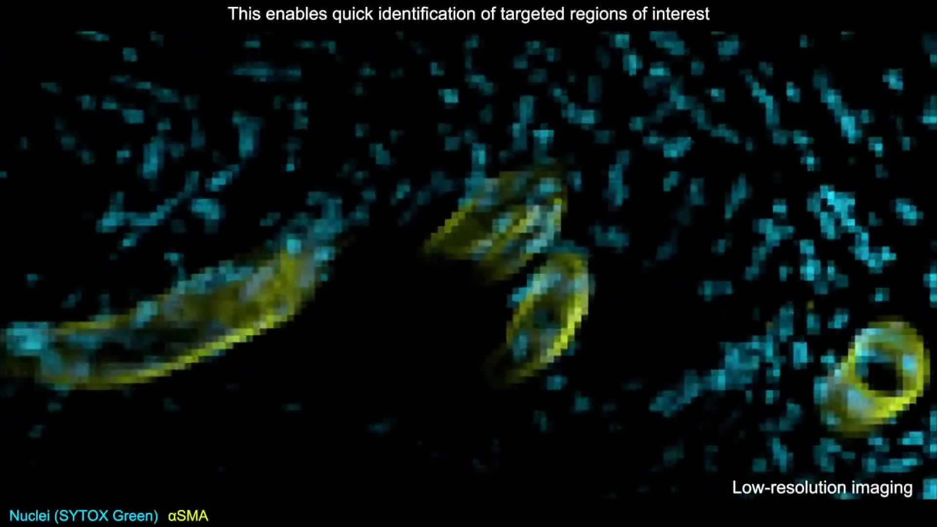
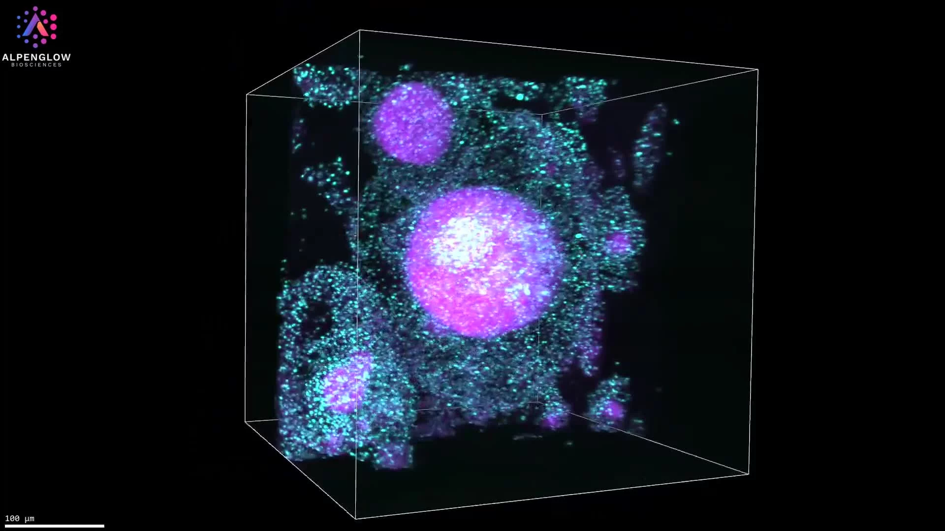
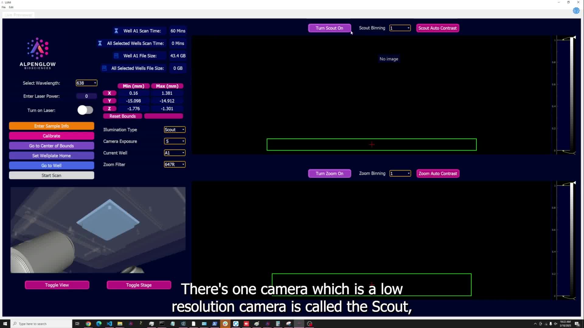
This video presents a compelling example of the complete Aurora Workflow, showcasing an advanced 3D imaging study of murine heart architecture. Using the Aurora™ 3Di Hybrid Open Top Light Sheet (HOTLS) microscope, the dataset highlights the complexity of cardiac vasculature and the power of volumetric imaging to capture intact morphology.
Key analyses include:
Cell counts across tissue volumes
Vessel diameter and length measurements
Distance calculations between structures
Branching pattern analysis of vascular networks
With seamless integration of3Dm data management and3Dai AI-powered segmentation, the Aurora Workflow transforms stunning visuals into meaningful, quantitative data. This end-to-end platform scales from whole-organ imaging to single-cell resolution, empowering precise analysis of tissue biology.
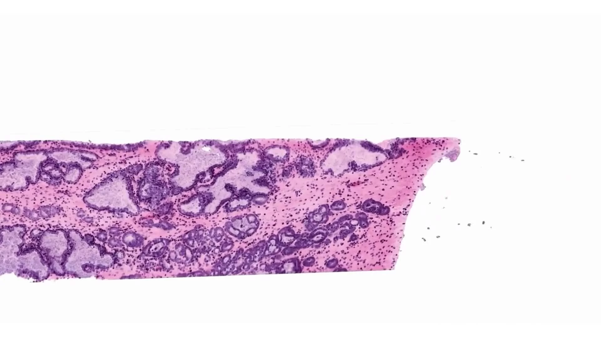
Applications include cardiovascular research, vascular biology, and translational studies where quantifying complex networks is essential. Each dataset demonstrates how Aurora delivers the ground truth in 3D histology, advancing digital pathology and spatial profiling.
Contact us to learn how the Aurora Workflow can be tailored to your research needs and help you lead the 3D imaging revolution.