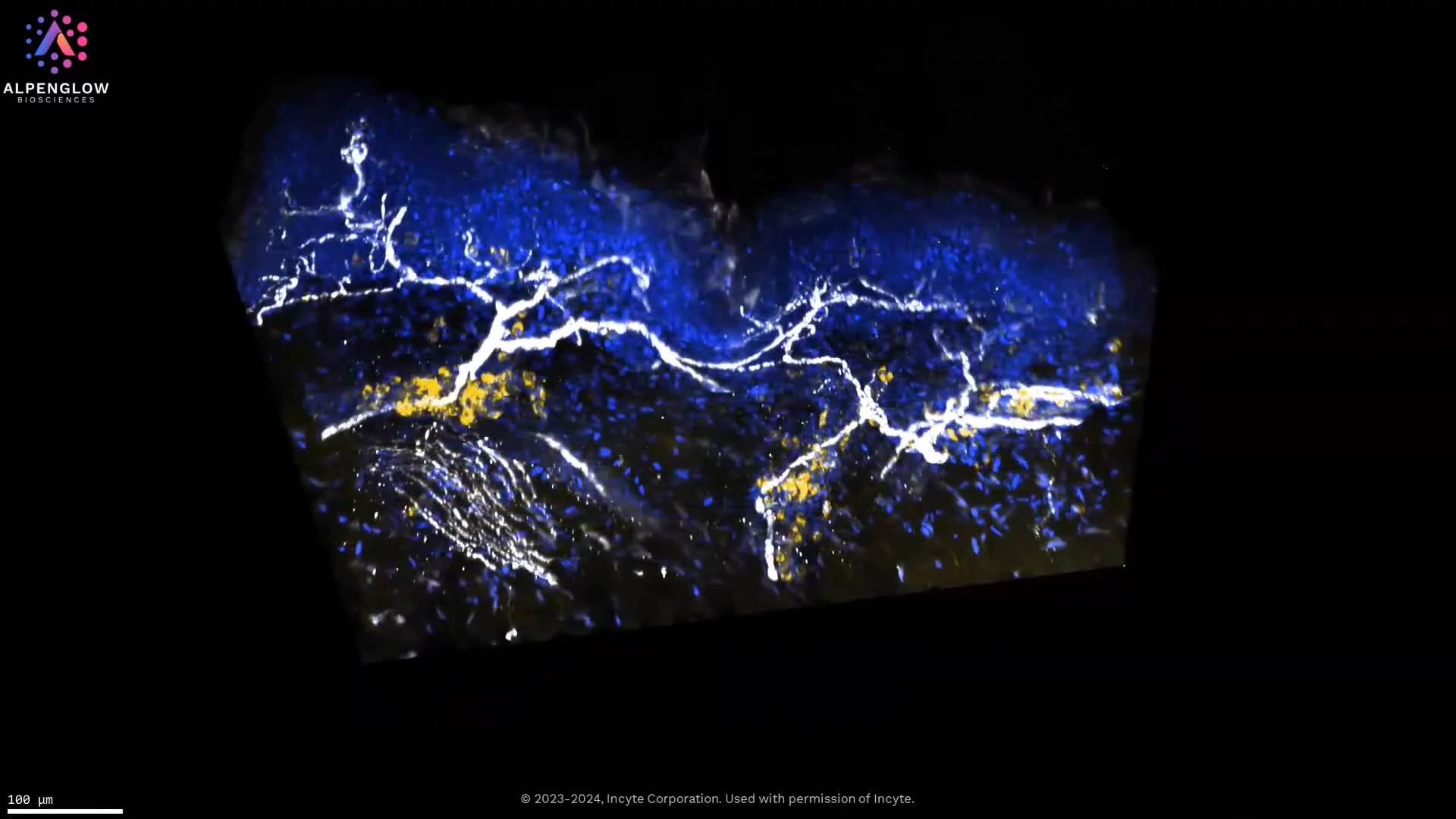
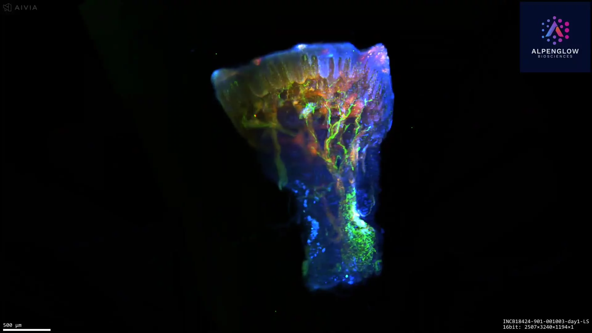
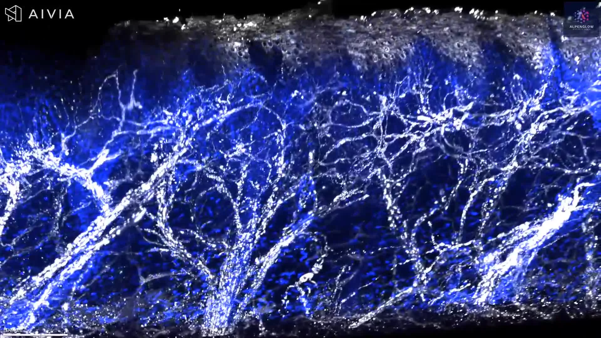
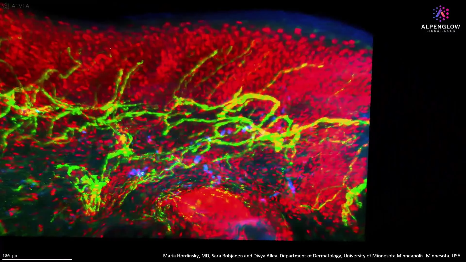
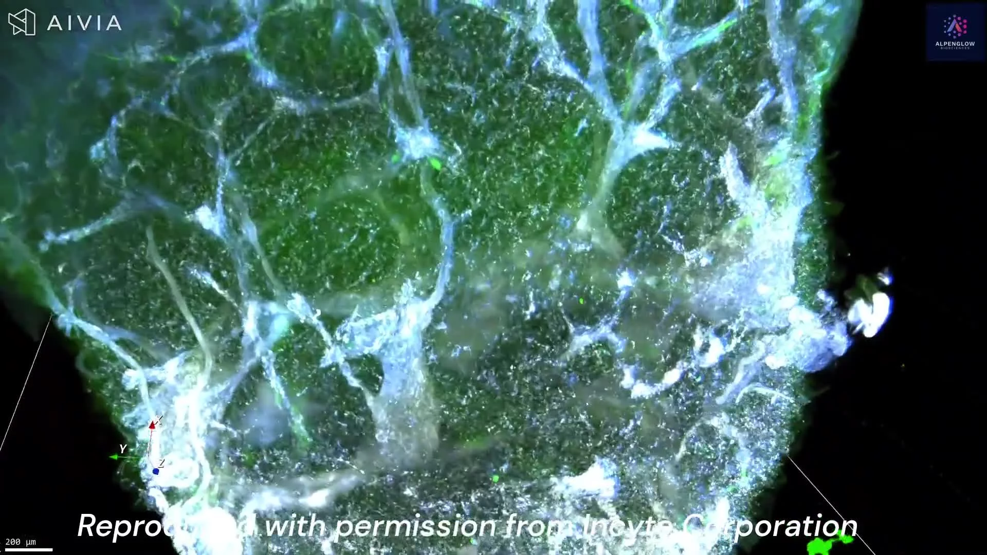
3D Imaging of Melanoma Tissue with Immune Markers
Through advanced staining and high-resolution 3D imaging, we move beyond the limitations of flat, 2D slides to capture the full spatial context of human melanoma tissue.
In this video, you can observe:
🟢 CD45 – highlighting immune cells
🔴 Neutrophil Elastase – marking neutrophils
🔵 TO-PRO-3 – illuminating nuclei
Each marker reveals cellular architecture with striking clarity, uncovering complex immune–tumor interactions previously hidden in 2D views.
This depth of visualization enhances our understanding of melanoma biology and establishes a new benchmark for research.
Previous
Whole-biopsy 3D segmentation of prostate glands
Next