High-Resolution 3D Imaging of Scalp Tissue for Hair and Skin Research
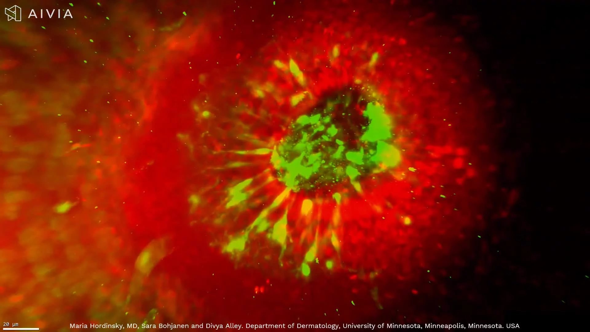
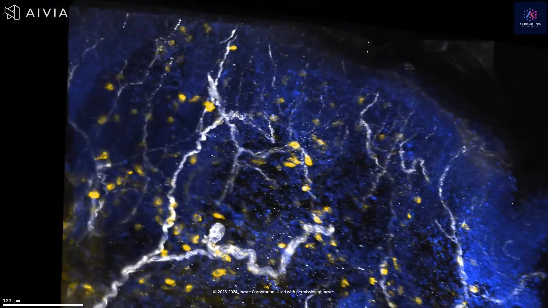
This high-resolution 3D dataset reveals scalp tissue architecture relevant to hair biology and inflammatory skin disease.
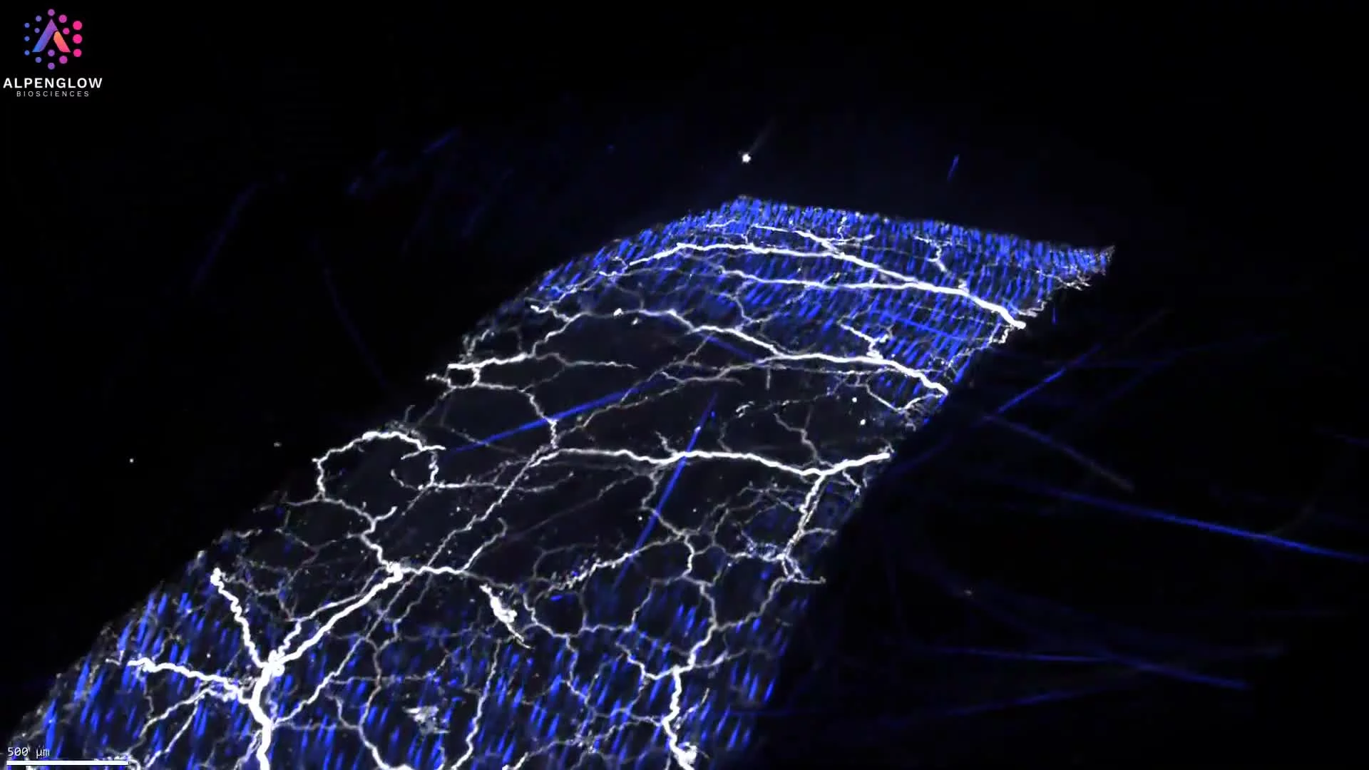
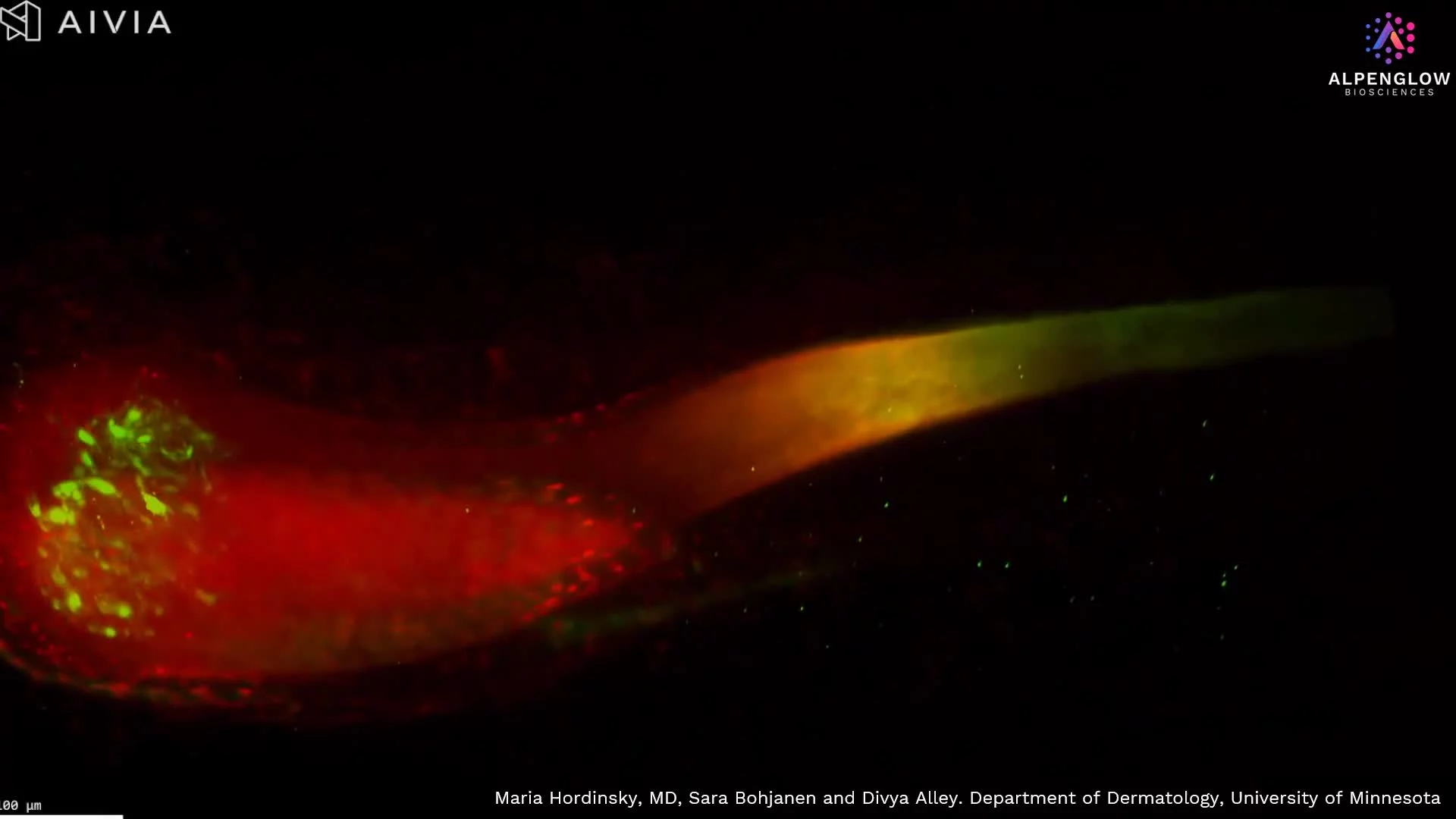
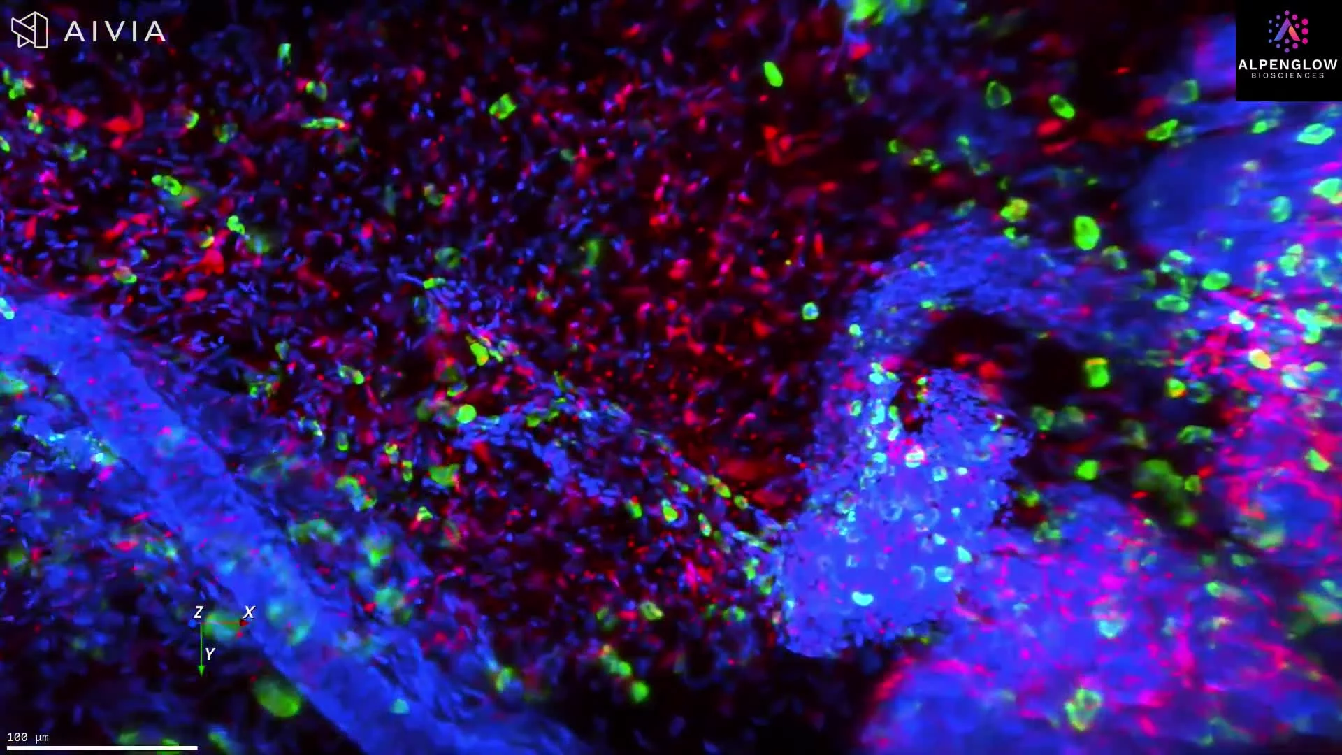
TO-PRO-3, shown in red, labels nuclei and highlights the epidermal and follicular cellular architecture. CD45, shown in blue, reveals the distribution of immune cells, while PGP9.5, shown in green, maps cutaneous nerve fibers across the tissue volume.
The volumetric view preserves intact hair follicles, including the hair bulb, along with surrounding immune cells and nerve networks. Following these structures through depth provides spatial information on follicle morphology, innervation, immune cell distribution, and relationships among skin compartments that may be fragmented in individual tissue sections.
The dataset supports quantitative analysis of follicle dimensions and orientation, immune-cell density and clustering, nerve density and branching, and distances between follicles, immune cells, and nerve fibers. These measurements are relevant to research on alopecia, atopic dermatitis, prurigo nodularis, hidradenitis suppurativa, and other inflammatory skin conditions.
Explore how 3D tissue imaging supports dermatology research across skin architecture, innervation, immune organization, and spatial relationships.
The tissue was imaged using the Aurora 3D™ Spatial Biology Solution, including the 3Di™ Hybrid Open-Top Light-Sheet microscope.