Unprecedented Detail in Atopic Dermatitis with 3D Imaging
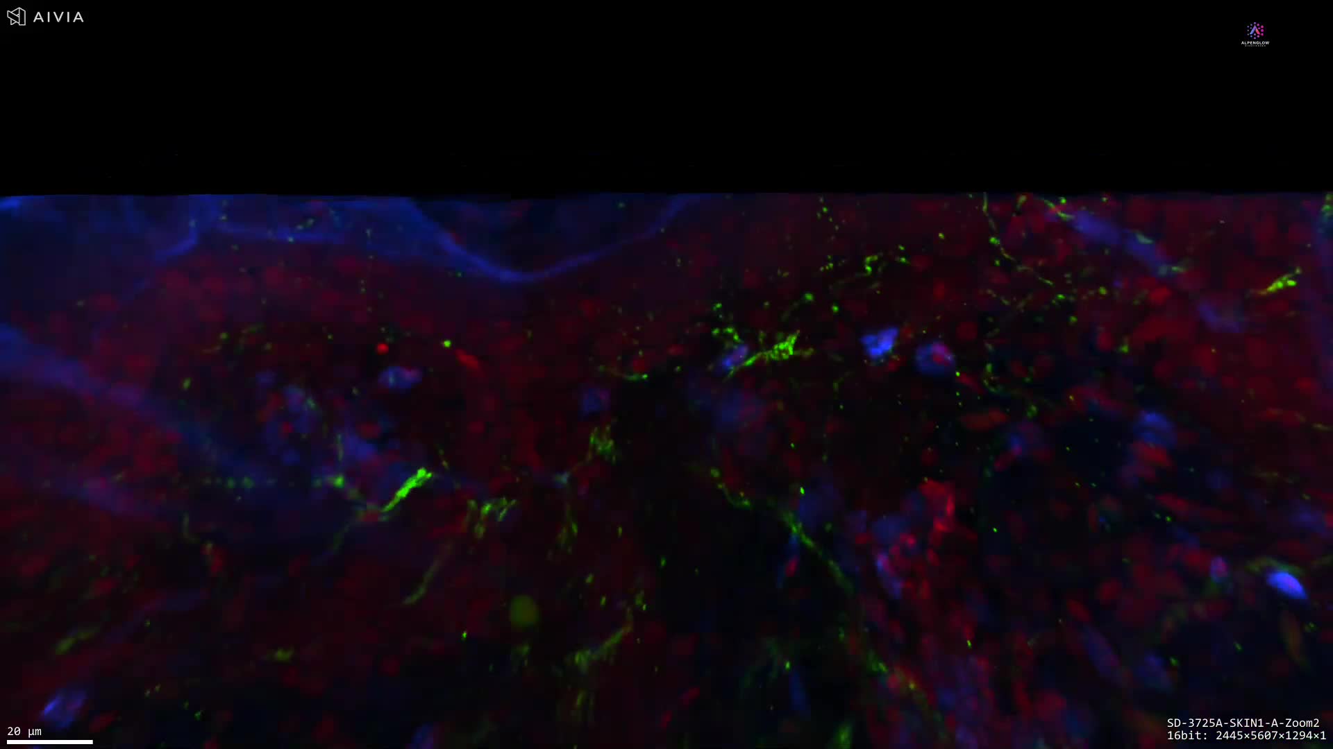
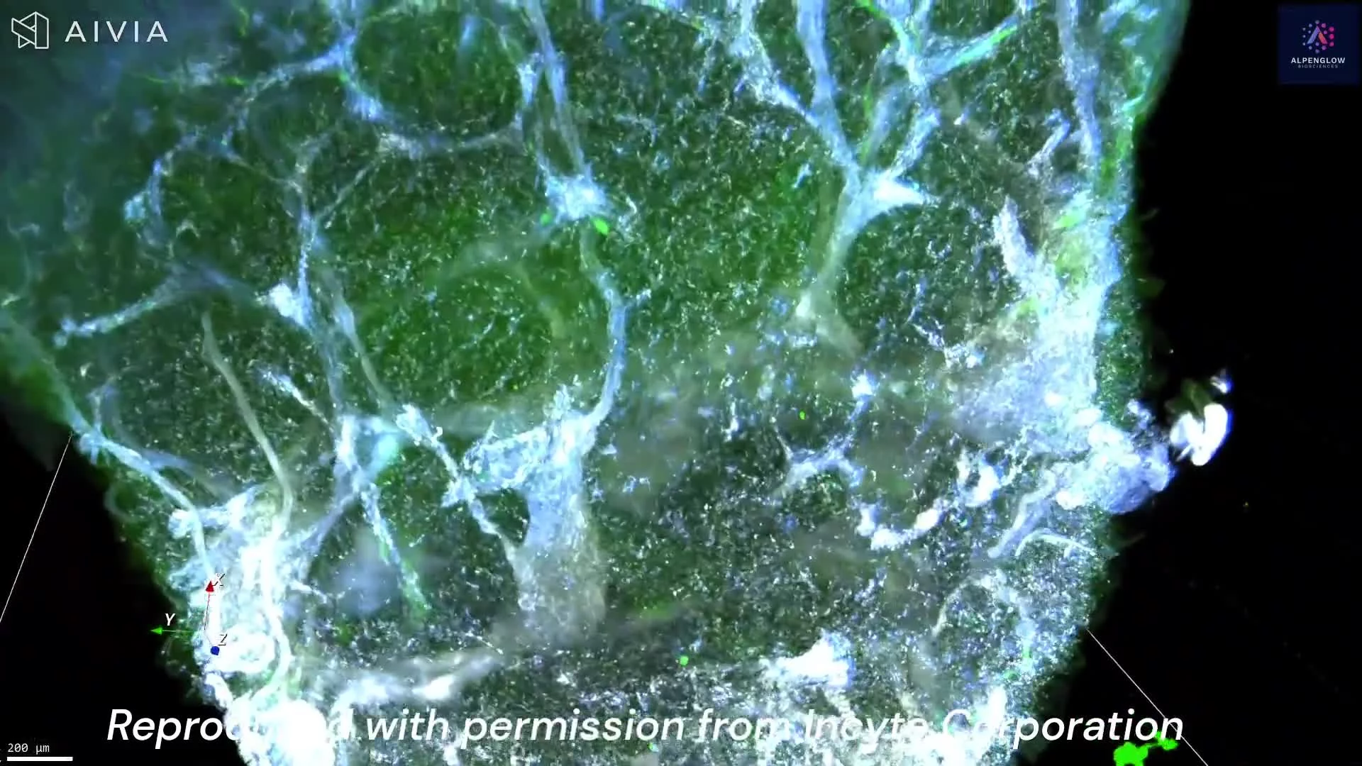
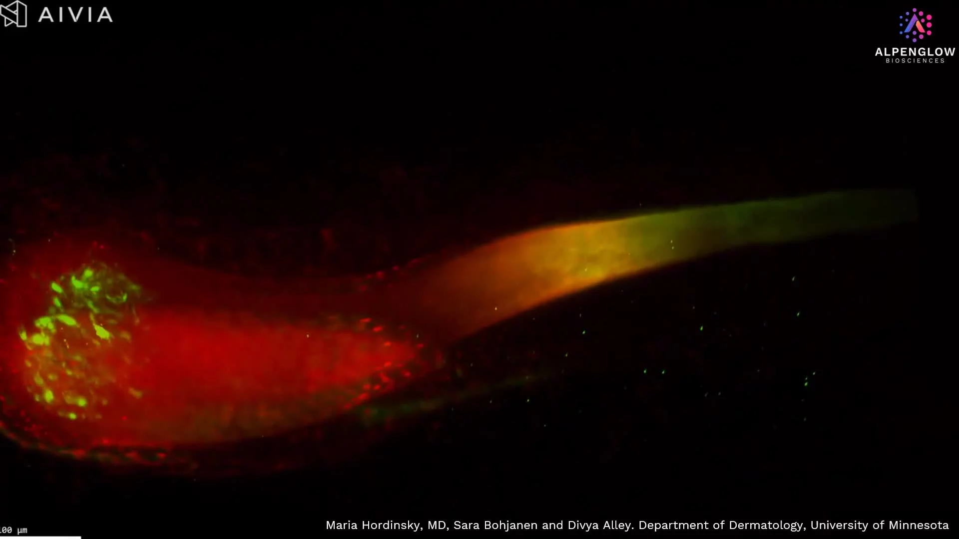
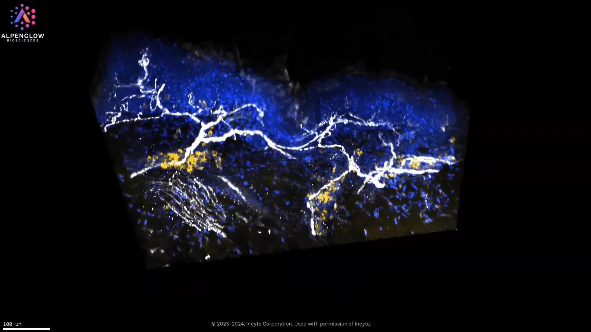
This high-resolution 3D visualization of an atopic dermatitis skin punch biopsy captures structural and cellular details that conventional tissue sections cannot achieve. Every nerve and immune cell is displayed in its true spatial context, offering clarity and depth into disease biology.
Stains used:
TO-PRO-3 (Blue): Highlights nuclear structures
PGP 9.5 (White): Maps precise innervation from larger nerves to finer branches
CD45 (Yellow): Clearly delineates immune cell distribution
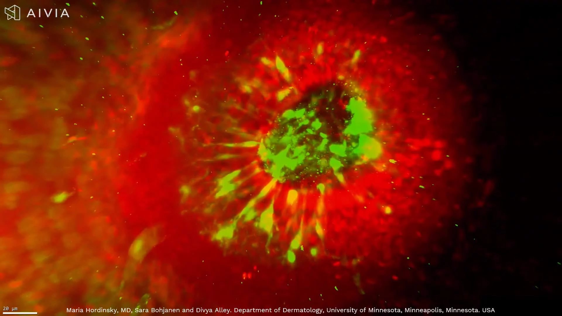
Beyond visualization, the data is fully quantifiable through advanced data management workflows and AI-powered segmentation, enabling precise measurements of immune clustering around nerves.
This capability opens new avenues in dermatology research, driving insights into immune–neural interactions and inflammatory skin disease.
By combining whole-tissue 3D imaging with quantification, the dataset demonstrates the transformative potential of digital pathology and spatial biology.