Whole-biopsy 3D segmentation of prostate glands
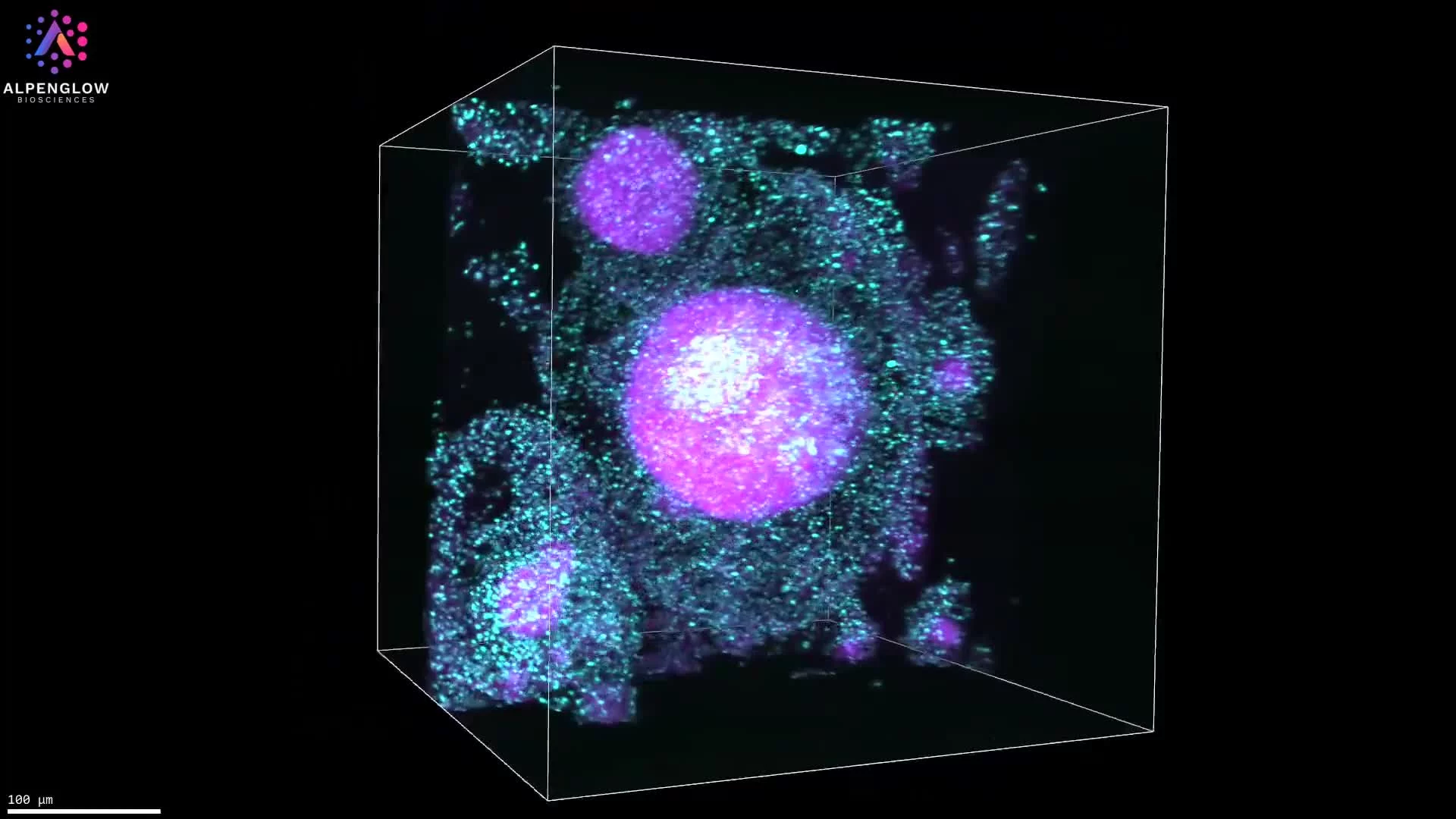
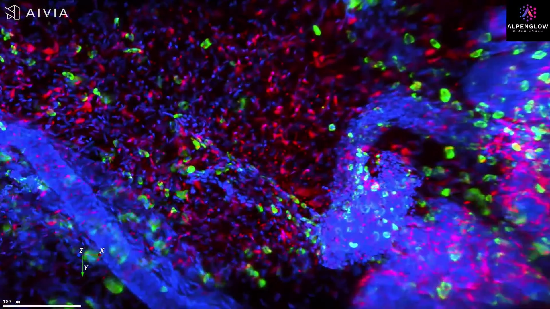
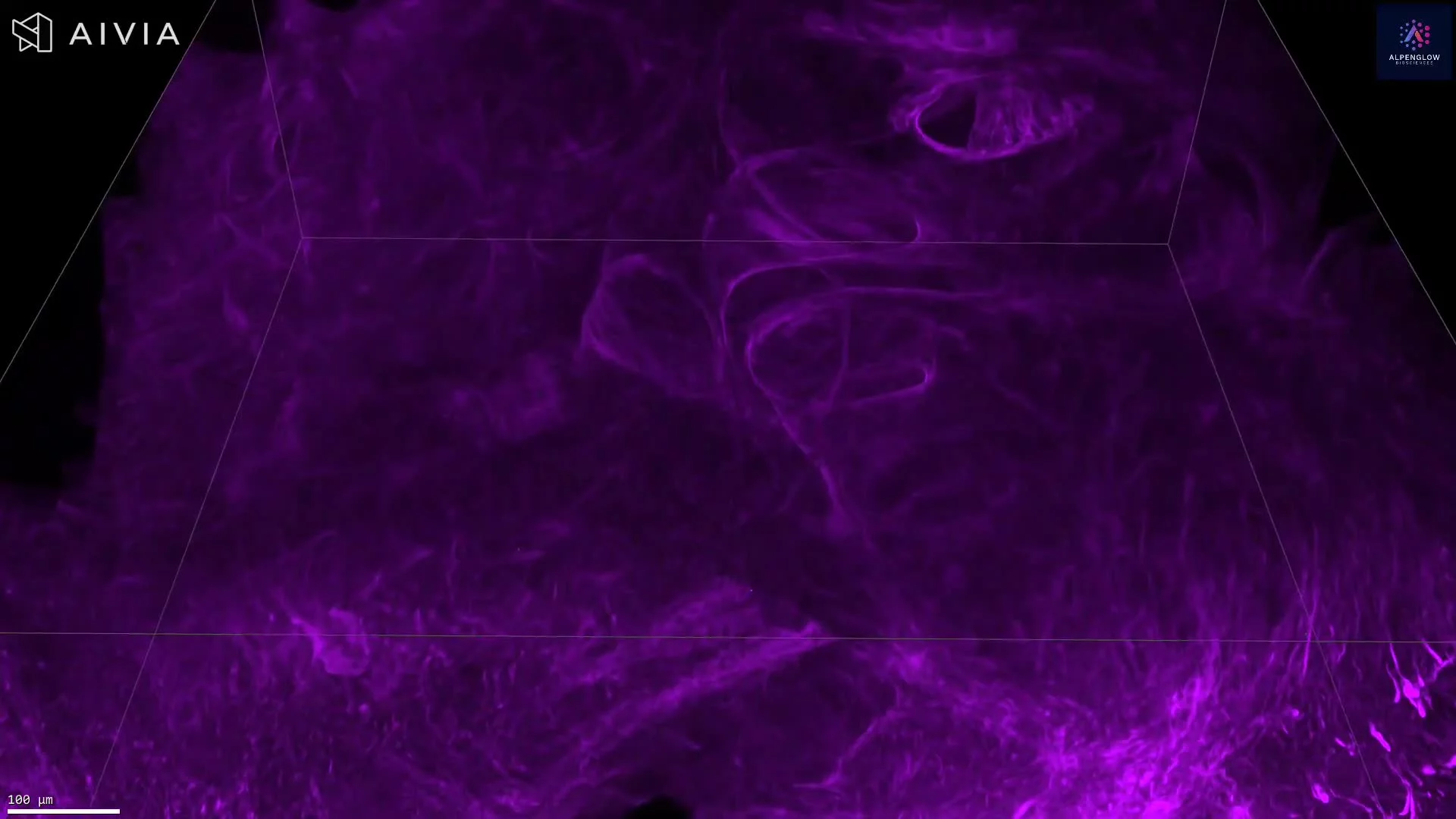
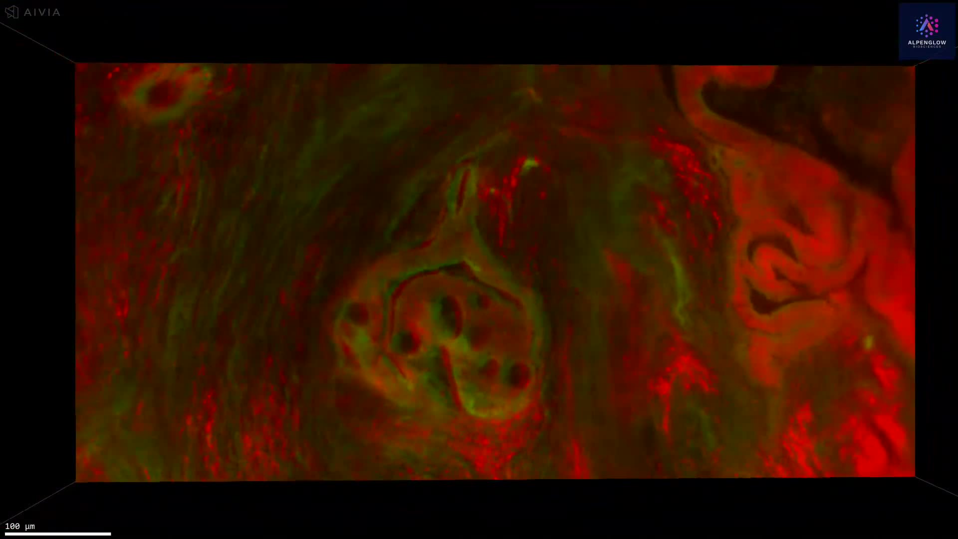
This study demonstrates annotation-free 3D segmentation of prostate glands using fluorescence-based microscopy and AI. A prostate specimen stained with a fluorescent analogue of H&E was converted into a synthetic CK8 immunofluorescence dataset via an image-sequence translation model trained on paired H&E analogue and real CK8 datasets. Traditional computer-vision algorithms were then applied to the synthetic CK8 images for segmentation of gland epithelium, lumen, and stromal regions. The synthetic CK8 image blocks were mosaicked to reconstruct a 3D CK8 dataset of the entire biopsy, enabling accurate gland segmentation. Gland lumen spaces were further segmented by filling regions enclosed by epithelia, with refinements from the cytoplasm (eosin) channel.
3D images acquired with the Aurora™ 3Di Hybrid Open Top Light Sheet (HOTLS) microscope.
With integration of 3Dm data management and 3Dai AI-powered segmentation, researchers can perform reproducible, high-content quantification. This 3D approach eliminates slice bias and ensures accurate analysis at scale.