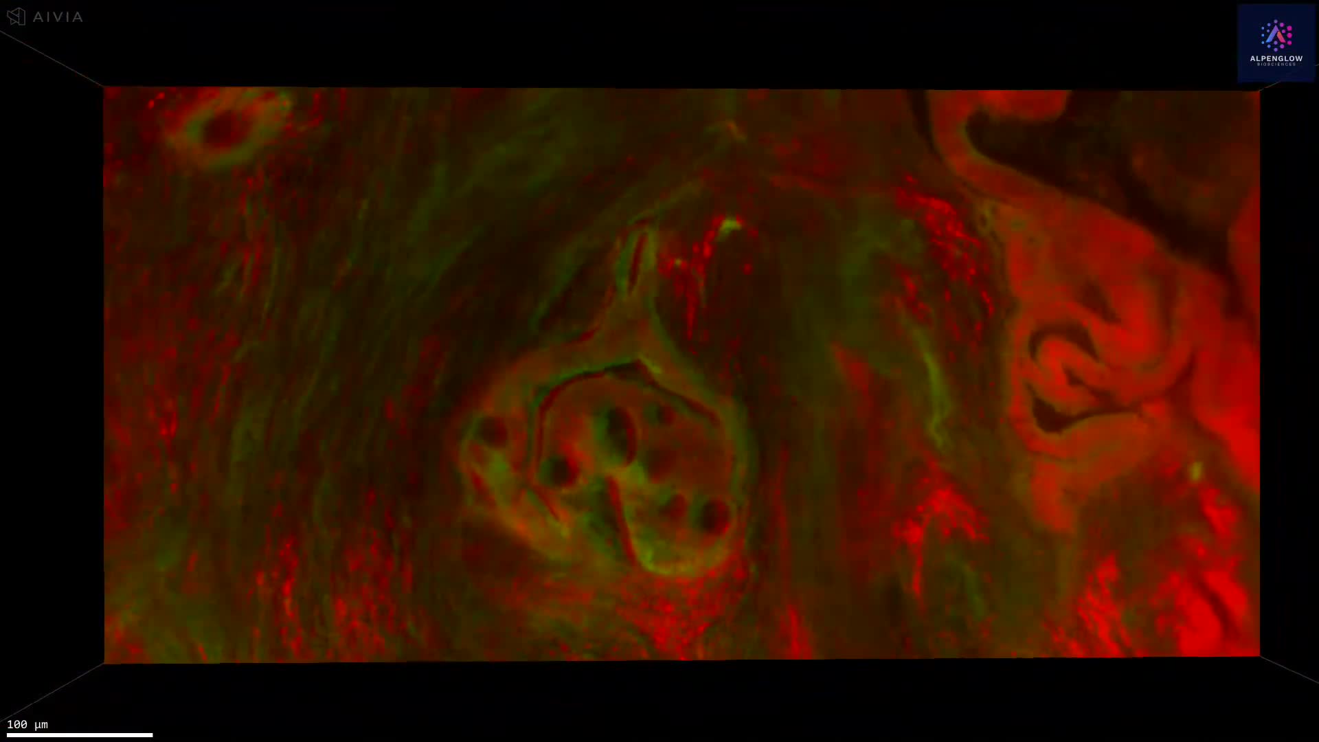
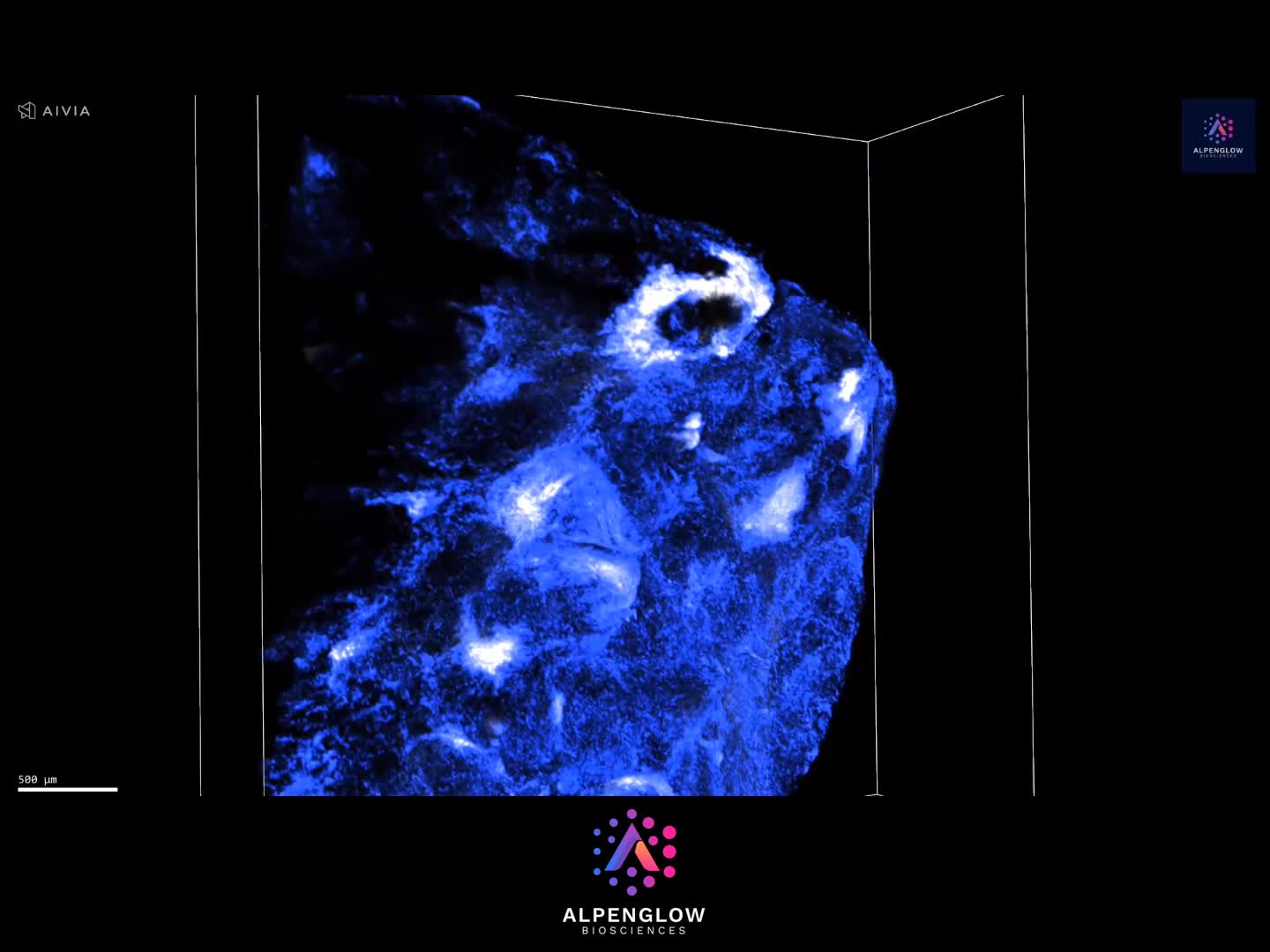
Volumetric 3D Imaging of Human Colon Using Calretinin
This large-scale 3D visualization highlights the human colon stained with Calretinin, a marker of enteric neurons. The dataset reveals the dense innervation of the mucosa and submucosa, preserving the native architecture of the enteric nervous system in full volumetric detail.
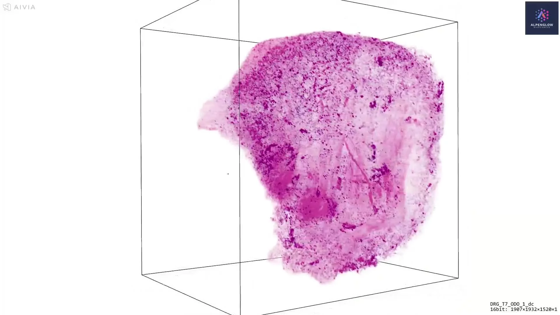
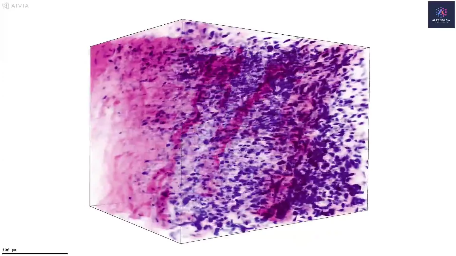
Captured on the Aurora™ 3Di Hybrid Open Top Light Sheet (HOTLS) microscope, the imaging demonstrates the power of 3D histology to overcome sampling limitations of thin sections and provide a comprehensive view of gastrointestinal neural networks. The volumetric dataset can be analyzed through 3Dm data management and 3Dai AI-powered segmentation workflows, enabling quantitative mapping of neuronal density and distribution.
Applications include gastrointestinal motility disorders and developmental conditions such as Hirschsprung disease, where the presence or absence of enteric innervation is central to diagnosis and research.
This approach exemplifies how digital pathology and spatial profiling with 3D imaging drive new insights into gut biology and disease mechanisms.
Contact us to learn how Alpenglow’s 3D spatial biology platform can advance your research with large-scale, quantitative imaging datasets.