
Alpenglow publications
Research from Alpenglow Biosciences and collaborators.
Explore publications connected to 3D tissue imaging, whole tissue imaging, digital pathology, spatial profiling, volumetric datasets, and quantitative tissue analysis.
Selected publications
Open-top light-sheet microscopy, 3D pathology, and quantitative analysis
Explore selected publications spanning the development of open-top light-sheet microscopy, translational and biological applications, and computational approaches for extracting quantitative information from volumetric tissue datasets.
Technology and platform
Foundational work establishing open-top and hybrid open-top light-sheet microscopy, multiscale imaging, tissue processing, and reproducible workflows for nondestructive 3D pathology.
-
View publication
Assessing the effects of a 3D pathology tissue-processing workflow on downstream molecular analyses
Baraznenok, E., et al. Laboratory Investigation 106, 106147, 2026.
-
View publication
An end-to-end workflow for nondestructive 3D pathology
Bishop, K. W., et al. Nature Protocols 19, 1122–1148, 2024.
-
View publication
A hybrid open-top light-sheet microscope for versatile multi-scale imaging of cleared tissues
Glaser, A. K., et al. Nature Methods 19, 613–619, 2022.
-
View publication
Harnessing non-destructive 3D pathology
Liu, J. T. C., et al. Nature Biomedical Engineering 5, 203–218, 2021.
-
View publication
Multi-resolution open-top light-sheet microscopy to enable efficient 3D pathology workflows
Barner, L. A., et al. Biomedical Optics Express 11, 6605–6619, 2020.
-
View publication
Multi-immersion open-top light-sheet microscope for high-throughput imaging of cleared tissues
Glaser, A. K., et al. Nature Communications 10, 2781, 2019.
-
View publication
Light-sheet microscopy for slide-free non-destructive pathology of large clinical specimens
Glaser, A. K., et al. Nature Biomedical Engineering 1, 0084, 2017.
Translational and biological applications
Studies using OTLS and nondestructive 3D imaging to examine intact human and preclinical tissues, reveal spatial biology, evaluate disease, and measure therapeutic effects.
-
View preprint
Spatially resolved multimodal hallmarks of response to neoadjuvant immunotherapies in the melanoma ecosystem in 2D and 3D
Liu, Z., Song, X., Chen, W.-S., et al. bioRxiv preprint, 2026.
-
View publication
3D pathology-guided microdissection
Hsieh, H.-C., Gao, G., Han, Q., et al. Nature Methods 23, 1313–1317, 2026.
-
View publication
Delivery of a fibrin-binding hemostatic polymer ameliorates neurovascular damage and neural tissue loss after traumatic brain injury
Han, Q., et al. Science Advances 11, eadw7425, 2025.
-
View publication
Nondestructive 3D Pathology Image Atlas of Barrett Esophagus With Open-Top Light-Sheet Microscopy
Reddi, D. M., et al. Archives of Pathology & Laboratory Medicine 147, 1164–1171, 2023.
-
View publication
Evaluation of initial prostate cancer biopsies utilizing 3D open-top light-sheet microscopy for detection of early disease
Mo, G., Reder, N. P., Schweizer, M. T. The Prostate 83, 1121–1124, 2023.
-
View publication
Comprehensive Surface Histology of Fresh Resection Margins With Rapid Open-Top Light-Sheet (OTLS) Microscopy
Gao, G., et al. IEEE Transactions on Biomedical Engineering 70, 2160–2171, 2023.
-
View publication
Multiresolution nondestructive 3D pathology of whole lymph nodes for breast cancer staging
Barner, L. A., et al. Journal of Biomedical Optics 27, 036501, 2022.
-
View publication
Ablating Lgr5-expressing prostatic stromal cells activates the ERK-mediated mechanosensory signaling and disrupts prostate tissue homeostasis
Wei, X., et al. Cell Reports 40, 111313, 2022.
-
View publication
Diagnosing 12 prostate needle cores within an hour of biopsy via open-top light-sheet microscopy
Xie, W., et al. Journal of Biomedical Optics 25, 126502, 2020.
-
View publication
Open-Top Light-Sheet Microscopy Image Atlas of Prostate Core Needle Biopsies
Reder, N. P., et al. Archives of Pathology & Laboratory Medicine 143, 1069–1075, 2019.
-
View publication
Rapid pathology of lumpectomy margins with open-top light-sheet (OTLS) microscopy
Chen, Y., et al. Biomedical Optics Express 10, 1257–1272, 2019.
3D analysis and AI
Computational methods for extracting quantitative features, segmenting complex tissue structures, identifying disease-relevant regions, and translating large 3D datasets into measurable outputs.
-
View publication
Deep-learning triage of three-dimensional pathology datasets for comprehensive and efficient pathologist assessments
Gao, G., Yan, R., Song, A. H., et al. Nature Biomedical Engineering, 2026.
-
View publication
AI-Informed Architectural Insights of Three-Dimensional Glandular Networks Identify Prostate Cancer Patients at a Higher Risk of Biochemical Recurrence
Salguero-Lopez, J., et al. Modern Pathology 39, 101018, 2026.
-
View publication
Detection of Prostate Cancer in 3-Dimensional Pathology Datasets via Generative Immunolabeling
Serafin, R. B., et al. Modern Pathology 39, 100975, 2026.
-
View preprint
Scalable 3D cell-interaction analysis via supercell graphs for prostate cancer risk stratification
Zhao, Y., et al. bioRxiv preprint, 2026.
-
View publication
Analysis of 3D pathology samples using weakly supervised AI
Song, A. H., et al. Cell 187, 2502–2520.e17, 2024.
-
View publication
Direct three-dimensional segmentation of prostate glands with nnU-Net
Wang, R., et al. Journal of Biomedical Optics 29, 036001, 2024.
-
View publication
Artificial Intelligence-Triaged 3-Dimensional Pathology to Improve Detection of Esophageal Neoplasia While Reducing Pathologist Workloads
Erion Barner, L. A., et al. Modern Pathology 36, 100322, 2023.
-
View publication
Prostate Cancer Risk Stratification via Nondestructive 3D Pathology with Deep Learning–Assisted Gland Analysis
Xie, W., et al. Cancer Research 82, 334–345, 2022.
Scientific posters
Selected 3D imaging and spatial biology studies
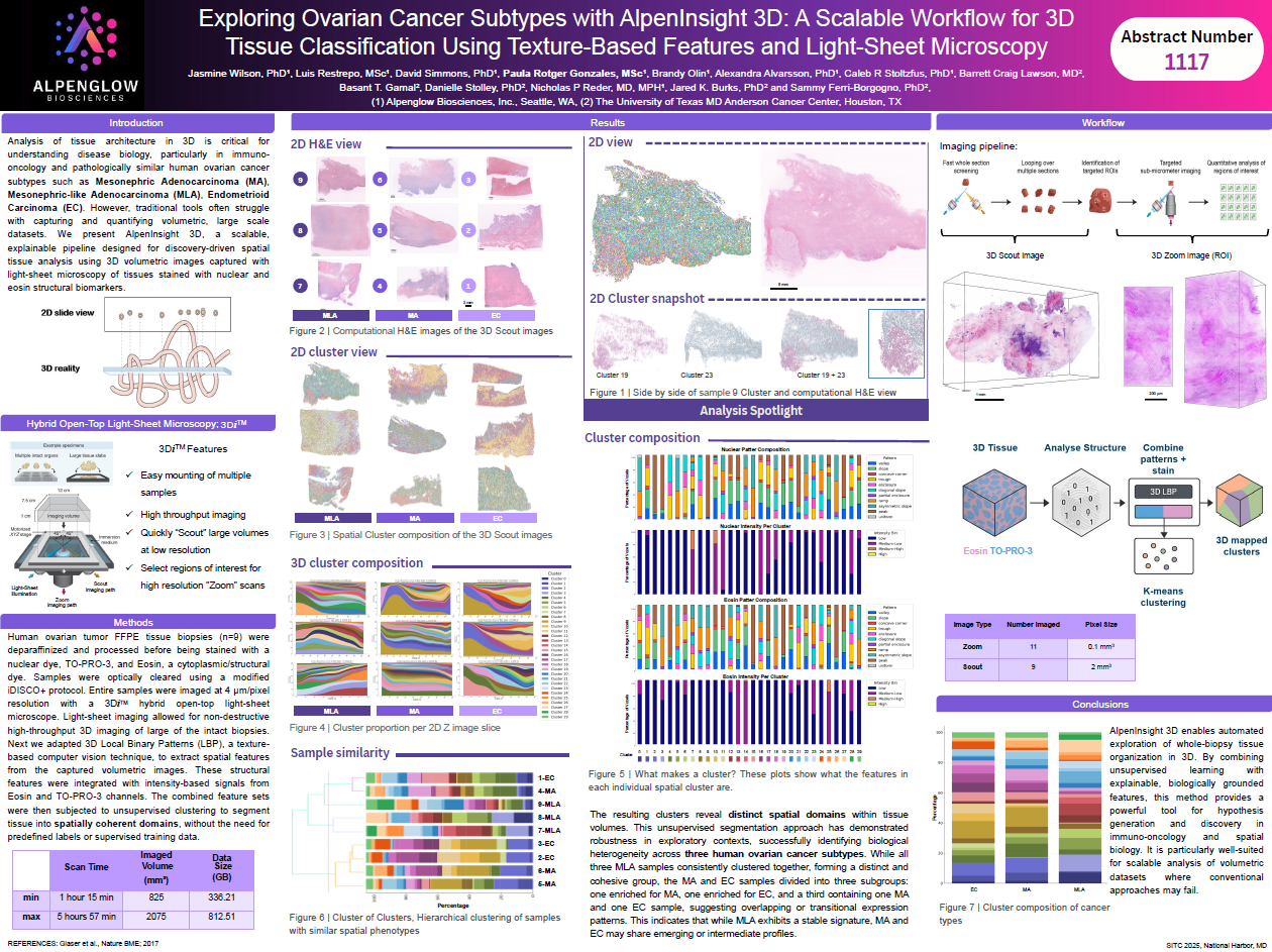
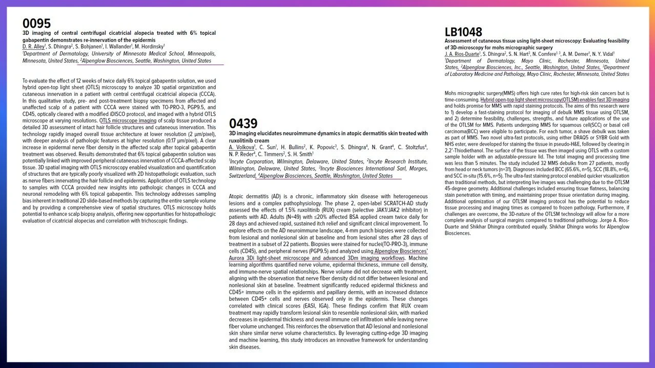
Explore posters and conference abstracts from Alpenglow Biosciences and collaborators covering immuno-oncology, dermatology, liver disease, 3D image analysis, and HOTLS microscopy.
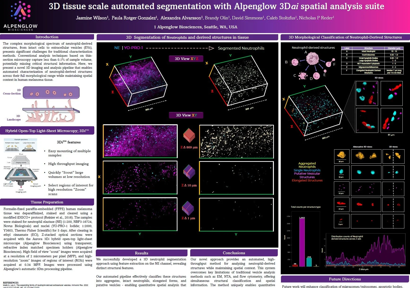
3D tissue-scale automated segmentation with Alpenglow 3Dai spatial analysis suite
View poster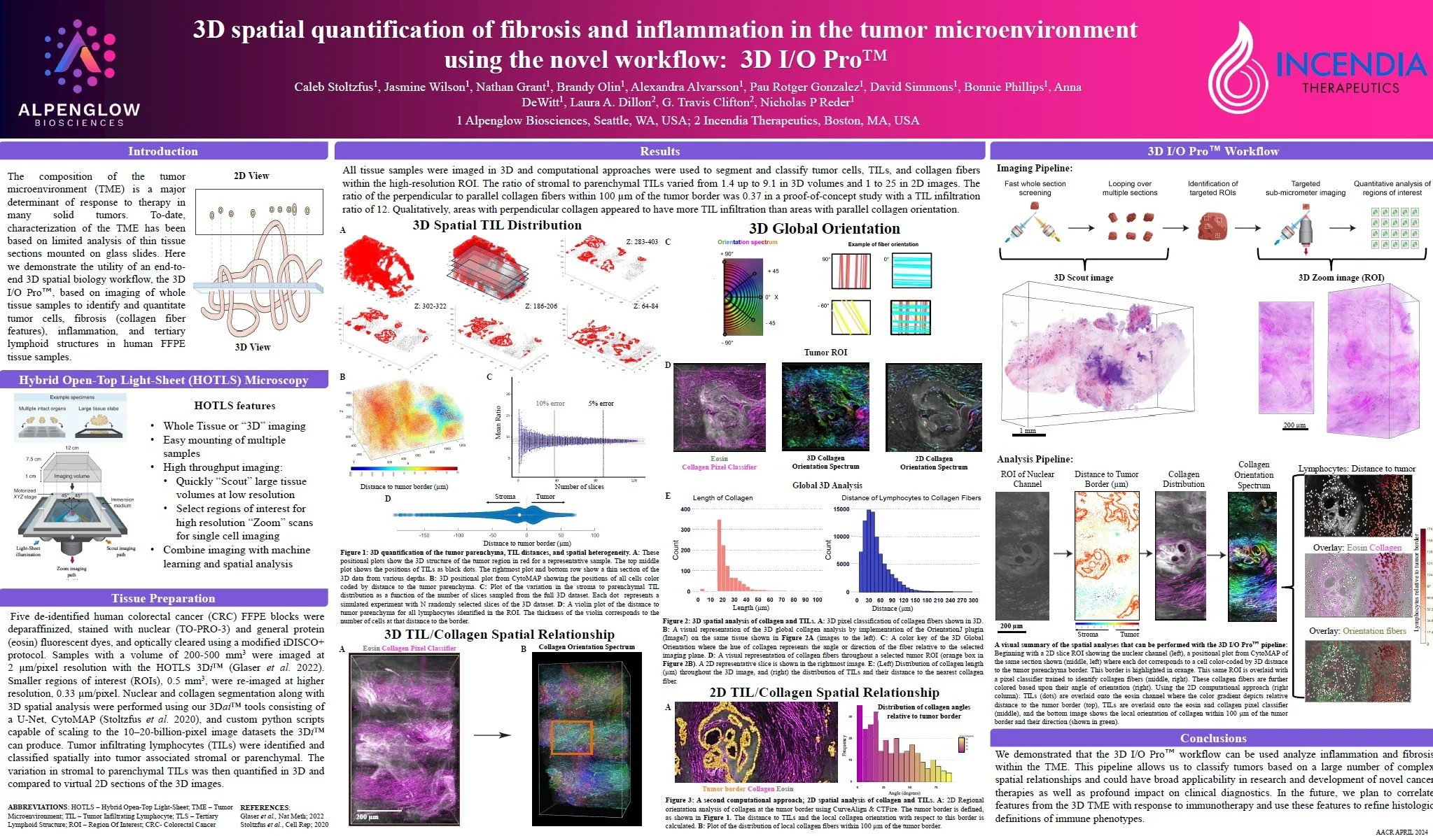


From publications to projects
Ready to apply Alpenglow’s 3D imaging workflows to your research?
Connect published examples to your own tissue questions with 3D tissue imaging, whole tissue imaging, volumetric datasets, spatial profiling, and AI-powered analysis.