3D histology imaging services
End-to-end 3D histology services for intact tissue analysis
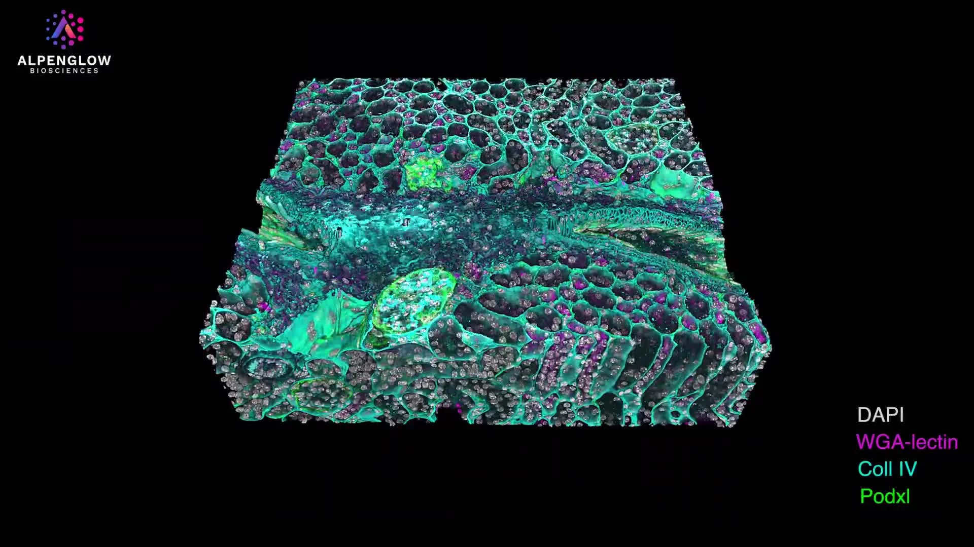
Alpenglow Biosciences supports 3D tissue imaging workflows from sample preparation to whole tissue imaging, volumetric datasets, AI-powered analysis, and quantitative tissue analysis for digital pathology and spatial profiling studies.
Built around Aurora 3Di, 3Dm, and 3Dai to connect imaging, data management, and analysis in one 3D spatial biology workflow.
3D Dataset of H&E-Stained Liver Biopsy
Service pathway
From tissue to quantitative 3D data
Alpenglow Biosciences helps teams move from intact tissue samples to volumetric datasets, spatial profiling, and quantitative tissue analysis without building the full 3D histology workflow internally.
Plan and prepare the sample
We support study planning, tissue handling, clearing, staining, and marker strategy for intact tissue workflows.
- Sample preparation guidance
- Marker and assay planning
- Workflow design for pilot or translational studies
Image the intact volume
Whole tissue imaging captures 3D tissue architecture and spatial relationships across depth, not just thin section views.
- 3D tissue imaging of intact samples
- Volumetric imaging for large tissue regions
- Digital pathology datasets with preserved spatial context
Process, analyze, and deliver readouts
Image processing and AI-powered analysis convert volumetric datasets into quantitative tissue analysis outputs for spatial biology studies.
- Large dataset processing and management
- AI-powered segmentation and quantification
- Study-specific spatial profiling readouts
The service workflow is built around Aurora 3Di, 3Dm, and 3Dai, connecting 3D imaging, data management, and quantitative analysis in one spatial biology workflow.
Start a service discussionDeliverables
What your team receives
The service is designed to give research, translational, biomarker, and pathology teams usable 3D tissue data, not just image files. Outputs can support visualization, spatial profiling, and quantitative tissue analysis.
Volumetric 3D datasets
Whole tissue imaging outputs preserve spatial context across intact tissue volumes, helping teams review tissue architecture, cellular organization, and biological structures in 3D.
Whole tissue imagingProcessed imaging files
Volumetric datasets can be processed for visualization, review, and downstream interpretation, reducing the burden of handling large 3D tissue imaging files internally.
Data processingQuantitative tissue analysis
Measurements tailored to the study, including cell counts, densities, distances, volumes, morphology, branching, and other spatial or structural metrics. Quantitative readouts
AI-powered analysis outputs
Reviewable segmentations, masks, classifications, and detected biological structures generated from intact 3D tissue datasets.
AI-powered analysisStudy-specific reporting
Organized results and visual summaries aligned with the biological question, study design, and selected quantitative endpoints.
Spatial biology supportFlexible study scale
Built for studies at every stage
Start with sample and marker evaluation, extend validated workflows across translational cohorts, and scale preparation, imaging, and quantitative analysis as study needs grow.
Pilot studies
Evaluate sample compatibility, staining strategies, imaging parameters, and initial quantitative readouts before expanding the study.
Translational studies
Apply consistent whole-tissue imaging, spatial profiling, and AI-powered analysis across study cohorts and biological conditions.
Scalable programs
Extend validated preparation, imaging, and quantitative tissue analysis workflows across larger programs while maintaining consistent outputs.
2D vs 3D histology
Add intact tissue context to slide-based insight
2D sections are useful, but many spatial biology questions depend on depth, continuity, and full-volume architecture.
A thin plane through tissue
- Limited tissue volume sampled
- Spatial relationships inferred across sections
- Complex structures can be harder to follow
A volumetric view of intact tissue
- Whole tissue imaging across depth
- Spatial profiling with preserved architecture
- AI-powered analysis and quantitative tissue analysis
Applications
Explore 3D histology services through real dataset examples
See how intact 3D tissue imaging supports spatial profiling, volumetric datasets, and quantitative tissue analysis across different sample types.
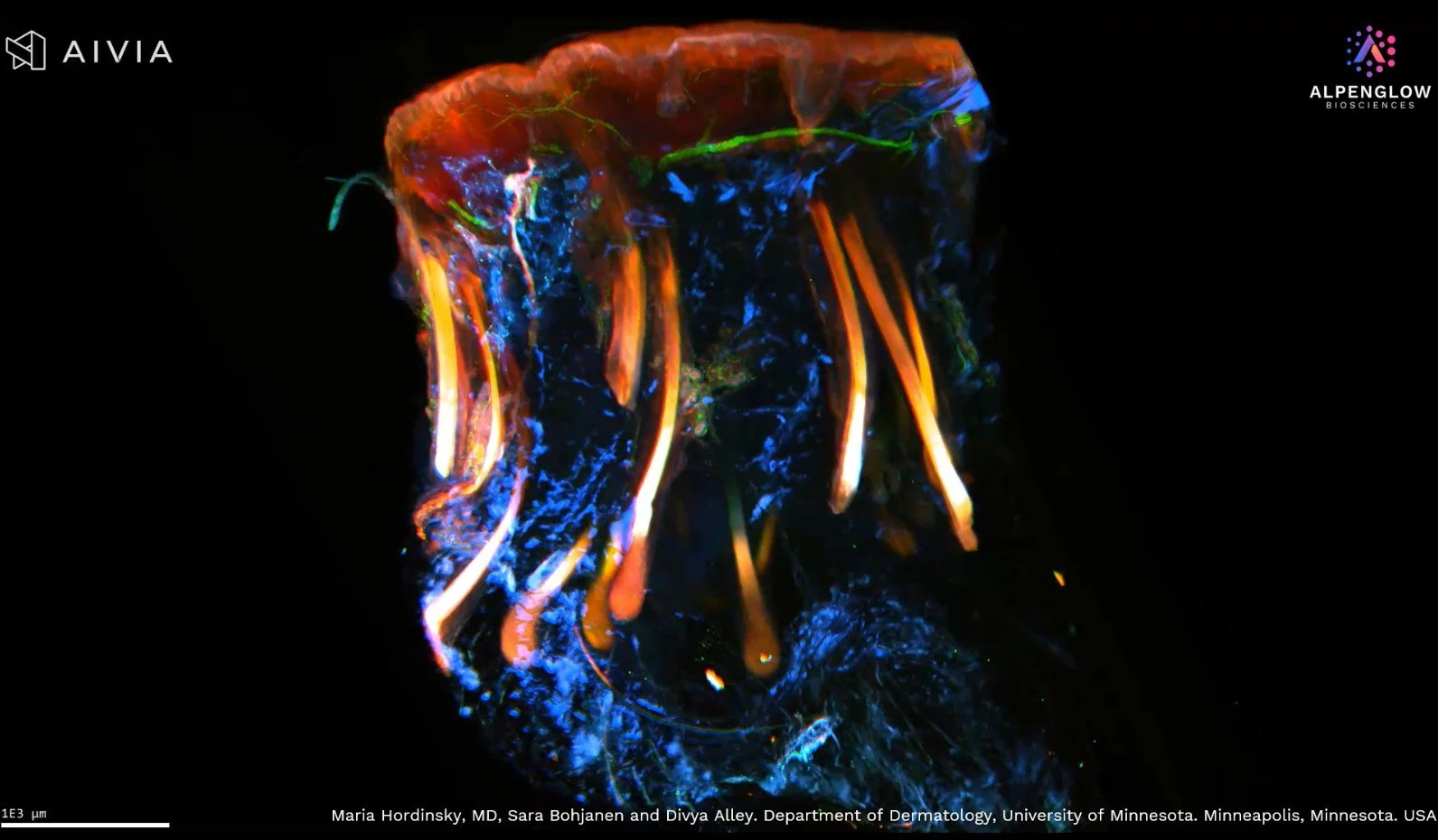
Skin and inflammation
Nerve branching and tissue architecture in intact skin.
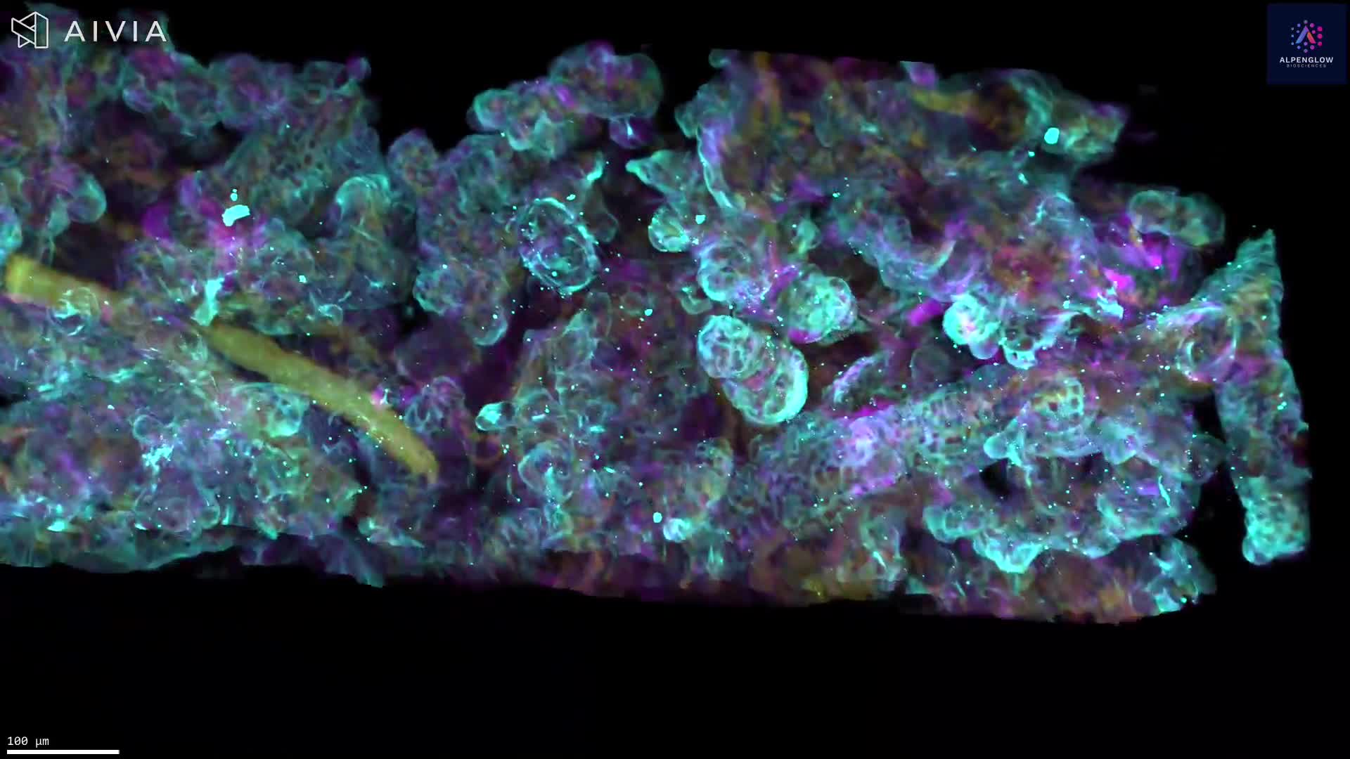
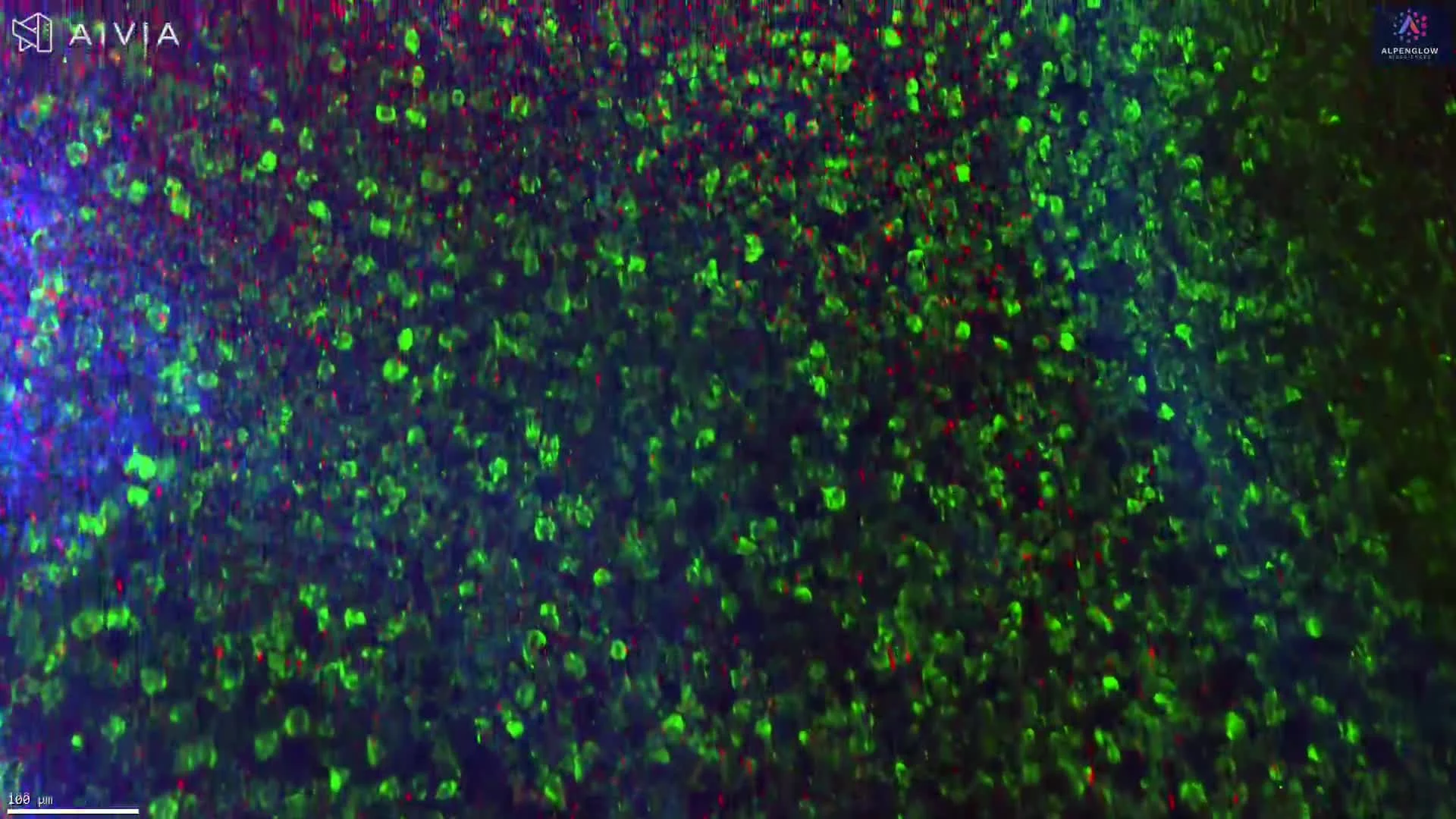
View skin dataset 02Tumor microenvironment
Immune organization and tertiary lymphoid structures in 3D.
View tumor dataset 03Fibrosis and remodeling
Collagen architecture and structural remodeling in 3D.
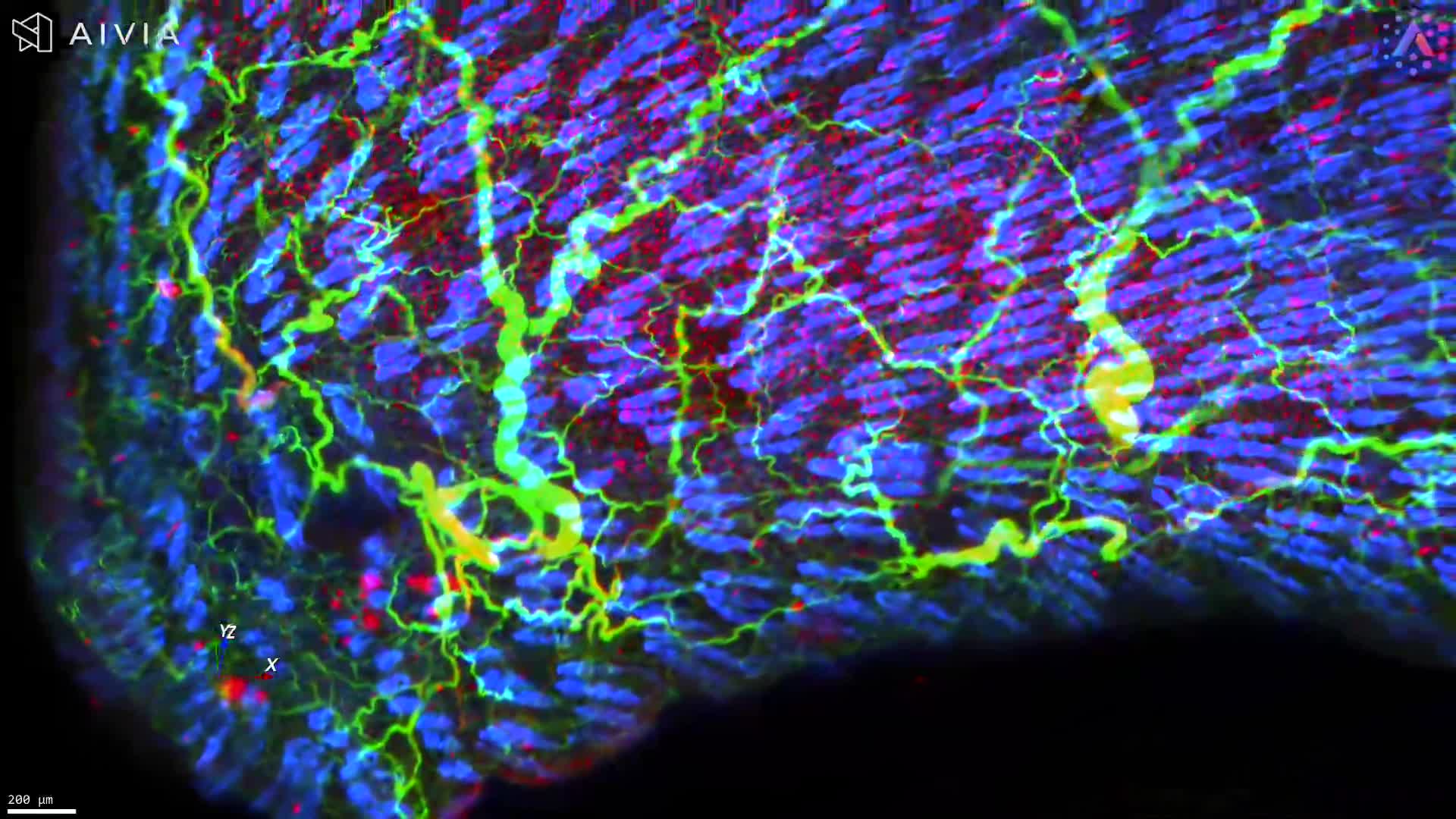
View fibrosis dataset 04Segmentation and spatial profiling
AI-powered analysis for quantitative tissue readouts.
View analysis datasetNeed a different tissue type or marker strategy? Alpenglow can help evaluate whether 3D histology imaging services fit your sample and study goals.
View more 3D datasets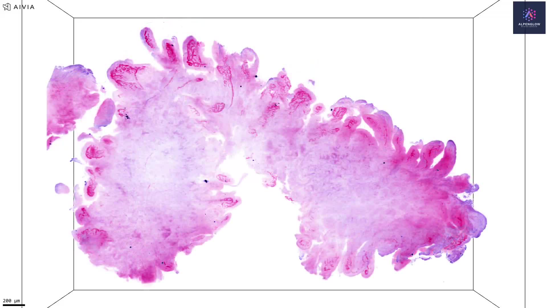

Start a service discussion
Ready to evaluate what 3D histology can reveal in your tissue?
Share your sample type, marker strategy, and study goals. Alpenglow Biosciences can help determine whether 3D tissue imaging, volumetric datasets, and quantitative tissue analysis are a fit for your program.