Frequently asked questions
Answers about 3D tissue imaging, whole tissue workflows, and AI-powered analysis.
Learn how Alpenglow Biosciences supports volumetric datasets, digital pathology, spatial profiling, and quantitative tissue analysis through the Aurora 3D Spatial Biology Solution, including 3Di, 3Dm, and 3Dai.
About Alpenglow
What Alpenglow Biosciences does
-
Both. We build custom hybrid open-top light-sheet microscopes, develop advanced imaging and analysis software, and provide full contract research services, offering an end-to-end solution for tissue processing, 3D imaging, and quantitative analysis.
-
Yes. We can tailor hardware configurations, staining protocols, imaging strategies, and analysis pipelines based on project needs. Customization is a key part of how we support a wide range of research and clinical applications.
-
Yes. Our platform is flexible enough for early-stage research, translational studies, and clinical sample imaging. We partner with groups across discovery, biomarker development, and pathology innovation.

3D tissue imaging
Whole tissue context beyond 2D sections
-
A laser is shaped into a thin blade of light to illuminate a large slice of the sample, allowing for fast, efficient 3D imaging.
-
The 3Di HOTLS is Alpenglow’s patented system combining open-top design and light-sheet microscopy for rapid, large-area 3D tissue imaging at cellular resolution.
-
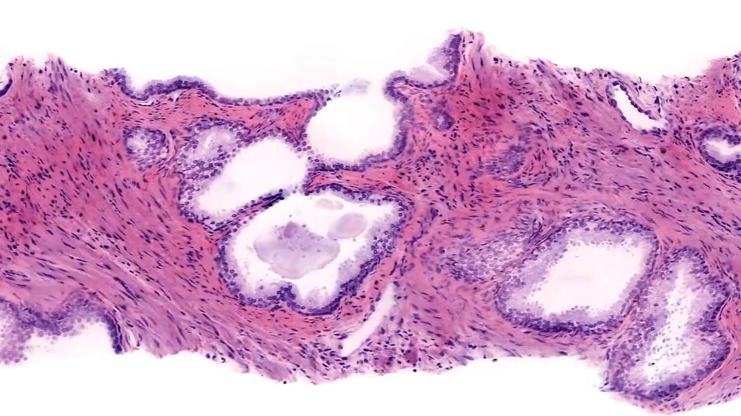
3D imaging preserves full spatial context, identifies complex tissue architectures, and improves detection of rare events often missed in 2D slices.
-
Our light-sheet system is purpose-built to image everything from organoids to large FFPE samples, using custom-designed sample holders. A large lateral scan area and dual imaging paths (low-resolution Scout and high-resolution Zoom) enable both overview and near-confocal scans in one system. ROI rescans can be performed without sample movement.
-
405 nm, 488 nm, 561 nm, 638 nm; custom configurations up to 785 nm available.
-
Alpenglow routinely images samples ranging from a few millimeters up to several centimeters in size, ideal for large biopsies and full tissue sections. Lateral up to 10-12 cm and depth up to 1 cm; no fixed lateral limit with custom stages.
-
Scout path: 2.021 µm/pixel.
Zoom path: 0.167 µm/pixel.
Samples and preparation
From tissue type to imaging-ready sample
-
Any fresh, frozen, formalin-fixed, PFA-fixed, or FFPE tissue. Optimal sections are 1 mm- 1 cm thick.
-
Yes, routinely.
-
Yes, after deparaffinization and antigen retrieval if needed.
-
We primarily use a modified DISCO/iDISCO clearing with ethyl cinnamate as the clearing agent. Other clearing methods can be used if needed for specific projects.
-
No, it preserves DNA and proteins; tissue shape is maintained with ~10% shrinkage. RNA preservation is under validation.
-
Minimal — distortion is comparable to FFPE processing. Tissue architecture is preserved with minimal artifact, validated across thousands of samples.
-
Currently, up to four fluorescent channels can be simultaneously imaged, depending on marker compatibility and experimental design.
-
Alpenglow’s platform supports a wide variety of tissues, including (but not limiting):
-
Most fluorescent stains, conventional fluorophores, antibodies, and nanobodies are compatible.

Data analysis
Turning volumetric datasets into quantitative tissue analysis

-
You’ll receive high-resolution, full-volume 3D datasets along with rich, publication-ready outputs, including:
3D image volumes
Interactive viewers for data exploration
Visualization videos
Summary tables of cell-level features and spatial metrics
These deliverables are formatted for seamless integration with downstream analysis and interpretation.
-
Our AI/ML pipeline delivers powerful spatial insights, including but not limited to:
Automated segmentation of cells and tissue structures
Spatial profiling (e.g., distances, clustering, neighborhood analysis)
Density mapping, volume quantification, and pattern detection.
These tools reveal organization, interaction, and complexity within intact tissues.
-
Yes, our platform is built for flexibility. We collaborate with you to tailor AI models to your markers, regions of interest, and study goals. This includes model training, validation, and adaptation for specific tissue types or biological questions.
-
Data is processed through a GPU‑accelerated bioinformatics pipeline optimized for large 3D datasets. Results are securely delivered via encrypted cloud link and include all analysis outputs, ready for use in research or publication.
Clinical and pathology applications
Applying 3D tissue imaging across tissues, diseases, and translational research programs
-
Enables full biopsy digitization without serial sectioning and supports faster, more comprehensive diagnostics.
-
Minimal — 3D images can be displayed as traditional 2D slices for familiar navigation, with the option to scroll through depth.
-
No, it enhances their capabilities by providing more complete tissue information, supporting better diagnosis and outcomes.
Applications and use cases
Where 3D tissue imaging can support deeper biological insight
-
Neuroscience
Toxicology
Fibrosis research
Vascular biology
Discover more on -Who We Help-
-
Yes. It supports efficacy evaluation, mechanism of action studies, toxicity profiling, biodistribution analysis, and predictive AI modeling for drug candidates.
Projects and services
Starting a project with the Alpenglow team
-
Interested researchers can connect via the website contact form to discuss project needs, sample submission guidelines, and workflow timelines.
-
Reach out through the Contact Page to schedule an initial discovery meeting.
-
We will set up a personalized virtual demo to show you the instrument and its capabilities, and discuss a pilot project tailored to your specific samples.