

3D tissue imaging of mouse muscle reveals neural architecture across full volumetric context. NF200 labels large-caliber axons, PGP9.5 captures the broader neuronal network, and YO-PRO-1 marks nuclei, enabling quantitative spatial profiling and digital pathology workflows.
3D expansion microscopy of mouse kidney tissue reveals glomerular and extracellular matrix architecture with preserved volumetric context and enhanced spatial resolution.
3D light-sheet imaging of an intact mouse colorectal tumor reveals B and T cells in their native spatial context, resolving single immune cells across the entire tissue volume.
3D imaging of atopic dermatitis skin punch biopsy; the tissue is stained with TO-PRO-3, PGP9.5, and CD45. Explore detailed innervation and immune-cell interactions in lesional skin.
3D imaging of mouse colorectal tumor stained with CD3, B220, and YoPro1 reveals tertiary lymphoid structures (TLS) in full spatial context.

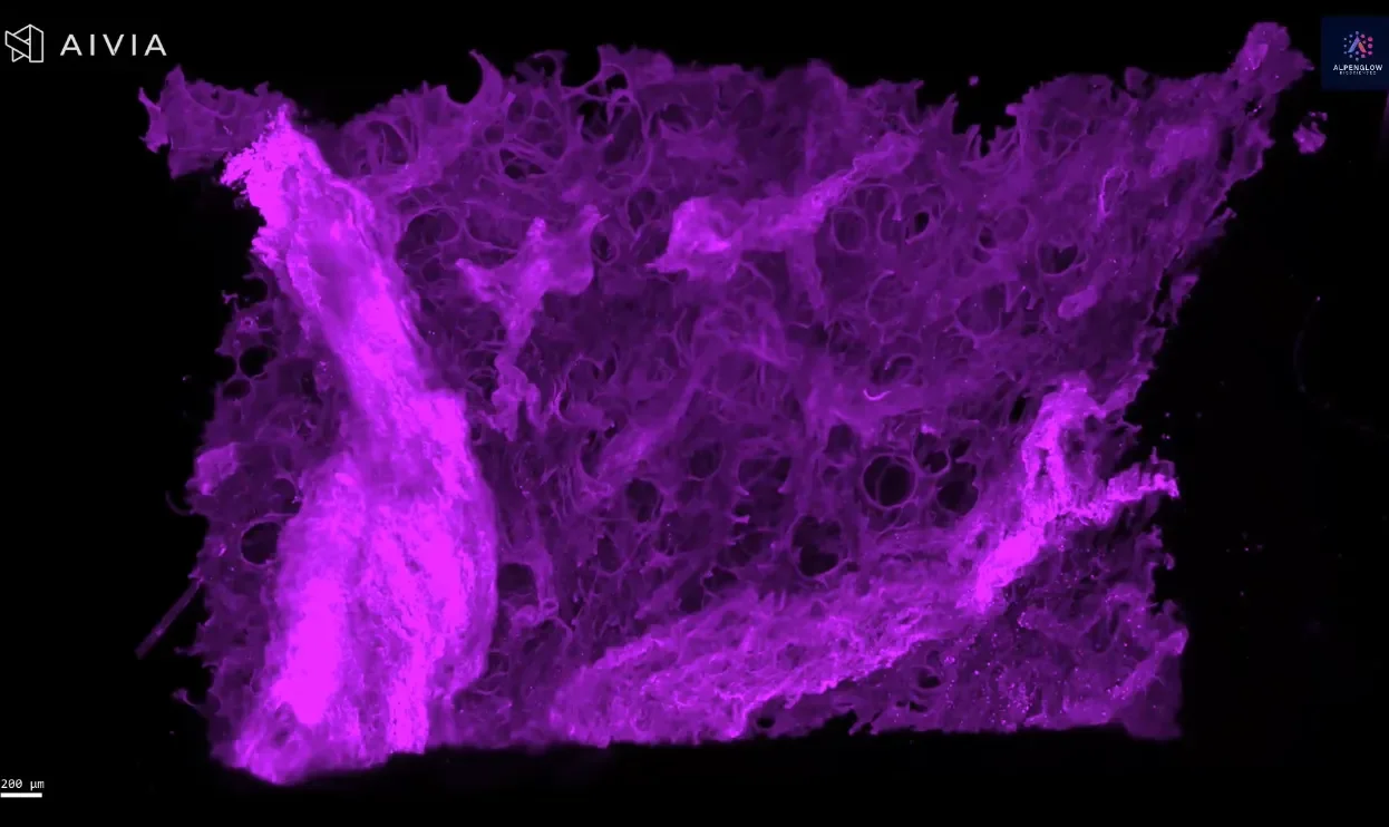
3D fluorescence imaging of human lung tissue stained with Fast Green reveals collagen architecture for fibrosis and tissue remodeling research.
3D imaging of human skin biopsy stained with tryptase, TO-PRO-3, and PGP9.5 reveals mast cell–nerve interactions for dermatology and oncology research.
High-resolution 3D imaging of a mouse pancreatic cyst stained with Ki67 and γ-H2AX reveals proliferation and DNA damage in full spatial context.
3D fluorescence imaging of prostate tissue stained with Fast Green reveals the collagen architecture, orientation, and density, providing insights beyond 2D histology.
High-resolution 3D imaging of human tonsil tissue stained with YO-PRO-1 for nuclei and tryptase for mast cells.
2 mm pig muscle section stained with CD31 and YO-PRO-1 imaged in 3D on Aurora HOTLS, revealing vascular networks with speed and precision.
FFPE colorectal tissue stained with YO-PRO-1 and anti-CD8 imaged in 3D at 2 μm/pixel on Aurora HOTLS. Quantification of CD8+ lymphocytes across whole section.
3D imaging of human placenta tissue stained with SMA, HLA-G, and CD31. From whole-tissue scans to single-cell resolution, uncover tissue complexity in detail.
3D imaging of a whole murine heart stained with SMA reveals vascular structures and mural cells, from large vessels down to pericytes.
3D imaging of cleared liver tissue stained with Collagen III, followed by segmentation analysis using a pixel classifier to highlight fibrosis.
3D imaging of human colon stained with Calretinin reveals dense innervation of the mucosa and submucosa.
3D imaging of cleared mouse fat pad reveals blood vessels, macrophages, and nerves labeled with lectin, CD68, and PGP9.5.
High-resolution 3D imaging of scalp epidermis reveals branching nerve structures and nuclear organization, advancing hair and dermatology research.
High-resolution 3D imaging of atopic dermatitis reveals lymphocyte clusters near nerves, stained with TO-PRO-3, PGP 9.5, and CD45. Explore detailed innervation and immune-cell interactions in lesional skin.
40X image of a lesional Atopic Dermatitis samples stained with PGP9.5 (white) to highlight fine epidermal innervation, TO-PRO-3 (blue) for nuclear detail, and CD3 (green) to illuminate T-Cell infiltration of the Epidermis and Dermis.
High-resolution 3D imaging of CD45-stained skin biopsy shows lymphocyte clustering around nerves, offering new insights into immune cell behavior.
3D imaging of cleared tonsil tissue stained with anti-CD21 and TO-PRO-3 reveals B cell and follicular dendritic cell distribution in FFPE samples.
3D imaging of cleared tonsil tissue stained with anti-CD45RO (UCHL1) and TO-PRO-3 reveals memory T cell distribution in thick FFPE samples.
3D imaging of cleared tonsil tissue stained with anti-CD3 (BC33) and TO-PRO-3 reveals the distribution of T cells in thick FFPE samples on the Aurora HOTLS platform.
3D imaging of cleared tonsil tissue stained with anti-CD8 (SP6) and TO-PRO-3 reveals cytotoxic T cells in thick, FFPE samples imaged on the Aurora platform.
High-resolution 3D imaging of human tonsil reveals cytotoxic T cells and macrophages stained with CD8 and CD68, mapped in their native spatial context.