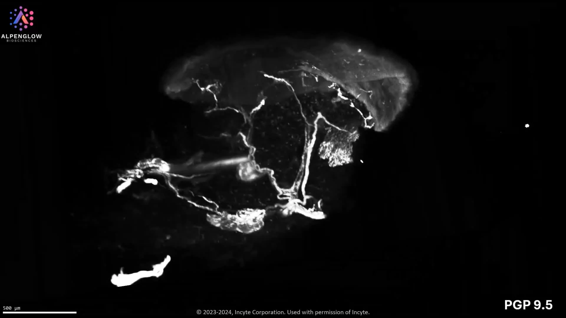
Whole Skin Punch Biopsy
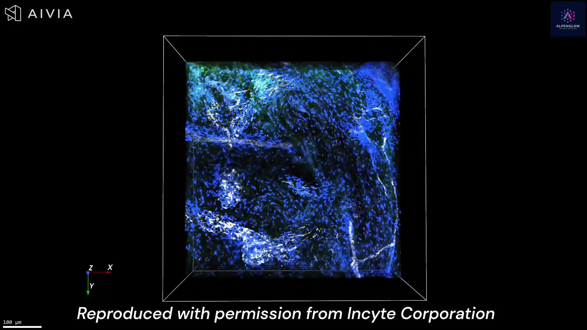
Low-resolution 3D imaging with Hybrid Open Top Light Sheet (HOTLS) microscopy of an entire lesional Atopic Dermatitis skin punch biopsy highlights epidermal and dermal innervation.
The tissue was stained with TO-PRO-3 (red, nuclei), PGP9.5 (green, nerves), and CD45 (blue, T cells), enabling comprehensive visualization of neuro-immune interactions and inflammatory changes across the full biopsy.
See our AD use case.
Image reproduced with permission from Incyte Corporation.
Previous
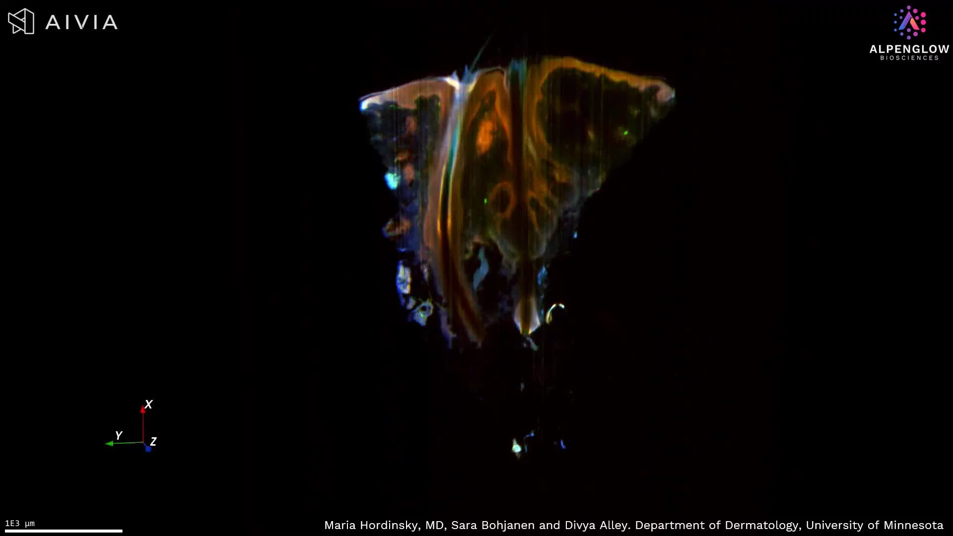
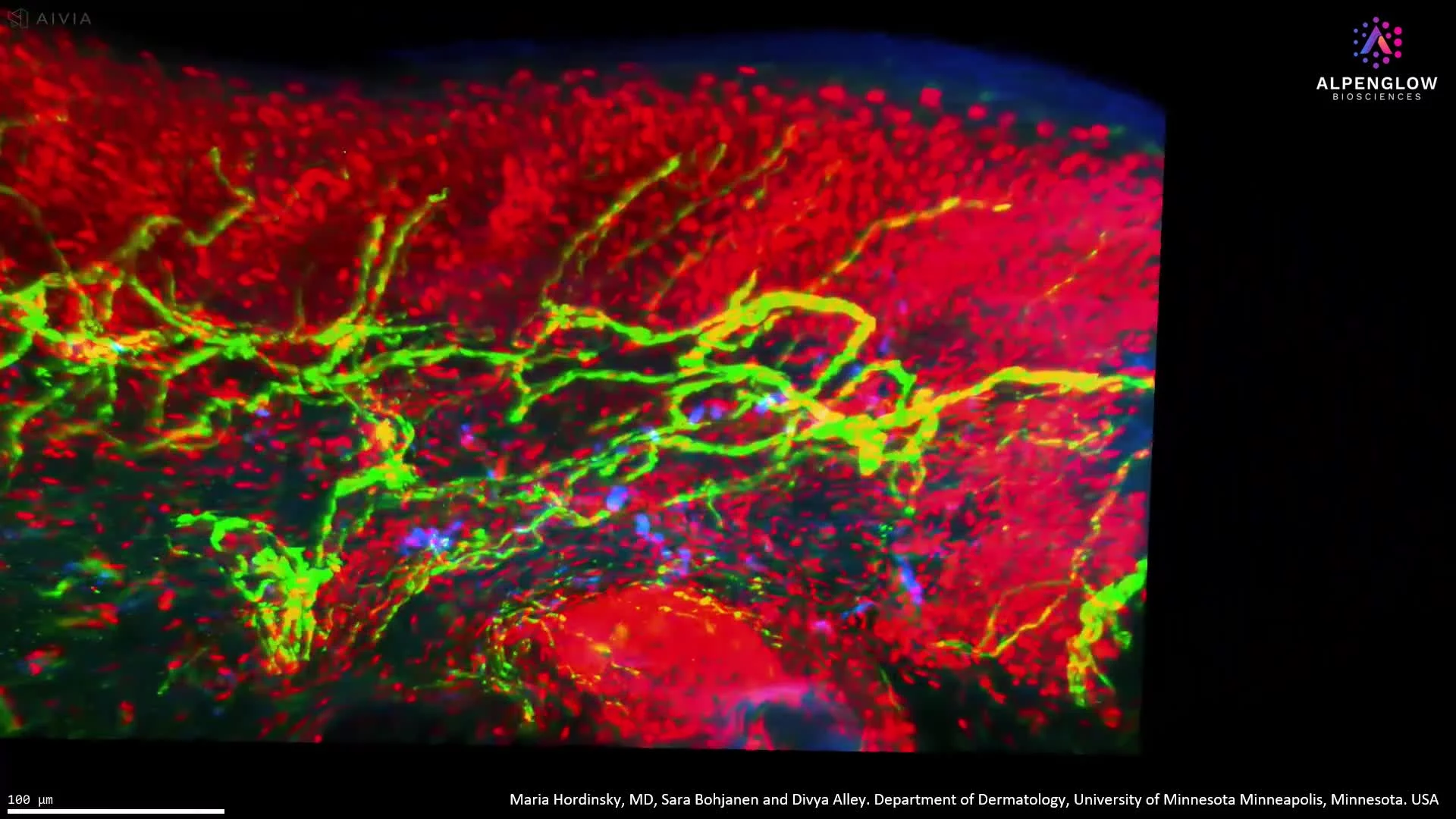
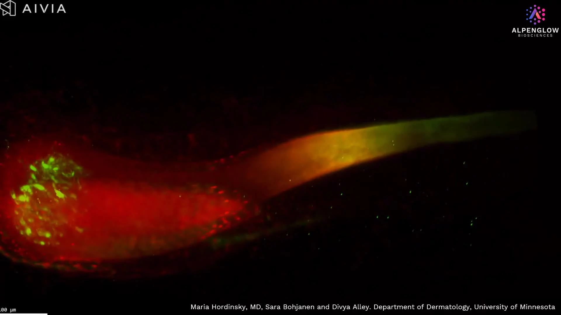
Unlocking the Secrets of Hair Follicles with 3D Imaging
Next