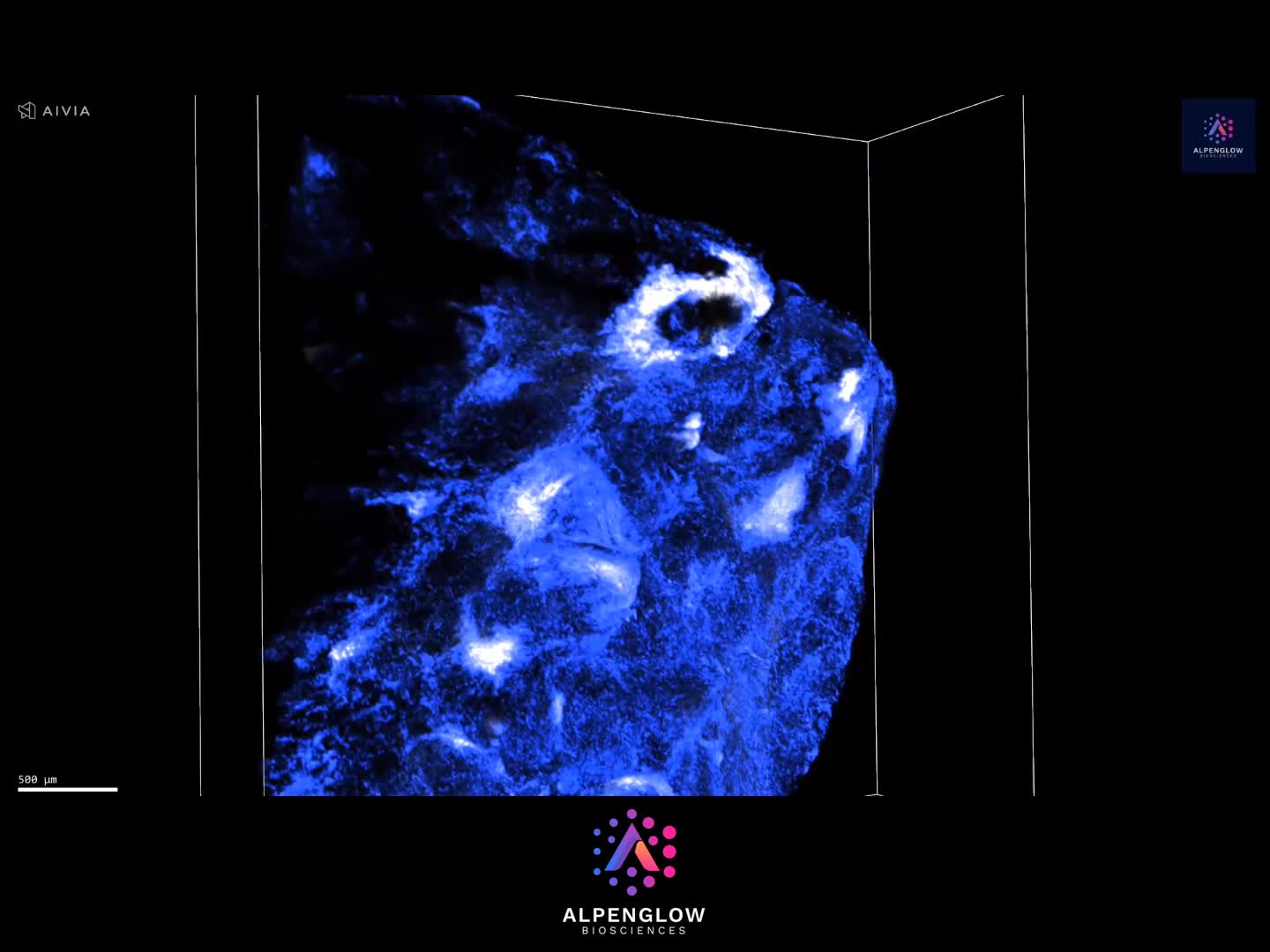
Whole Human Brain Slice Cleared and Imaged in 3D
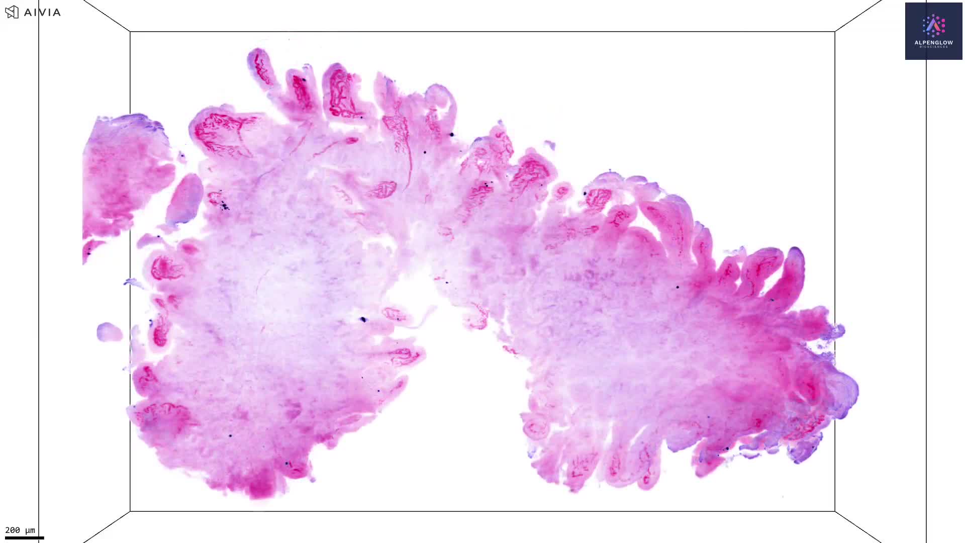
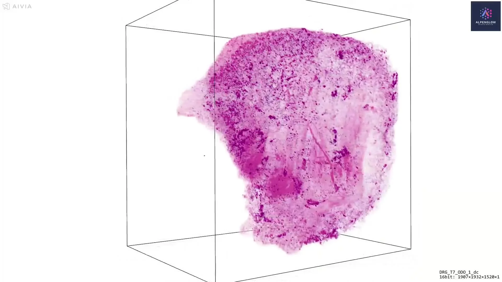
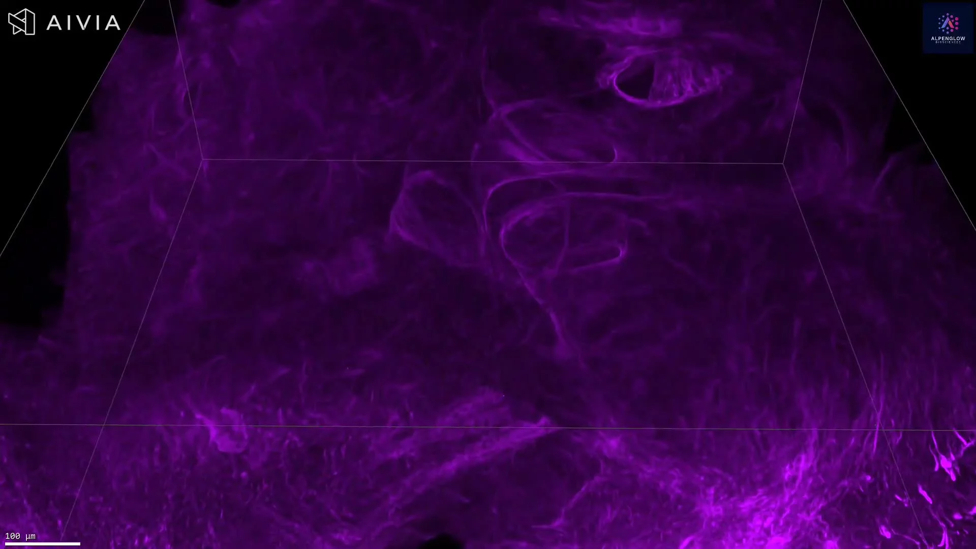
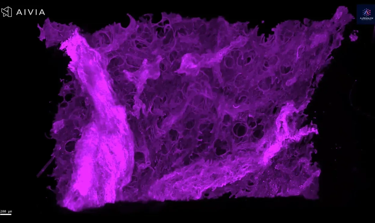
This dataset presents a whole cleared human brain slice, measuring approximately 10 cm × 7 cm × 0.3 cm, prepared with a CUBIC tissue clearing protocol and imaged at 0.17 microns per pixel. The high-resolution scan preserves tissue integrity while enabling volumetric imaging of large-scale brain architecture.
The scale of this dataset demonstrates the capacity of the Aurora™ 3Di Hybrid Open Top Light Sheet (HOTLS) microscope to handle large-format cleared samples with uniform clarity.
By maintaining intact morphology, this workflow enables comprehensive 3D histology of brain structures, overcoming the sampling limitations of thin 2D slices.
Applications of large-volume imaging extend to neuroscience research, brain mapping, and neurodegenerative disease studies, where quantifying tissue-scale structure is essential for translational discovery.