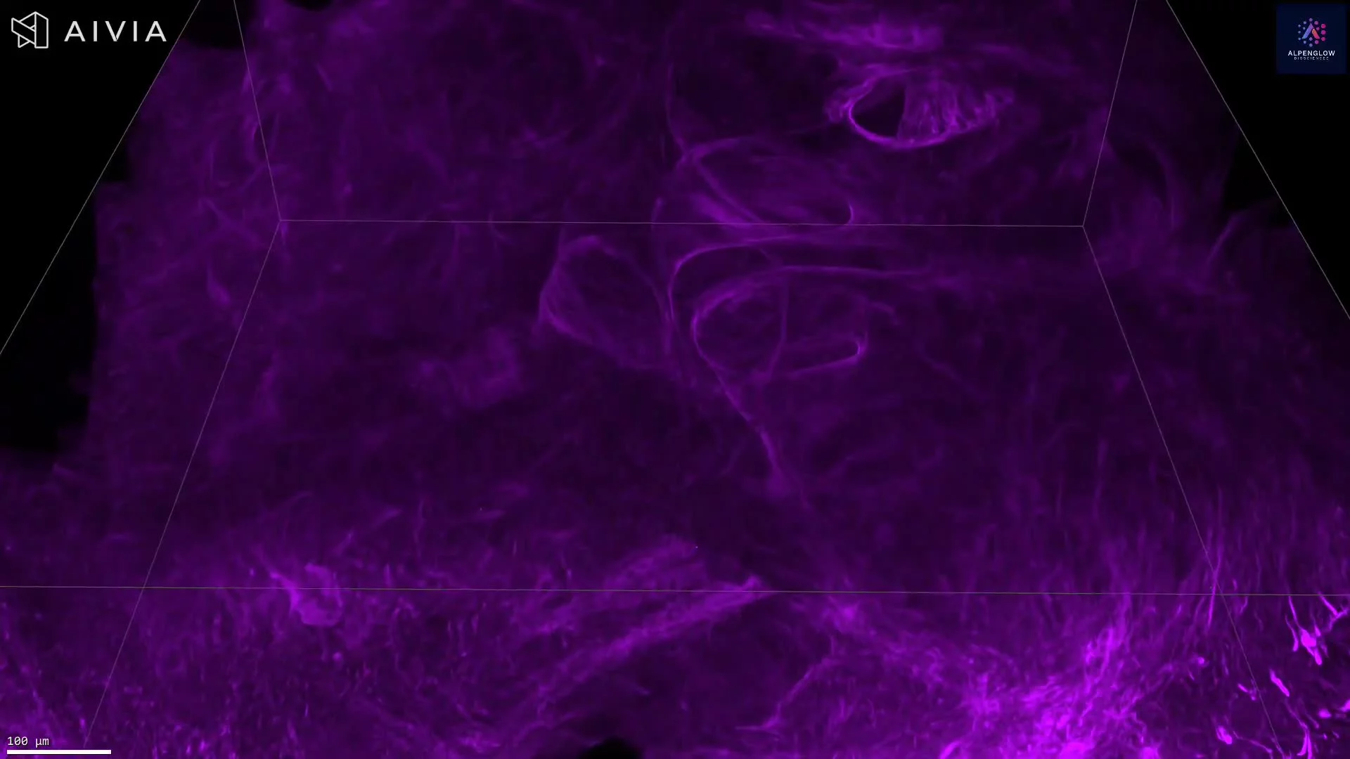
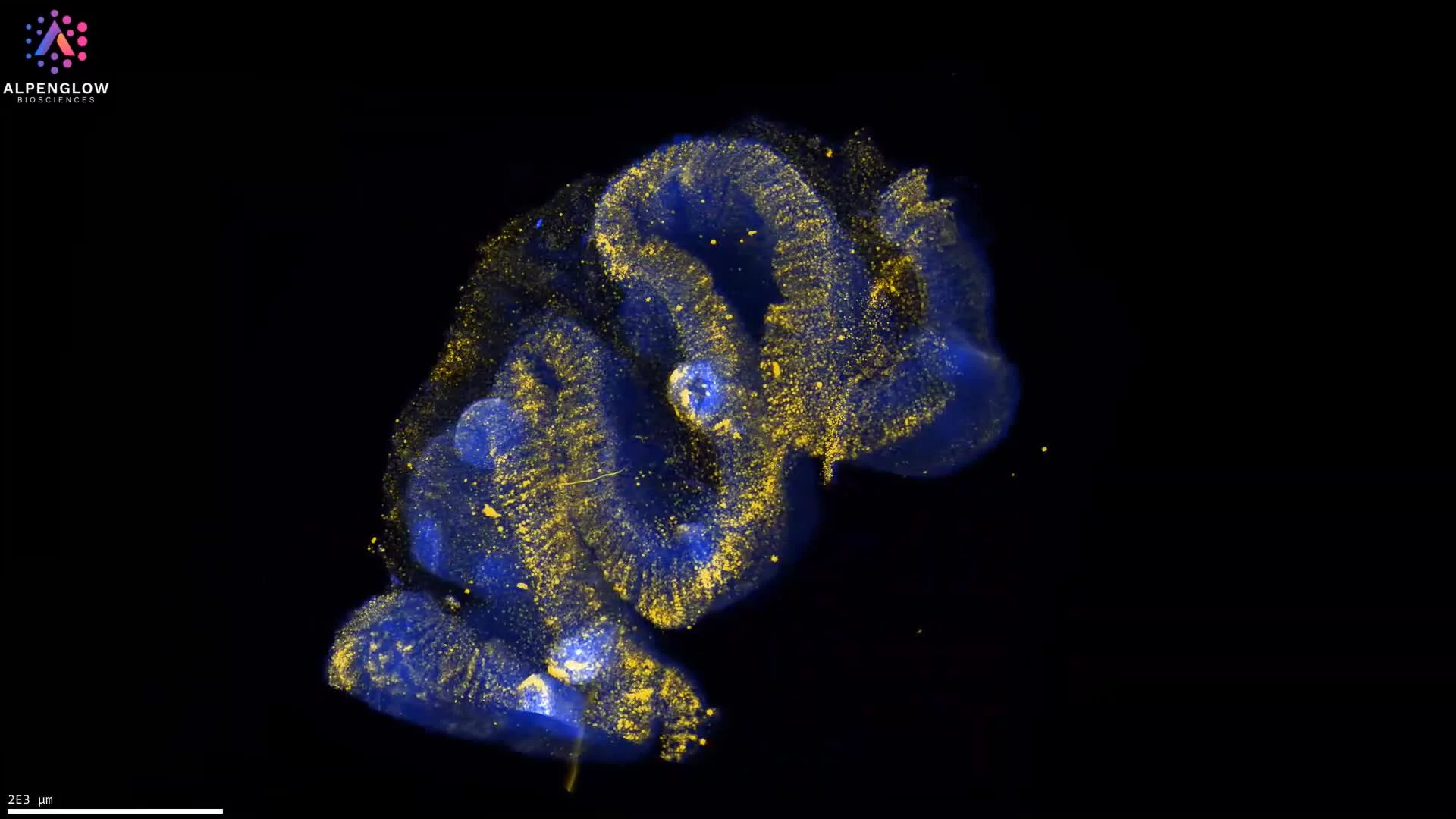
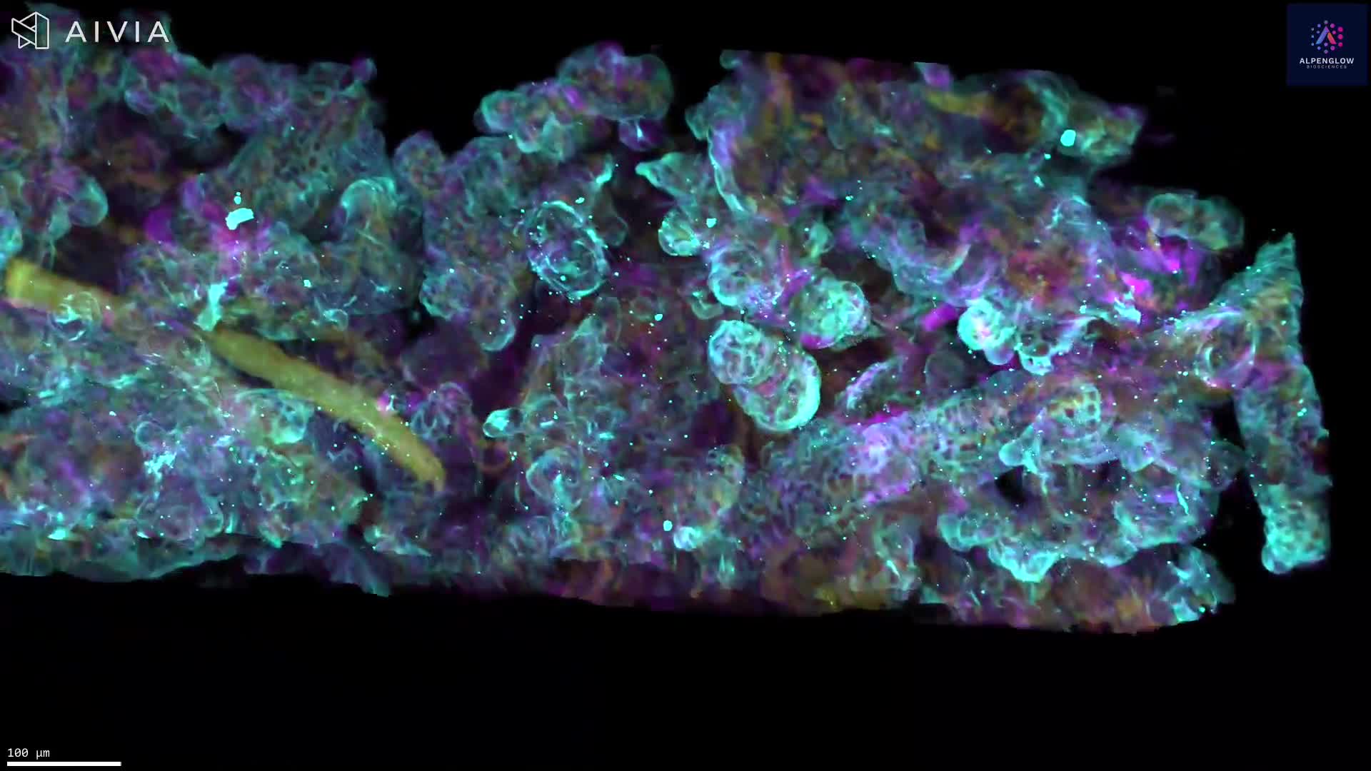
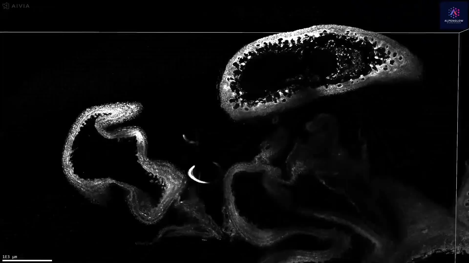
Fly-Through 3D Image of H&E-Stained Liver Biopsy
This dataset presents a 3D fly-through visualization of a human liver biopsy digitally stained with computational hematoxylin and eosin (H&E). Unlike traditional thin-section histology, this approach preserves the intact architecture of the tissue, enabling 3D histology that reveals spatial relationships and structures hidden in 2D slides.
By leveraging the Aurora™ 3Di Hybrid Open Top Light Sheet (HOTLS) microscope and advanced staining methods, researchers can visualize tissue morphology in full volumetric context. Integration with 3Dm data management and 3Dai AI-powered analysis enables quantitative assessment of hepatocyte organization, vascular networks, and microanatomical features critical to liver pathology.
This application demonstrates how digital pathology combined with computational H&E expands conventional workflows, delivering reproducible, high-content imaging datasets for translational research, drug development, and disease modeling.
Contact us to learn how Alpenglow’s 3D spatial biology platform can advance your research with large-scale, quantitative imaging datasets.