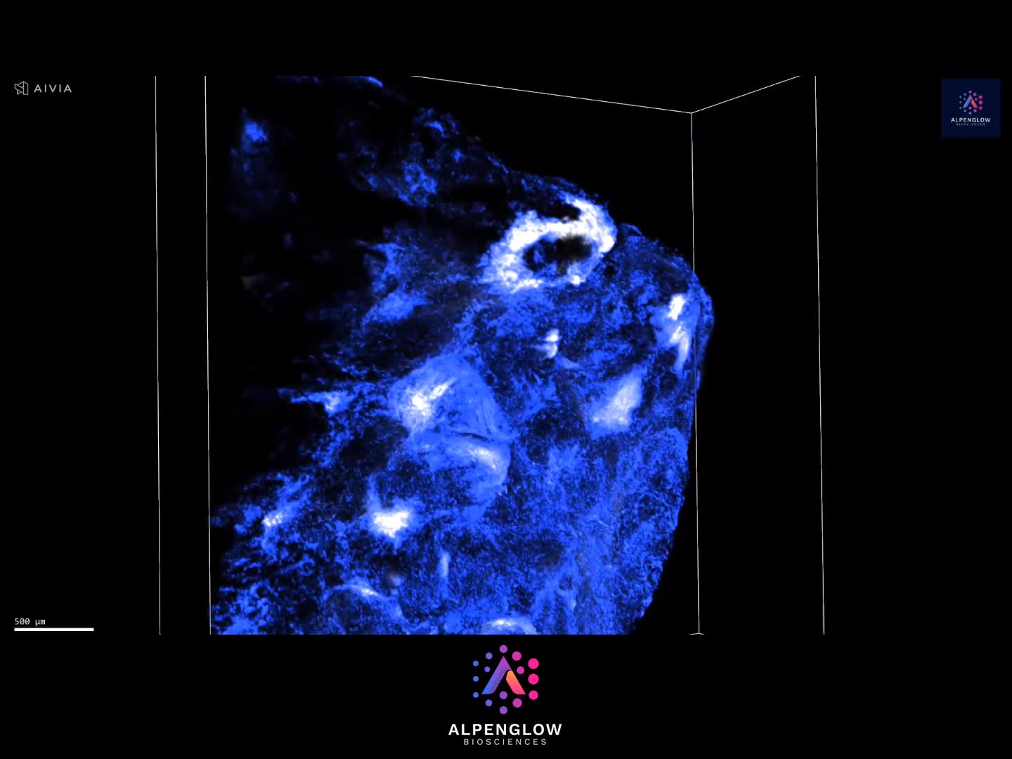
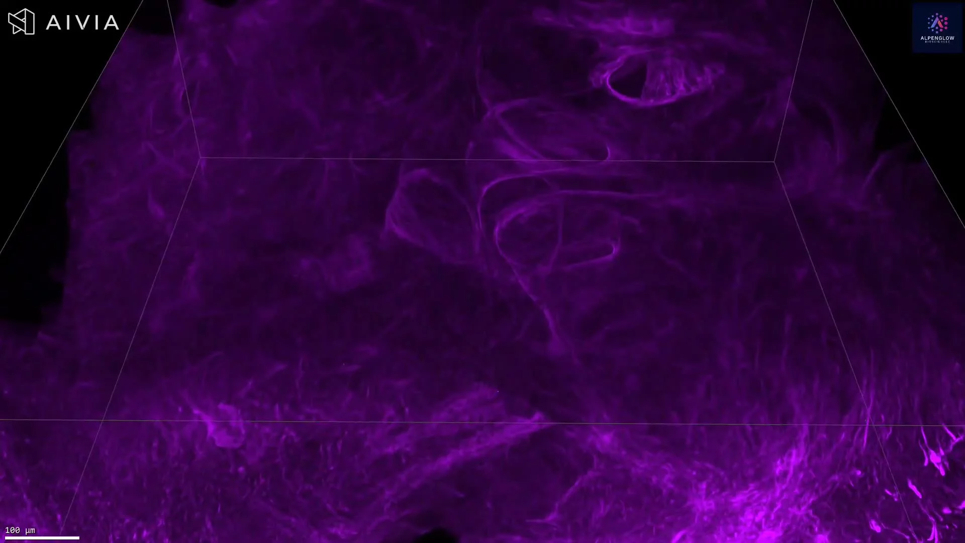
3D Imaging of Human Lung Collagen Architecture
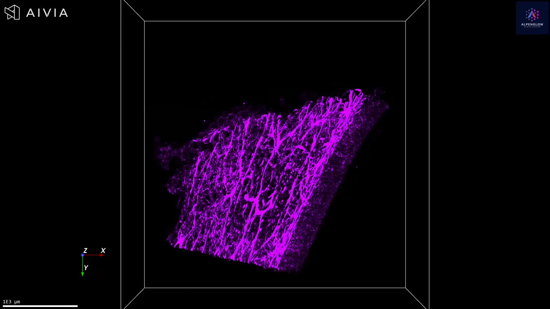
This 3D dataset reveals the intricate extracellular matrix of the human lung, captured with the Aurora™ 3Di Hybrid Open Top Light Sheet (HOTLS) microscope on Alpenglow’s 3D digital pathology platform. Collagen is stained with Fast Green and rendered in vivid detail, exposing the structural complexity and functional organization of lung tissue.
Unlike conventional 2D histology, this workflow images thick, intact tissue with no slicing or sectioning. The result is a distortion-free, spatially preserved view of the lung microenvironment, enabling accurate analysis of tissue architecture in its native state.
Through integration with 3Dm data management and 3Dai AI-powered segmentation, collagen orientation, density, and organization can be quantified across the tissue volume. These insights provide a deeper understanding of how collagen remodeling drives fibrosis, structural changes in tissue remodeling, and disease progression.
This dataset demonstrates the power of 3D histology and spatial profiling in lung research and translational studies.
Tissue provided by AnaBios.