Blog
Insights on 3D spatial biology and tissue imaging
Explore Alpenglow Biosciences updates, scientific perspectives, technology insights, and application stories across 3D tissue imaging, digital pathology, AI-powered analysis, spatial profiling, and quantitative tissue analysis.
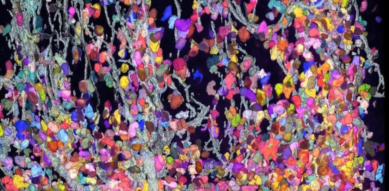
Quantifying Biology at Scale with AI and 3D Tissue Imaging
Advances in 3D tissue imaging and AI-powered analysis are enabling researchers to quantify biological systems at scale. By transforming volumetric data into measurable structures, these approaches reveal spatial relationships and tissue organization that are difficult to capture with traditional 2D methods.
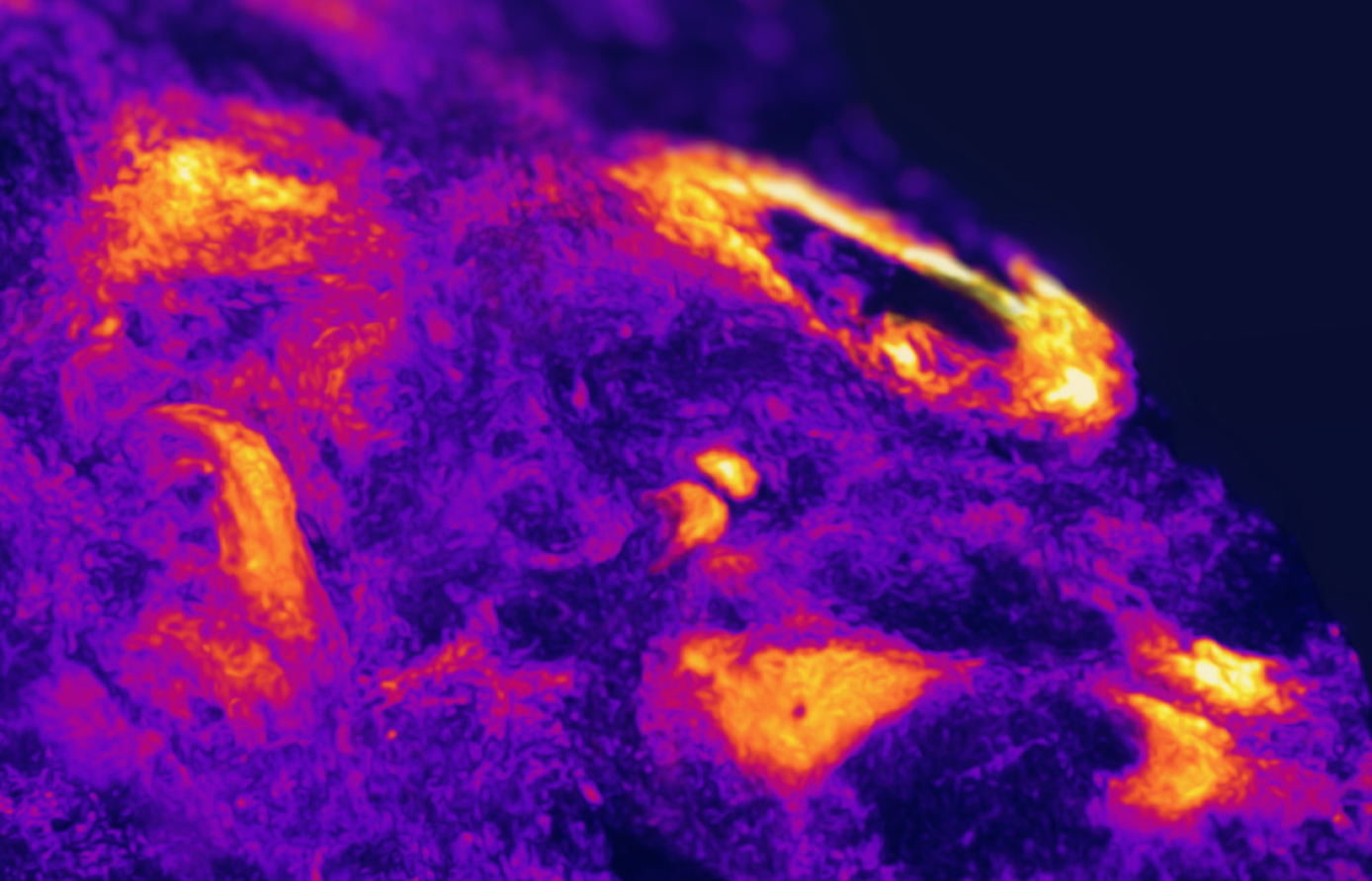
3D light-sheet imaging: how to avoid artifacts and ensure high-quality volumetric data
3D light-sheet imaging enables high-resolution volumetric analysis, but image quality depends on careful control of acquisition and processing. Learn how to identify and correct common artifacts to generate reliable 3D datasets.
When is light-sheet microscopy preferred over section-based confocal imaging?
Section based confocal imaging can approximate 3D, but it can introduce sampling gaps, distortion, and registration artifacts. Light sheet microscopy is preferred when you need intact tissue context, whole tissue imaging, and quantitative 3D measurements across depth.
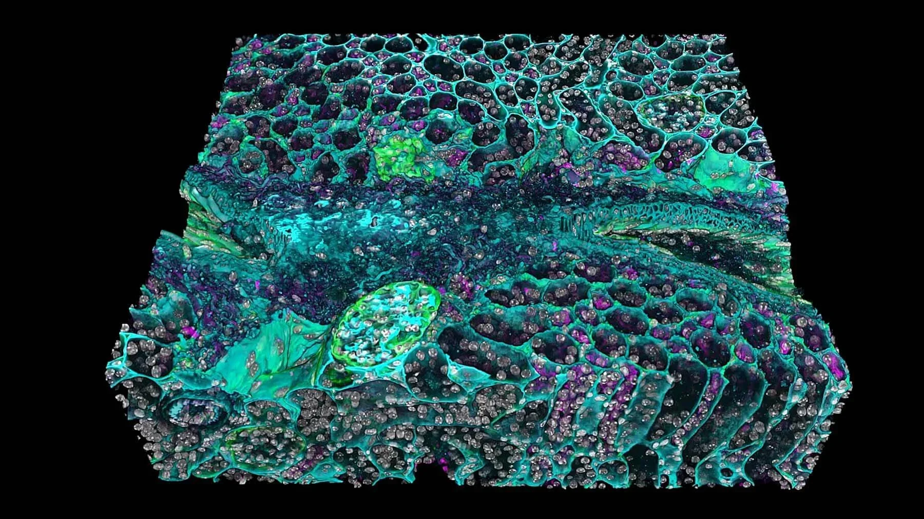
Tissue clearing for 3D tissue imaging: revealing hidden structures in intact samples
Tissue clearing enables high-resolution 3D tissue imaging by rendering samples transparent. This allows researchers to visualize intact biological structures and analyze spatial relationships that are difficult to capture with traditional 2D methods.
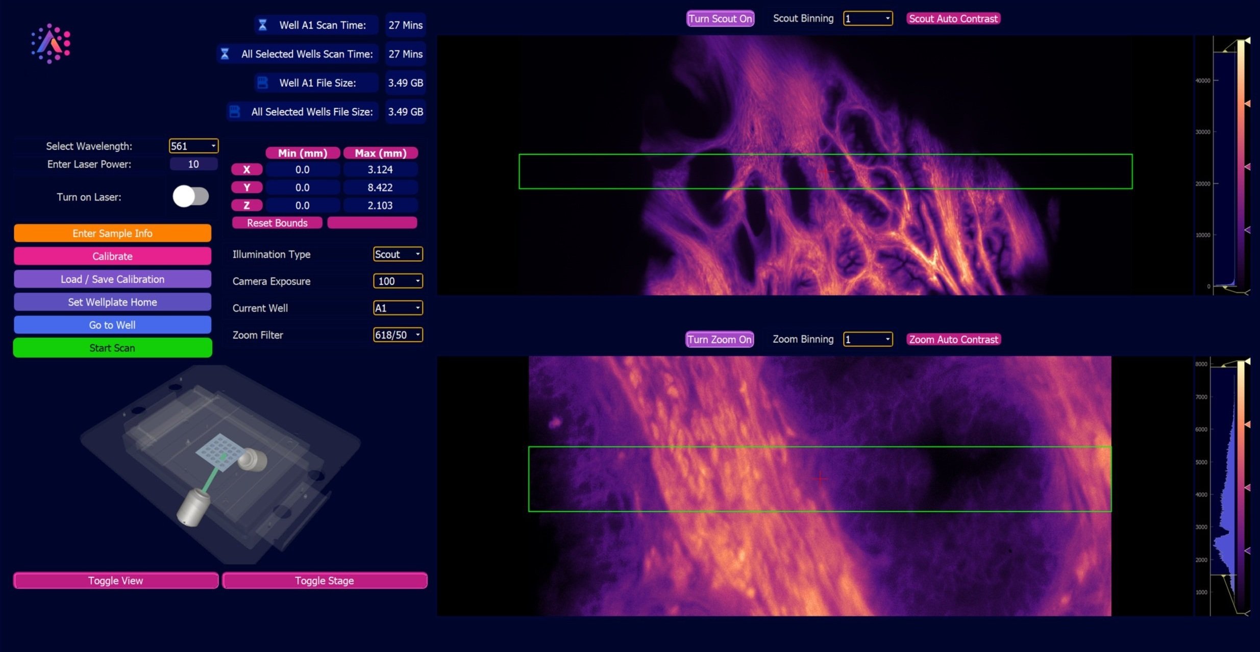
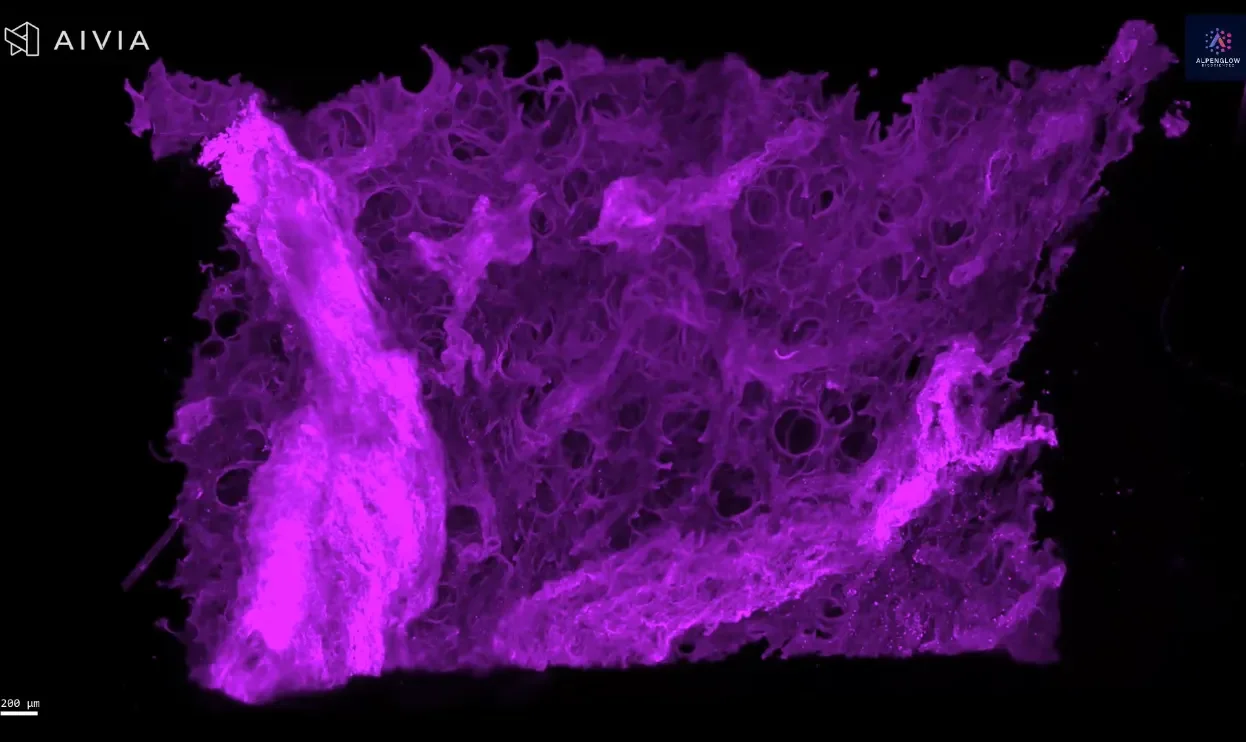
Benefits and advantages of Light-Sheet Microscopy for fast, high-resolution tissue imaging
Alpenglow’s 3Di™ Hybrid Open-Top Light-Sheet Microscope merges whole-sample speed with sub-micrometer detail in a single system. Designed for cleared tissues, it enables seamless switching between fast Scout mode and high-resolution Zoom mode. With an open-top design, multi-immersion optics, and AI-ready 3D data outputs, the 3Di HOTLS delivers true volumetric imaging at scale, revealing complete tissue architecture without physical sectioning.

3D Tissue Imaging for Dermatology: Seeing Skin Biology As It Truly Is
Skin is a 3D organ with complex nerves, structures, and patchy inflammation. 3D tissue imaging reveals intact architecture and spatial biology that 2D slides miss. From innervation in atopic dermatitis to vascular mapping in keloids and melanoma, whole-biopsy imaging with AI-powered analysis delivers reproducible, quantitative insights for dermatology research and clinical translation.

Why 2D Slides Miss Critical Insights: The Case for 3D Tissue Imaging
3D tissue imaging reveals what 2D slides often miss, intact tissue architecture, rare features, and the true complexity of the microenvironment. By combining high-resolution, non-destructive 3D histology with AI-powered analysis, researchers gain deeper insights in digital pathology, spatial profiling, and quantitative tissue analysis that accelerate discovery and development.
Continue exploring
Apply 3D imaging to your research
Explore use cases across intact-tissue imaging, digital pathology, spatial profiling, and AI-powered quantitative analysis, or discuss the requirements of your next study.