Multi-field capabilities
Multi-field 3D tissue imaging across research areas.
The Aurora 3D™ Spatial Biology Solution supports 3D tissue imaging, AI-powered analysis, spatial profiling, digital pathology, and quantitative tissue analysis across diverse tissues, disease areas, and research programs.
Dermatology
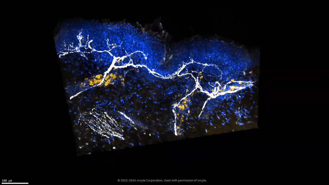
3D dermatology tissue imaging
Alpenglow Biosciences applies 3D tissue imaging and AI-powered analysis to inflammatory and immune mediated skin diseases, focusing on nerve immune interactions and skin architecture.
Indications
- Atopic dermatitis (AD)
- Alopecia Areata (AA)
- Hidradenitis Suppurativa (HS)
- Chronic Spontaneous Urticaria (CSU)
Tissue inputs
- FFPE skin punch biopsies
- Freshly fixed tissues
- Fresh, fixed or frozen skin samples
Analytics examples
Immuno-oncology
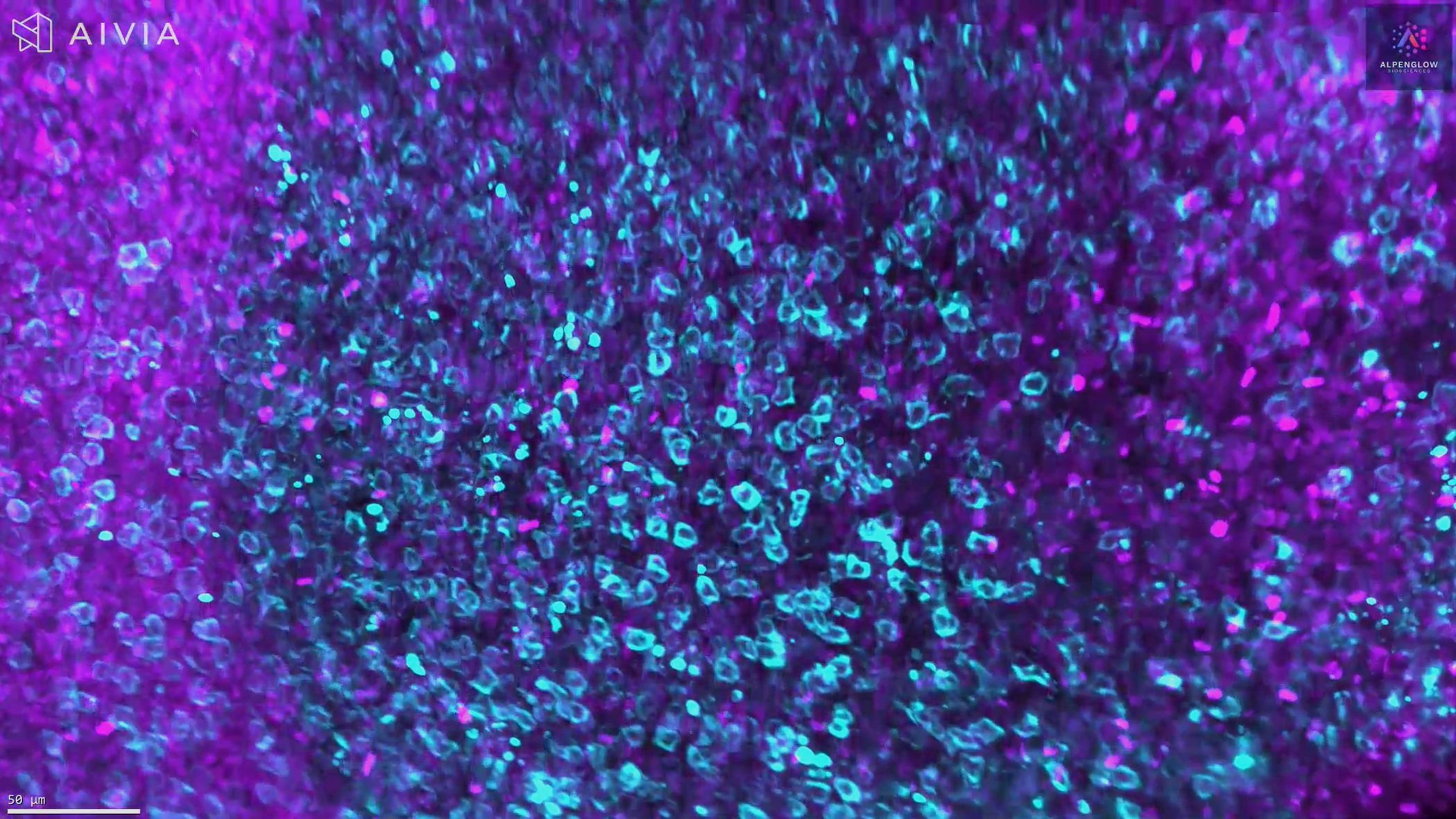
3D immuno-oncology profiling
The multi-field capabilities of Aurora 3D™ include immuno-oncology profiling across solid tumors, complementing dedicated 3D immuno-oncology assays for tumor immune microenvironment analysis.
Indications
- Colon tumors
- Lung tumors
- Skin tumors
- Pancreas tumors
Tissue inputs
- Tumor biopsies
- Resected tumor material
- Fresh, frozen, or FFPE tumor samples
Analytics examples
- TIL tumor/TIL stroma ratio
- TLS count and volume
- % tumor or tumor volume
- Immune cell phenotypes and spatial distribution
3D immuno-oncology profiling supports spatial profiling, immune contexture analysis, tumor immune microenvironment review, and quantitative tissue analysis from volumetric datasets.
Gastrointestinal
3D gastrointestinal tissue imaging
3D gastrointestinal imaging captures villi, crypts, mucosal architecture, and innervation in a single volume, supporting studies in intestinal disease.
Indications
- Celiac Disease
- Inflammatory bowel disease (IBD)
- Ulcerative Colitis
- Hirschsprung’s Disease
Tissue inputs
- Large resected gastrointestinal specimens
- Colonoscopy or endoscopy biopsies
- Fresh, frozen, or FFPE GI tissues
Analytics examples
- Villus height to crypt depth ratios
- Mucosal thickness and architecture
- Innervation patterns in the intestinal wall

Lung
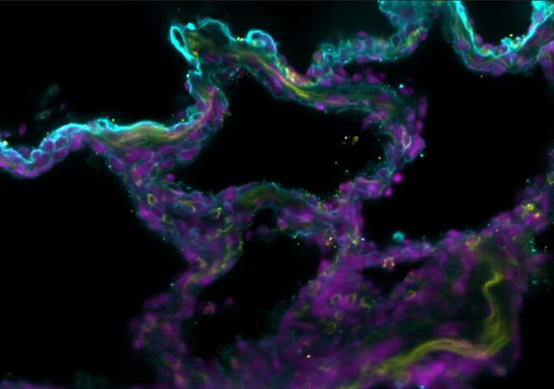
3D lung tissue imaging
3D lung imaging reveals fibrosis patterns, airway structure, and immune infiltration across the full tissue volume, which can be difficult to appreciate in 2D.
Indications
- Idiopathic Pulmonary Fibrosis (IPF)
- Chronic Obstructive Pulmonary Disease (COPD)
Tissue inputs
- Lung bronchial biopsies
- Resected material
- Fresh, frozen, or FFPE samples
Examples
Vascular and angiogenesis
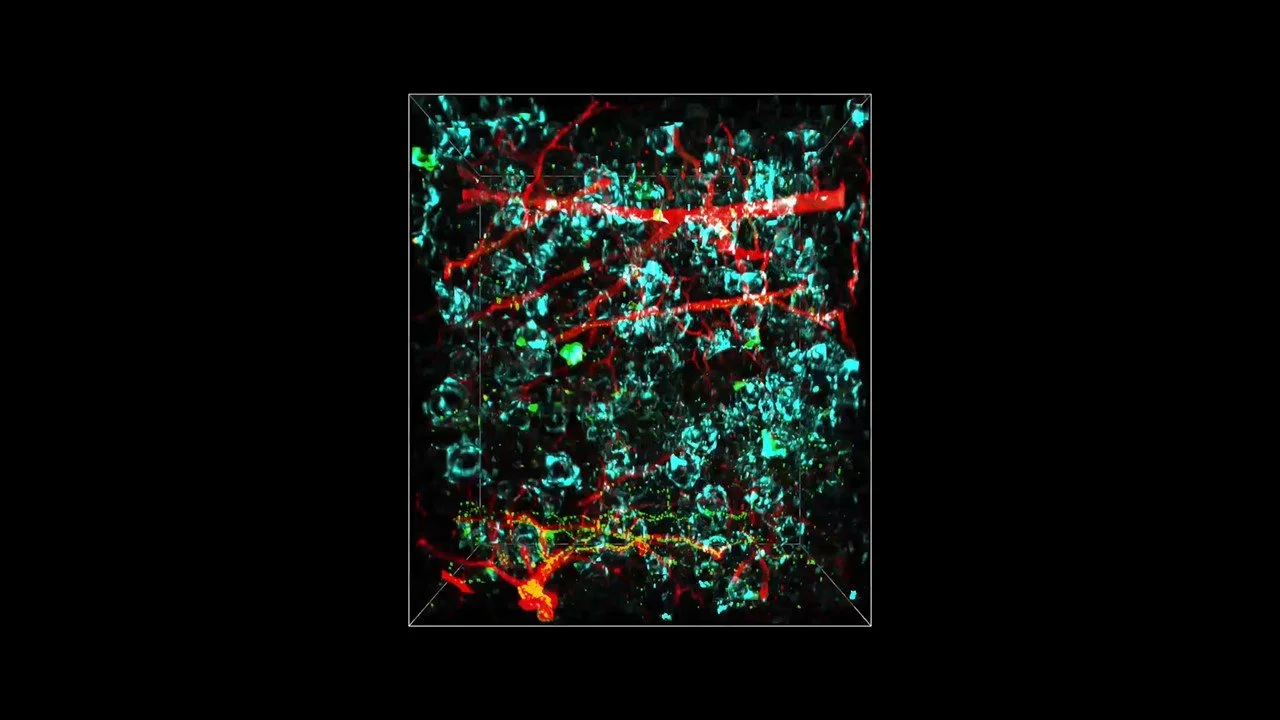
3D vascular and angiogenesis imaging
3D imaging of vasculature captures vessels, branching patterns, and relationships to surrounding tissue, enabling quantitative vascular analysis.
Indications
- Heart development
- Heart disorders
- Tumor angiogenesis
- Placental vascularization
Tissue inputs
- Tissue-specific biopsies
- Resected heart, tumor, or placental tissue
- Fresh, frozen, or FFPE samples
Analytics examples
- Cell count within and around vessels
- Vessel dimensions, for example, diameter and length
- Distance measurements between vessels and other structures
- Branching patterns and vascular network complexity
Explore 3D tissue imaging across fields
Have a tissue, disease model, or spatial biology question in mind?
Alpenglow supports multi-field 3D tissue imaging, digital pathology, spatial profiling, AI-powered analysis, and quantitative tissue analysis across tissue types, indications, and translational research programs.