

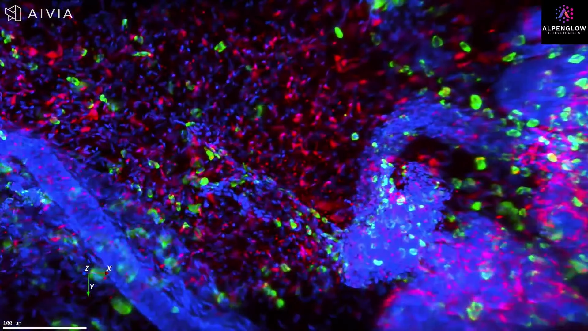
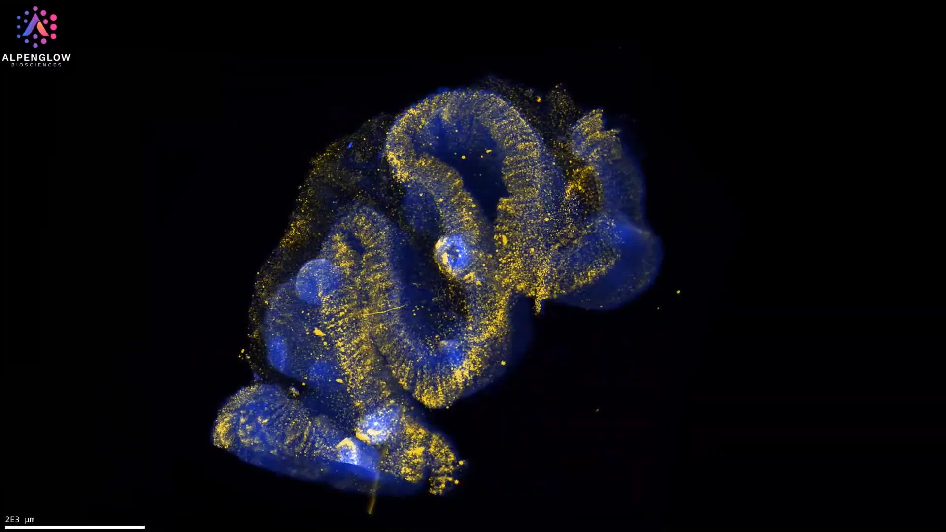
High-resolution 3D imaging of an intact mouse colorectal tumor reveals B and T cell organization and measurable tertiary lymphoid structures across the full tissue volume.
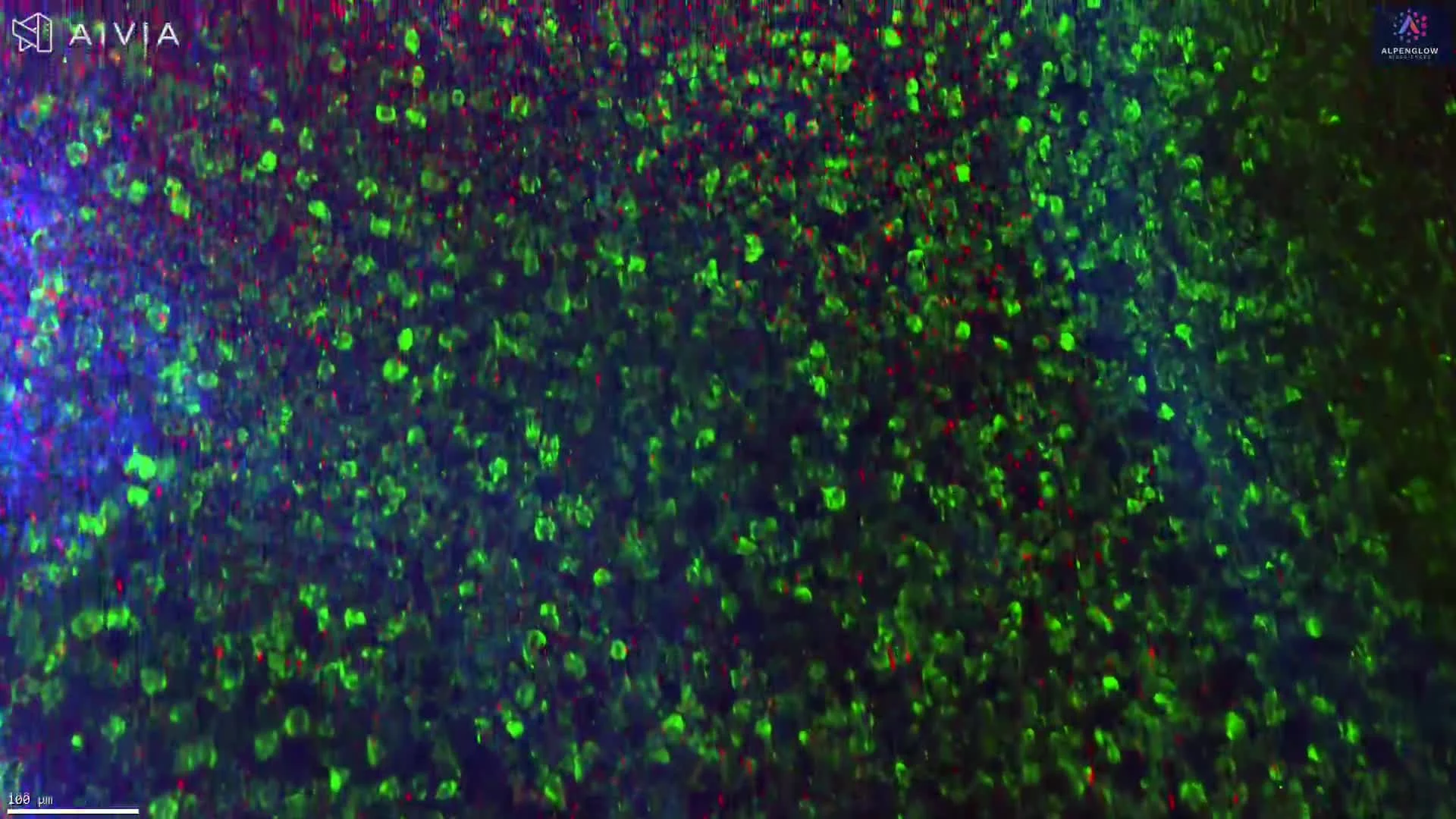
High-resolution 3D imaging of an intact mouse colorectal tumor reveals B and T cell organization and tertiary lymphoid structures across the full tissue volume.
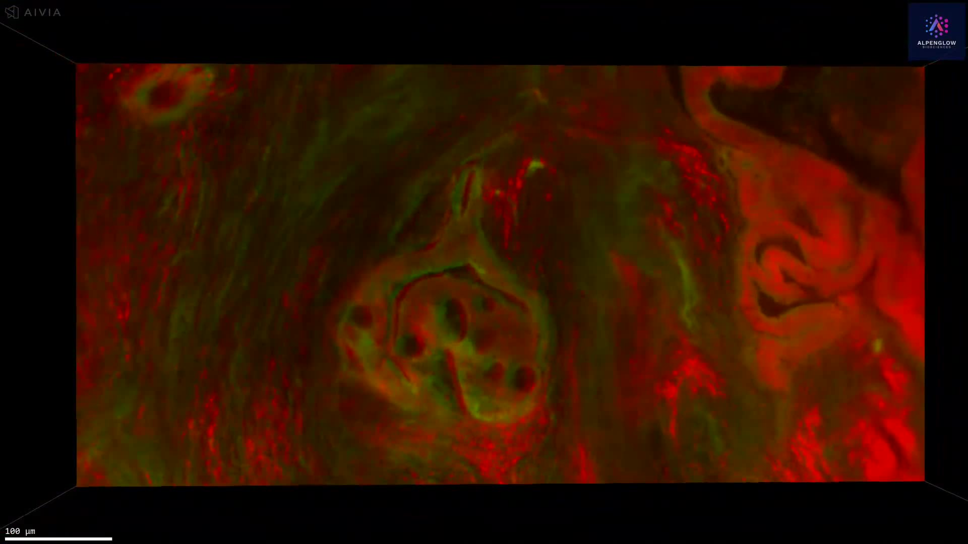
3D fluorescence imaging of human colorectal cancer FFPE tissue reveals spatial organization across depth that cannot be captured with single 2D histology sections.
Computational H&E staining, combined with 3D imaging, reveals the full structure of tertiary lymphoid structures (TLS) in NSCLC, overcoming 2D sampling bias and enabling the accurate quantification of volume, maturity, and spatial context.

Computational H&E staining combined with non-destructive 3D imaging reveals the true complexity of tertiary lymphoid structures (TLS) in NSCLC. Unlike thin 2D sections that risk misclassification, Alpenglow’s 3Di platform captures entire TLS morphology, volume, and cellular composition, delivering accurate insights into immune architecture and tumor context.
3D segmentation of prostate glands using synthetic CK8 immunofluorescence derived from fluorescent H&E analogues. By combining image-translation models with traditional computer-vision methods, researchers achieved whole-biopsy 3D gland segmentation without manual labeling.

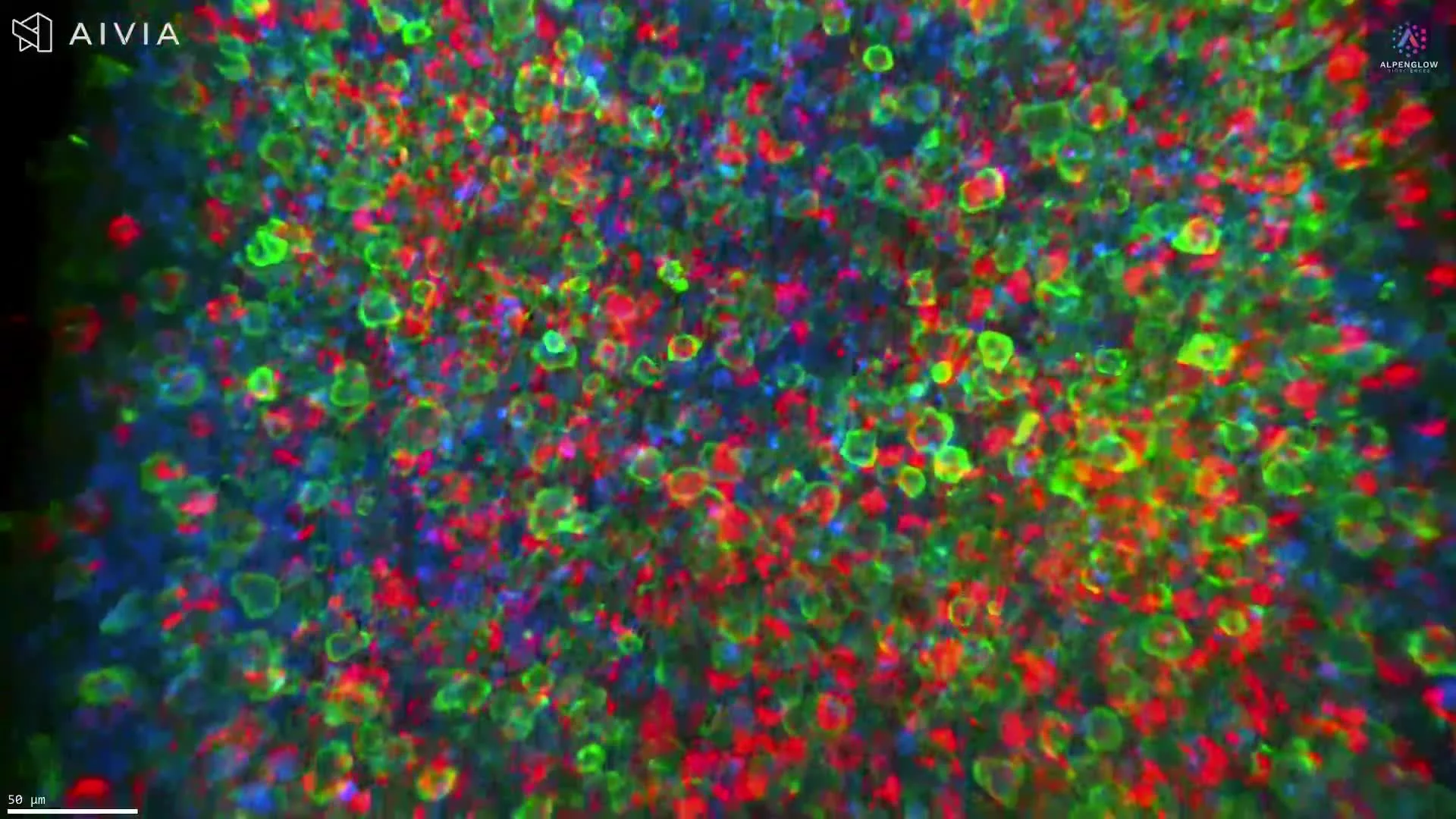
3D imaging of mouse colorectal tumor stained with CD3, B220, and YoPro1 reveals tertiary lymphoid structures (TLS) in full spatial context.
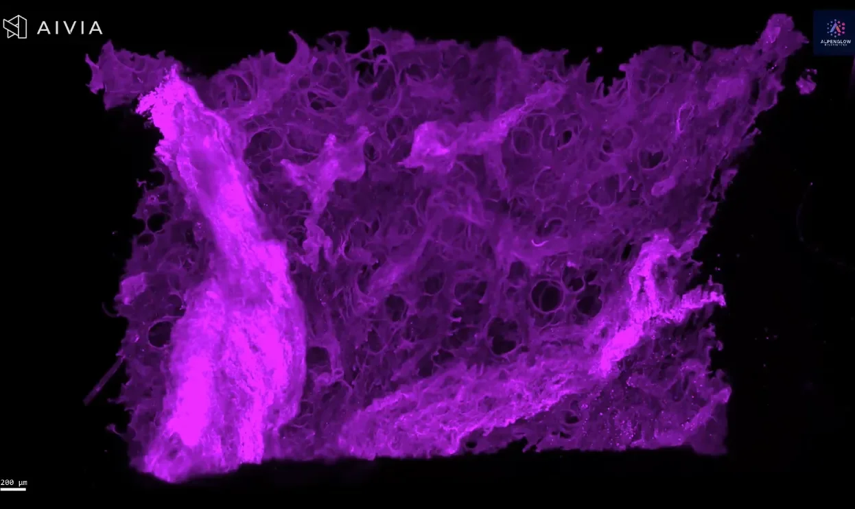
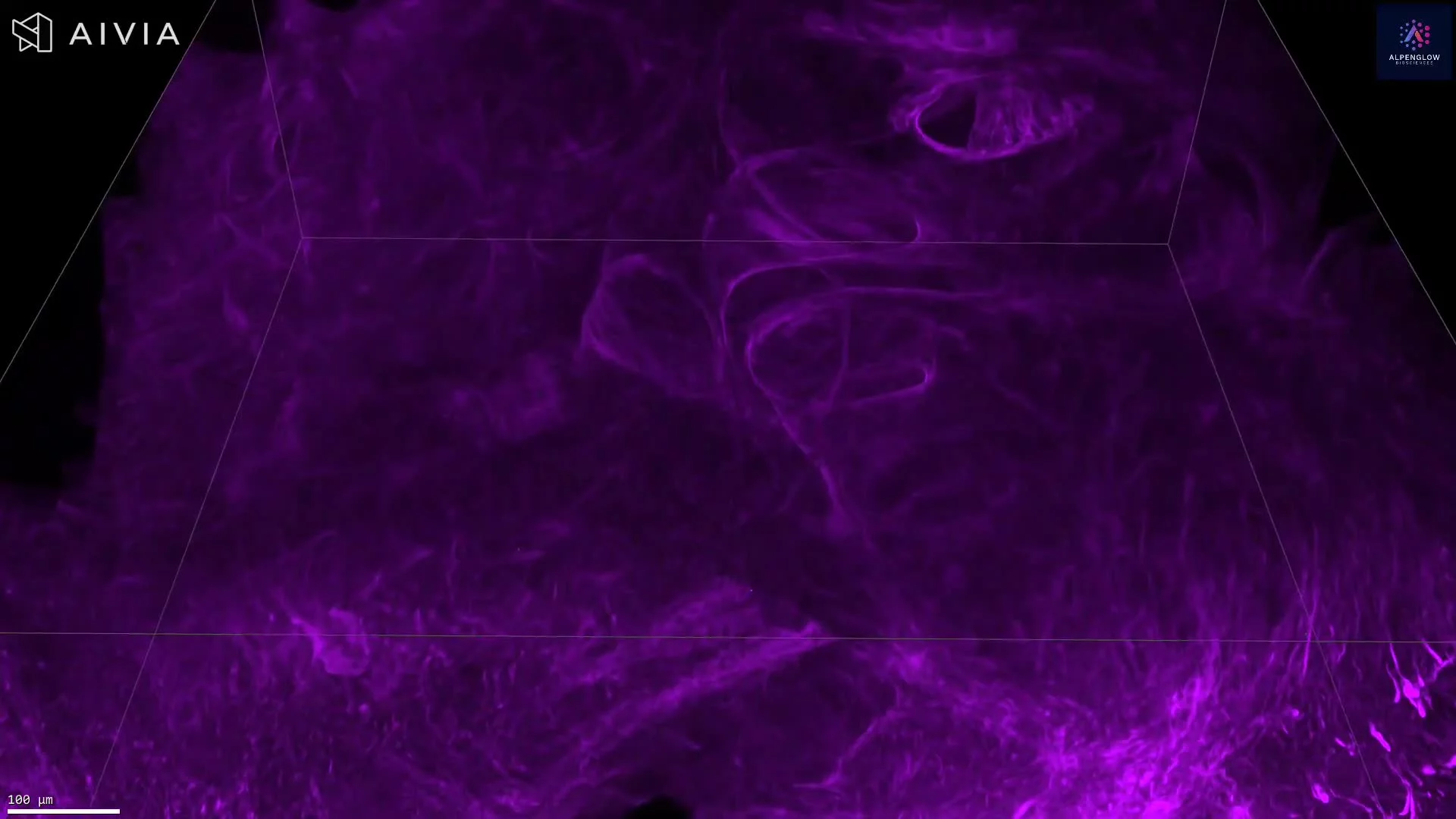
3D fluorescence imaging of human lung tissue stained with Fast Green reveals collagen architecture for fibrosis and tissue remodeling research.
3D imaging of human skin biopsy stained with tryptase, TO-PRO-3, and PGP9.5 reveals mast cell–nerve interactions for dermatology and oncology research.
3D imaging of prostate organoids stained with TO-PRO-3 and eosin reveals spatial heterogeneity, cellular interactions, and microenvironmental detail.
3D fluorescence imaging of prostate tissue stained with Fast Green reveals the collagen architecture, orientation, and density, providing insights beyond 2D histology.
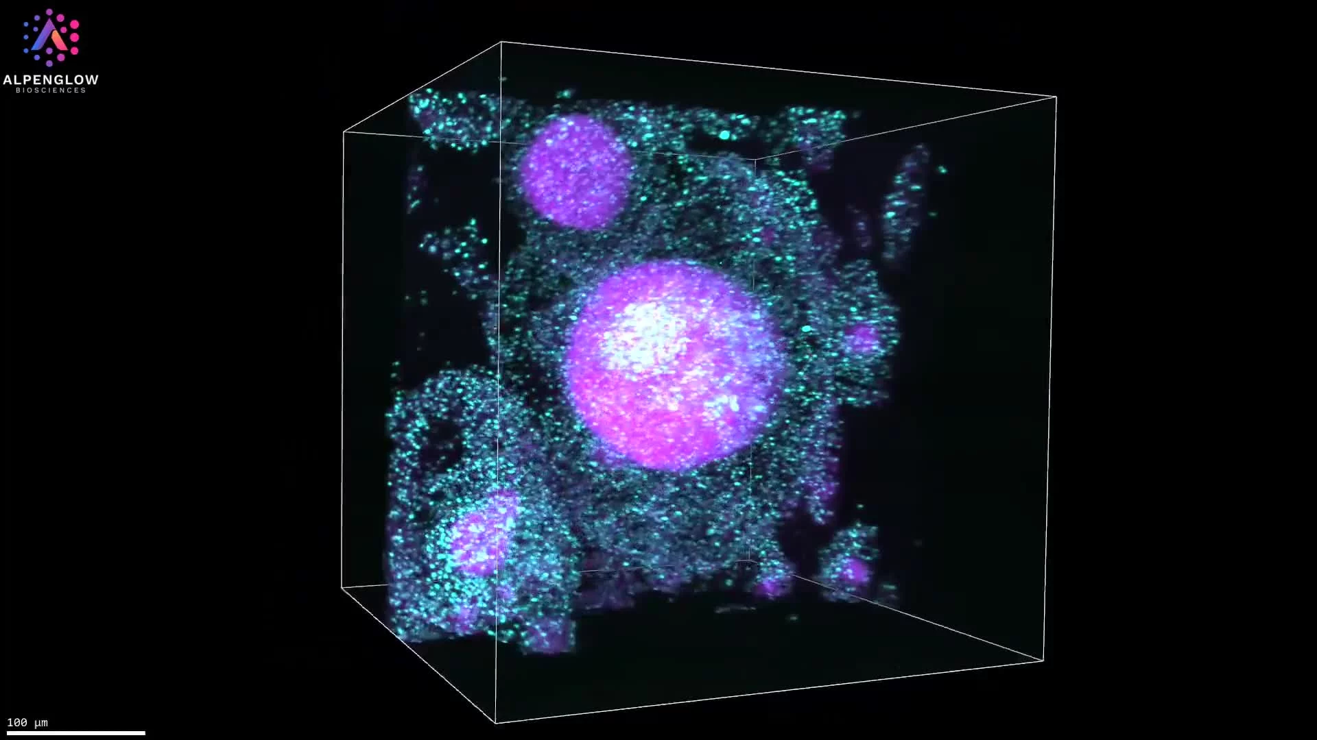
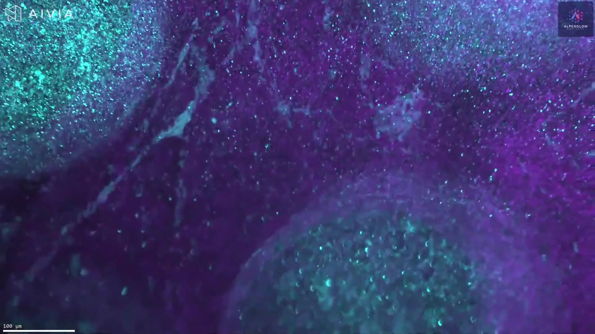
High-resolution 3D imaging of human tonsil tissue stained with YO-PRO-1 for nuclei and tryptase for mast cells.
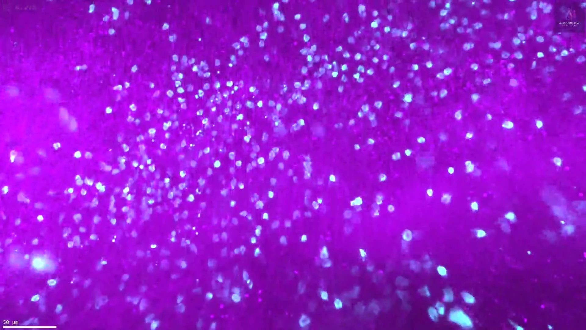
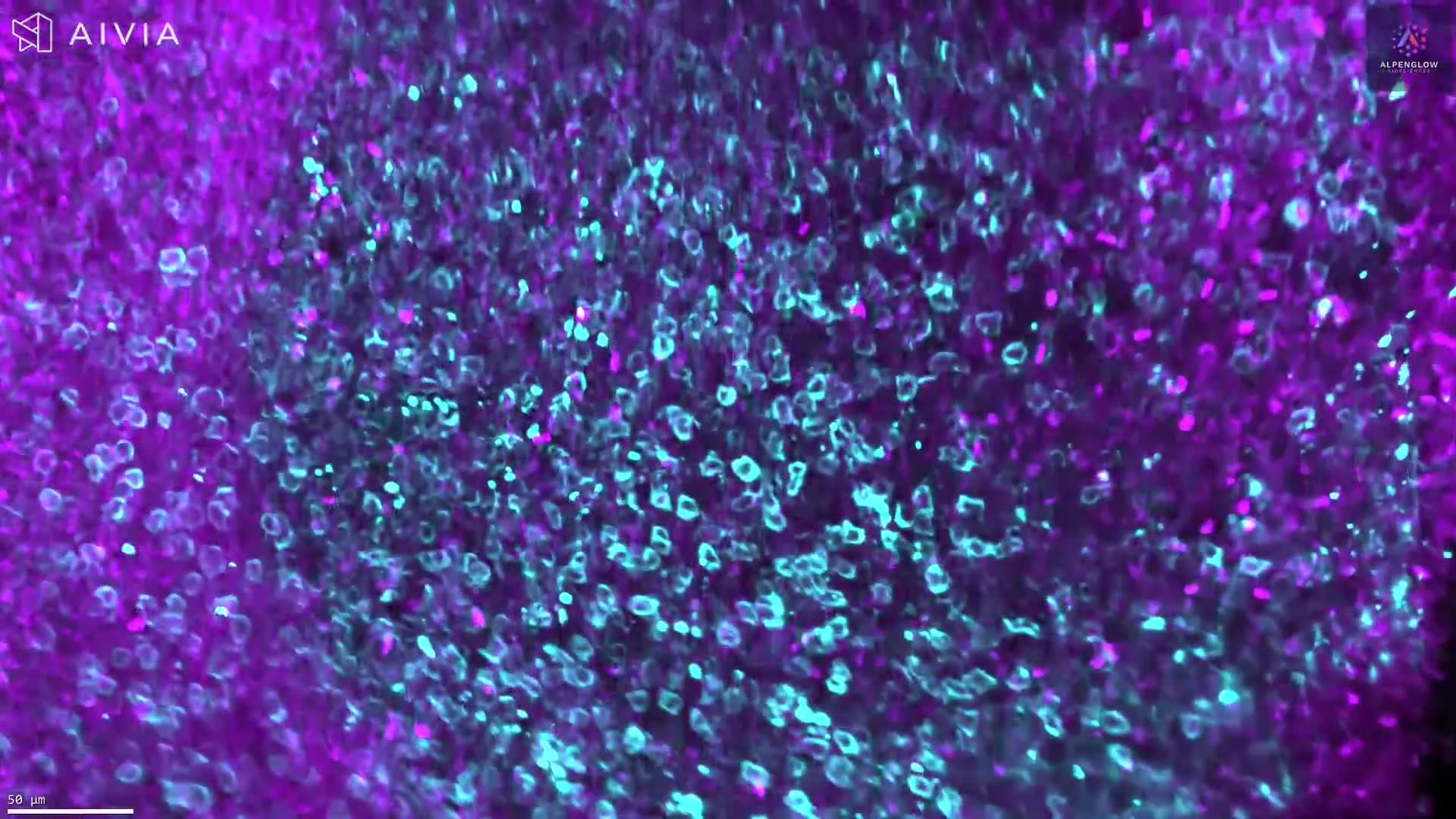
FFPE colorectal tissue stained with YO-PRO-1 and anti-CD8 imaged in 3D at 2 μm/pixel on Aurora HOTLS. Quantification of CD8+ lymphocytes across whole section.

3D imaging of cleared mouse fat pad reveals blood vessels, macrophages, and nerves labeled with lectin, CD68, and PGP9.5.
3D imaging of cleared tonsil tissue stained with anti-CD21 and TO-PRO-3 reveals B cell and follicular dendritic cell distribution in FFPE samples.
3D imaging of cleared tonsil tissue stained with anti-CD45RO (UCHL1) and TO-PRO-3 reveals memory T cell distribution in thick FFPE samples.