3D fluorescence imaging of colorectal cancer FFPE tissue
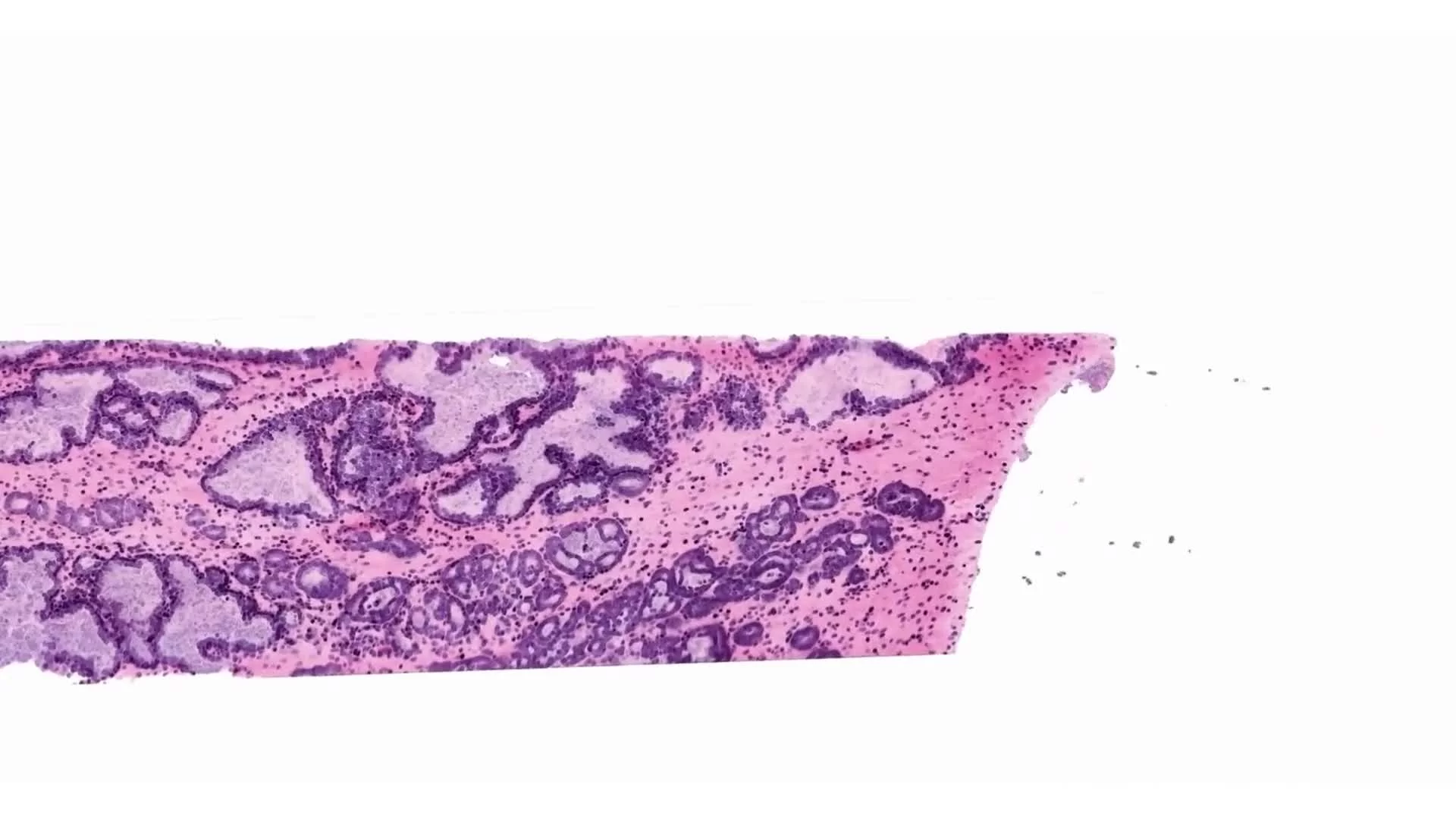
This video demonstrates three-dimensional fluorescence imaging of human colorectal cancer (CRC) FFPE tissue, acquired using a volumetric workflow designed to preserve tissue architecture across depth.
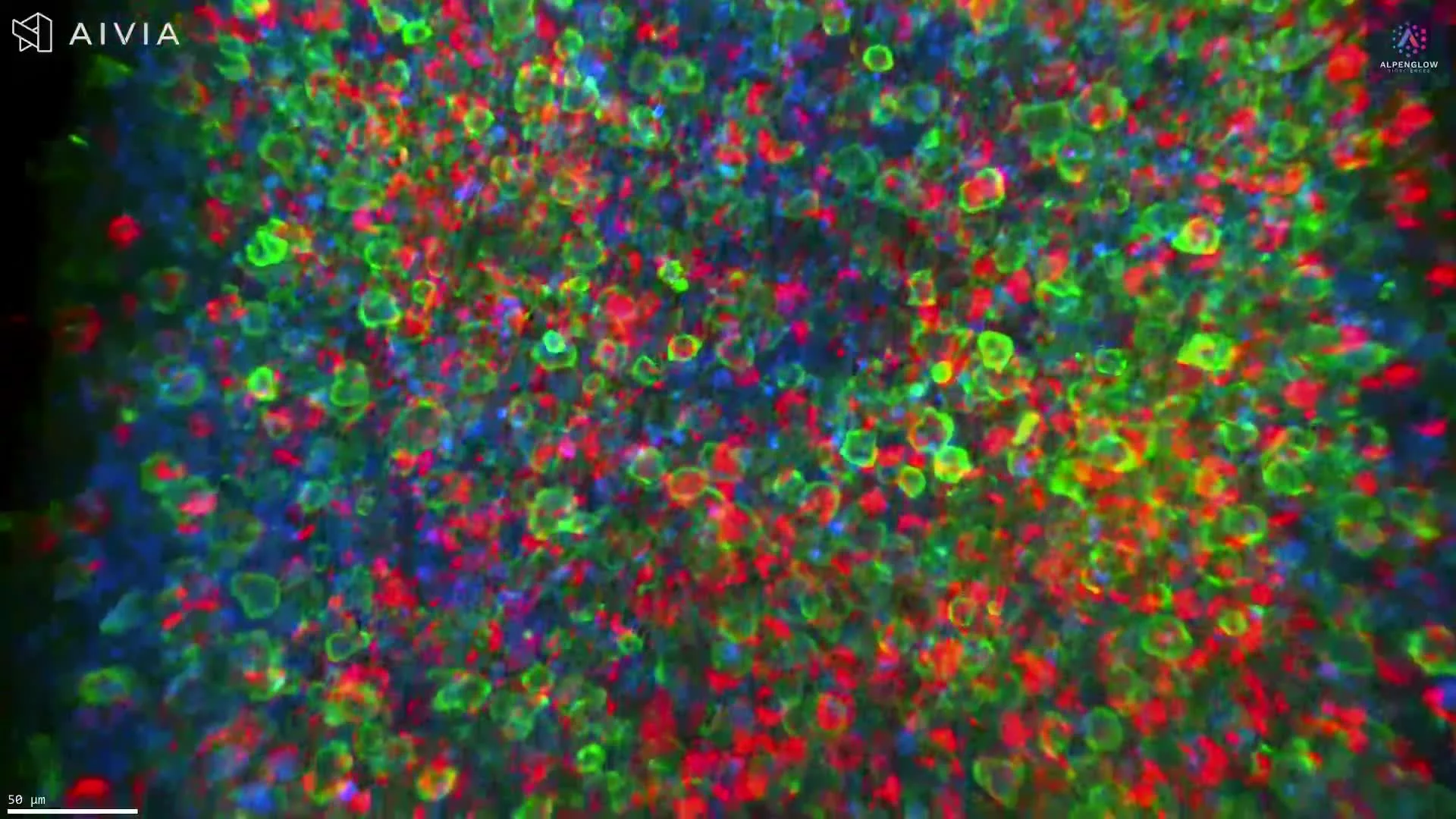
As described in the linked CRC immune-exclusion study, intact CRC FFPE blocks are deparaffinized, fluorescently labeled, optically cleared, and imaged in three dimensions to avoid sampling bias inherent to conventional histology.
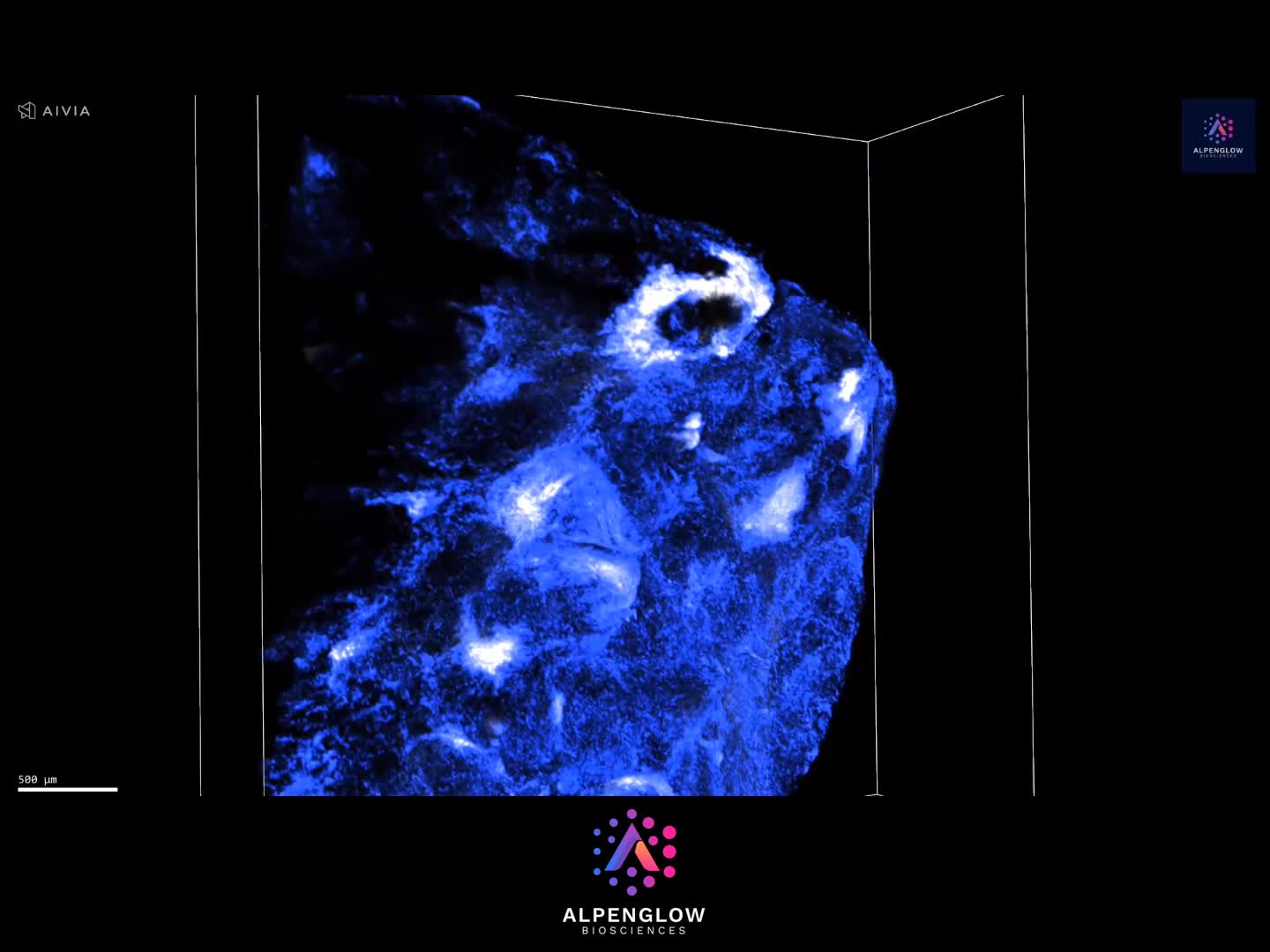
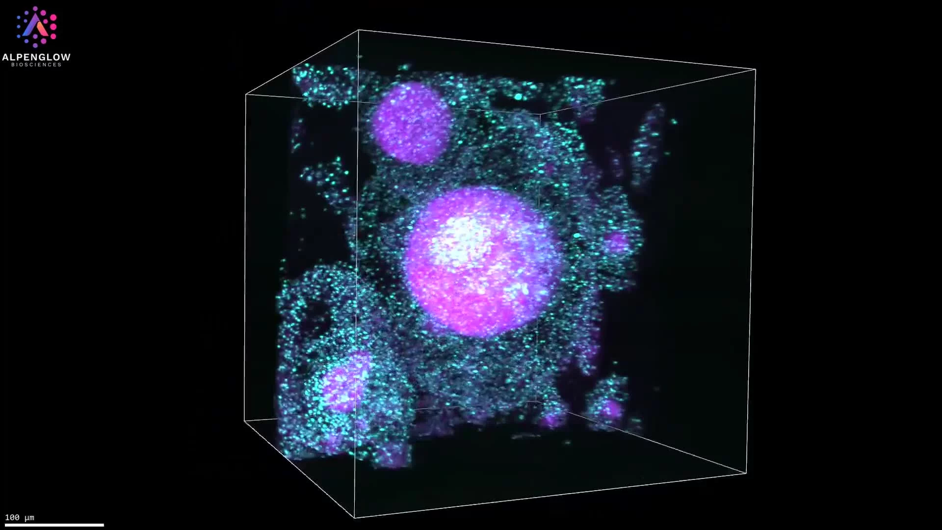
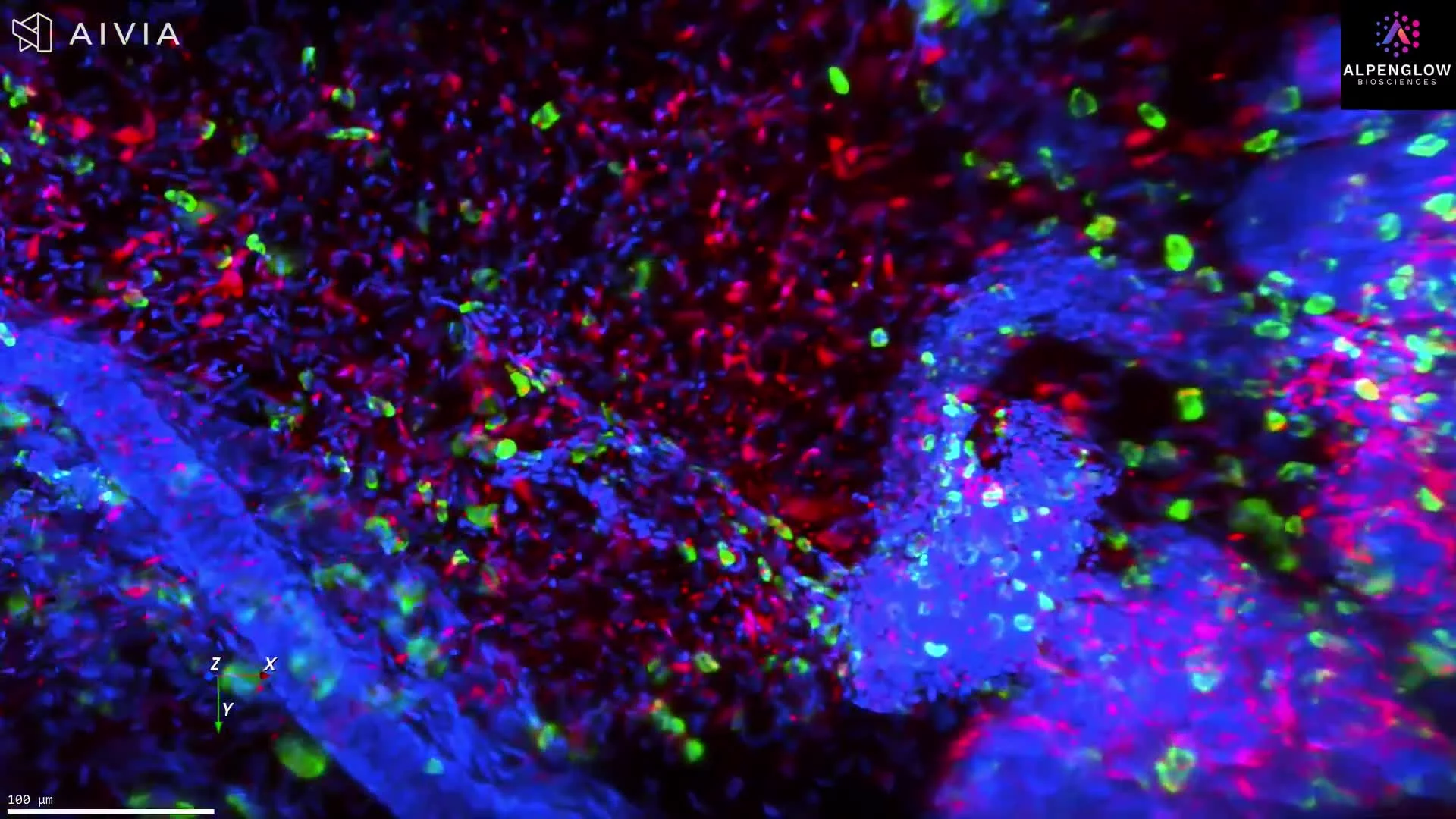
Large tissue volumes on the order of ~300 mm³ are first captured at 2 µm per pixel to retain full spatial context. From this dataset, a focused region of interest (~0.5 mm³) is selected and re-imaged at 0.33 µm per pixel, enabling high-resolution visualization of cellular and structural features within their native environment.
In this example, nuclei are labeled with TO-PRO-3, while eosin provides general protein contrast, allowing clear differentiation between tumor parenchyma and stromal compartments.
By traversing the full depth of the tissue, this volumetric view reveals spatial heterogeneity, compartmental organization, and boundary shifts that are not detectable in a single 2D section. As shown in the linked analysis, accurate characterization of stromal and parenchymal distributions in CRC can require dozens of 2D sections, whereas a single 3D dataset preserves this information inherently.
This example illustrates why 3D tissue imaging is critical for studying tumor microenvironments and immune phenotypes, and why volumetric data provide a more reliable foundation for quantitative spatial analysis than traditional slide-based workflows.