
Why 3D Tissue Imaging?
Because Biology Isn’t Flat.
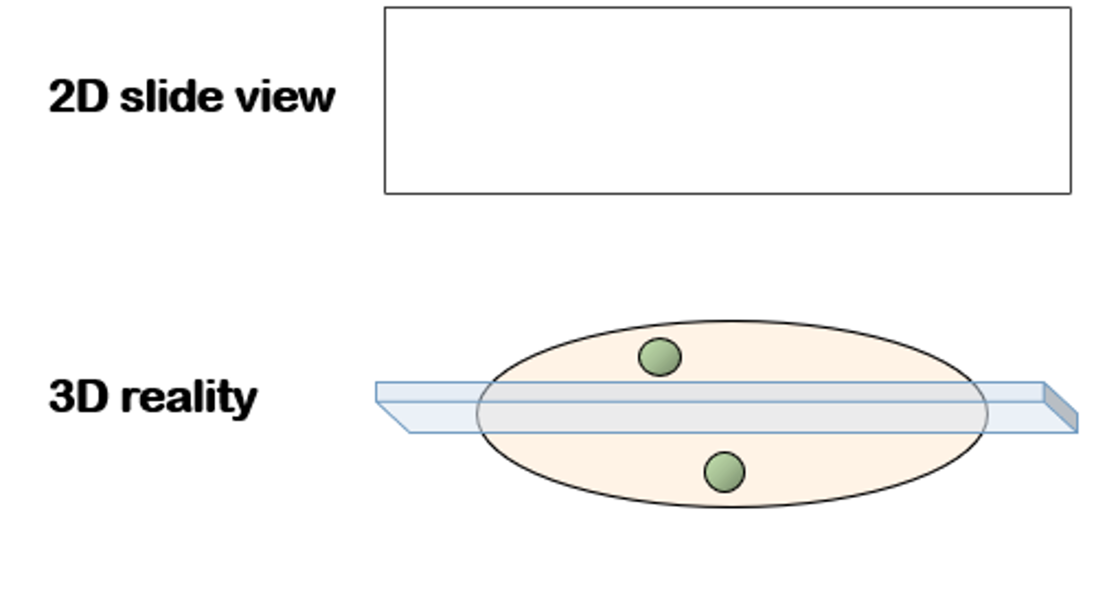
Clinical and translational decisions often rely on two-dimensional tissue sections. That approach collapses depth, fragments spatial relationships, and samples a small fraction of the specimen. As biology becomes increasingly spatial, this limitation increasingly leads to missed signals and uncertain conclusions.
Three-dimensional tissue imaging preserves intact architecture across depth. It captures how cells, structures, and microenvironments are truly organized, transforming tissue from a static slice into a quantitative volumetric dataset that supports confident decision-making.
Why traditional 2D histology breaks spatial biology
Too little tissue is sampled
Thin sections represent a small fraction of the specimen. Rare cells and localized structures are easily missed.
Depth and proximity are lost
Cell neighborhoods and gradients across tissue depth cannot be measured when biology is flattened.
Reconstruction introduces distortion
Serial section alignment and interpolation add artifacts, making true volumetric measurement unreliable.
Limits of 2D histology for spatial biology
Convoluted structures
Nerves in inflammatory skin disease follow tortuous paths that cannot be reconstructed from isolated sections.

Complex cell distributions
Immune organization in tumors varies dramatically across depth, affecting density, clustering, and proximity.

Sparse biological features
Tertiary lymphoid structures and other rare features may exist entirely between sections.
Tissue-scale architecture
Vasculature, ducts, and follicles depend on continuity and connectivity that are inherently three-dimensional.

Key biological applications where 3D tissue imaging is required
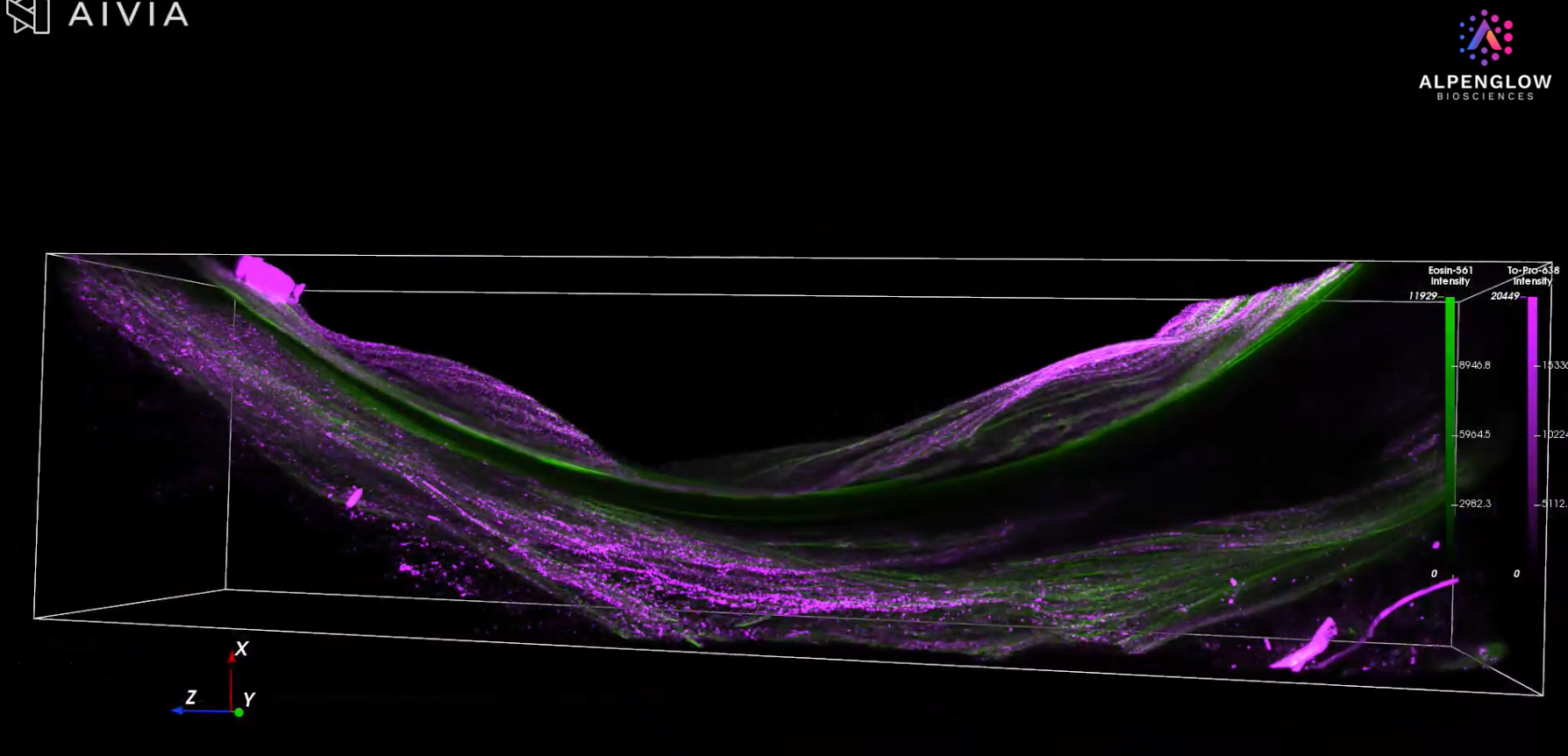
3D imaging of nerves in dermatology
Neural networks in skin follow tortuous, branching paths across depth. Single sections capture disconnected fragments, making it difficult to assess innervation density and spatial relationships in inflammatory skin disease.
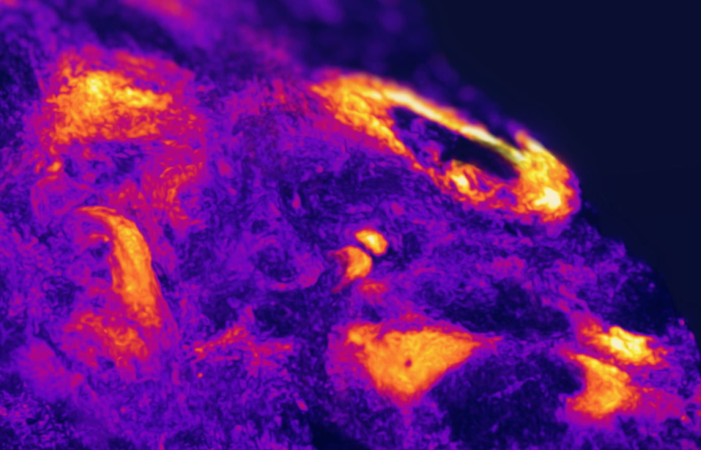
3D analysis of immune cell organization in cancer
Immune cell density, clustering, and proximity to tumor and stroma vary substantially across tissue depth. Two-dimensional sections miss this variation, resulting in an incomplete characterization of immune architecture.
3D detection of tertiary lymphoid structures
Tertiary lymphoid structures are sparse and spatially localized. Whole-tissue 3D imaging reduces false absence caused by sectioning gaps and limited sampling.
3D visualization of tissue-scale architecture
Vasculature, ducts, and follicles depend on continuity and connectivity that cannot be reliably inferred from isolated slices.
What changes when tissue stays intact
Whole-tissue imaging captures the full specimen volume, reducing false negatives without increasing sample numbers. Measurements of cell counts, distances, and spatial organization are made directly in three dimensions rather than inferred from partial views.
Preserving spatial context improves biological interpretation and supports more confident experimental and translational decisions.
How light-sheet microscopy enables whole-tissue imaging
Light-sheet microscopy scans a thin optical plane through intact tissue, enabling volumetric imaging without physical sectioning. This preserves native architecture, avoids cutting artifacts, and supports efficient imaging of large tissue samples.
From intact tissue to quantitative 3D data
Alpenglow’s Aurora™ 3D Spatial Biology Solution enables non-destructive whole-tissue imaging using light-sheet microscopy, followed by AI-powered analysis. Intact samples are converted into quantitative volumetric datasets while remaining compatible with downstream assays.
Who benefits from 3D tissue imaging
Translational research
Reveal heterogeneity and spatial drivers that are invisible in two dimensions.
Drug discovery and development
Capture full tissue context from limited samples to reduce uncertainty and missed biology.
Spatial biology and digital pathology innovation
Generate ground-truth 3D datasets that support validation, benchmarking, and scalable analysis.
From 2D slides to 3D ground truth tissue data
The Aurora™ 3D Spatial Biology Solution unifies three modules so you can work with ground truth 3D data.
3Di™ HOTLS microscope. Hybrid open top light sheet system for intact tissue imaging and multi-scale 3D histology.
3Dm™ data management. Automated data handling and processing workflows that keep large 3D volumes organized and accessible.
3Dai™ image analysis. AI-powered analysis and spatial statistics that extract quantitative insight from gigabyte to terabyte-scale datasets.
You can access Aurora™ as a complete end-to-end platform, or you can begin with 3D histology imaging services to generate 3D tissue data and AI-ready volumes without installing new hardware.