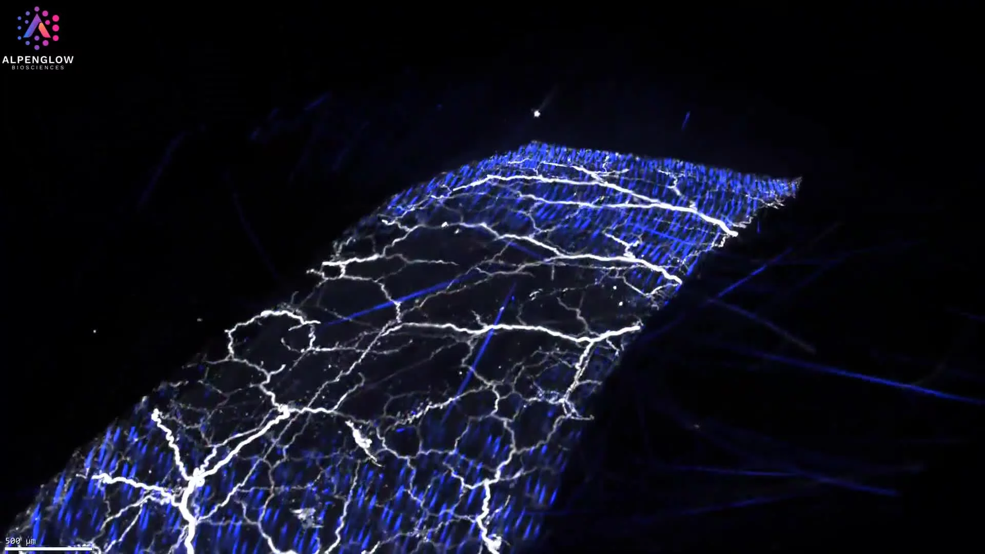
High-Resolution 3D Imaging of Hair Follicle and Gland Architecture
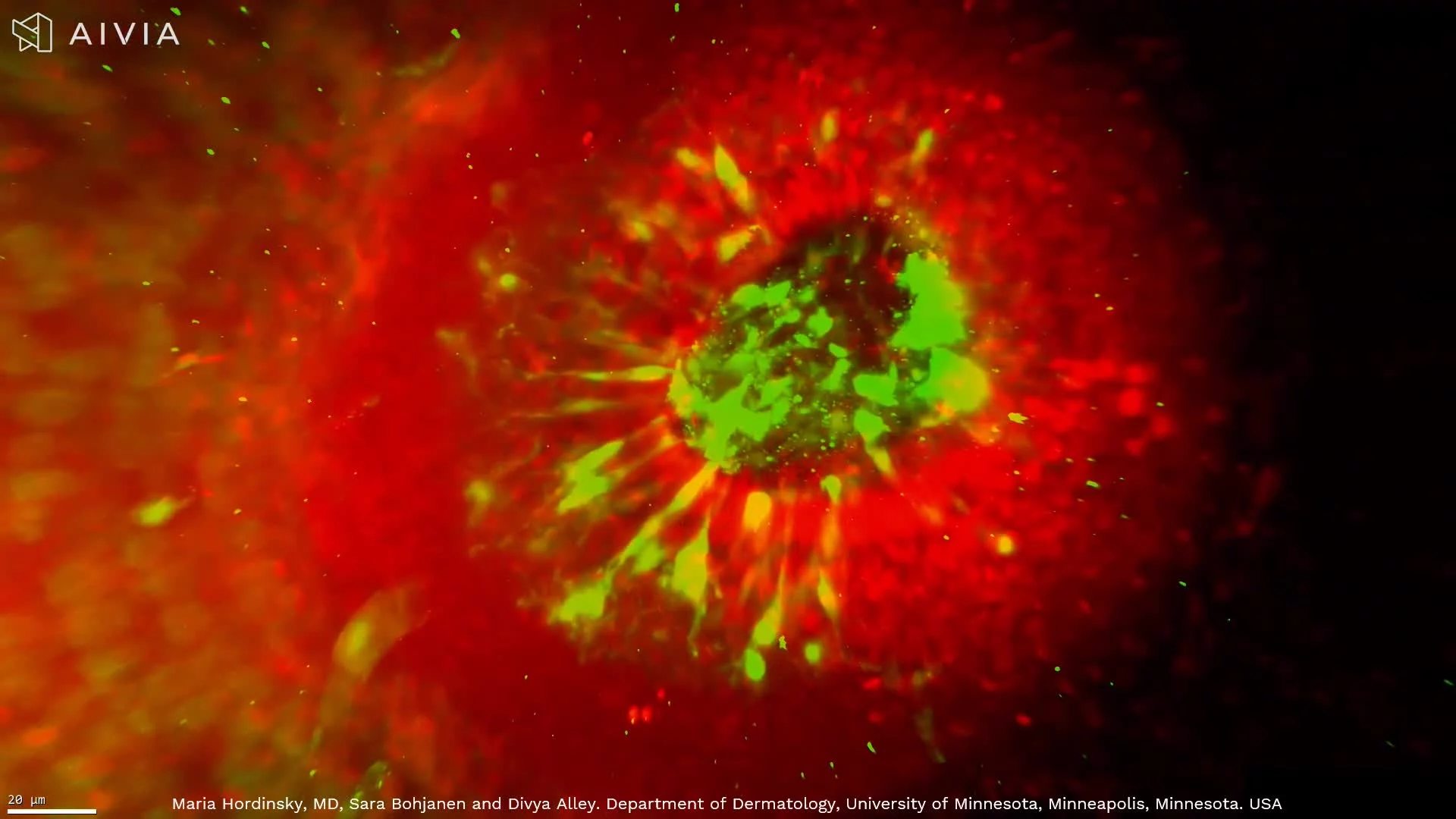
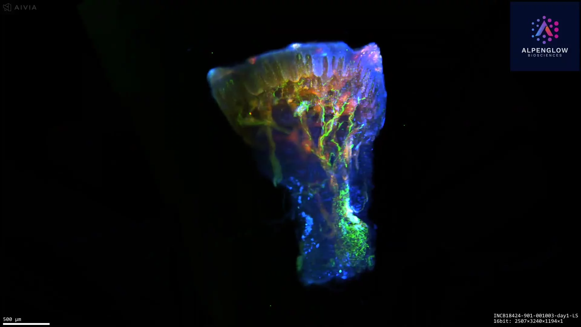
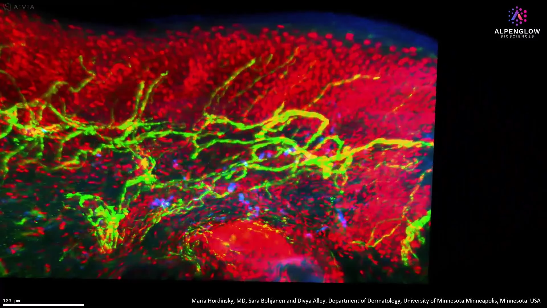
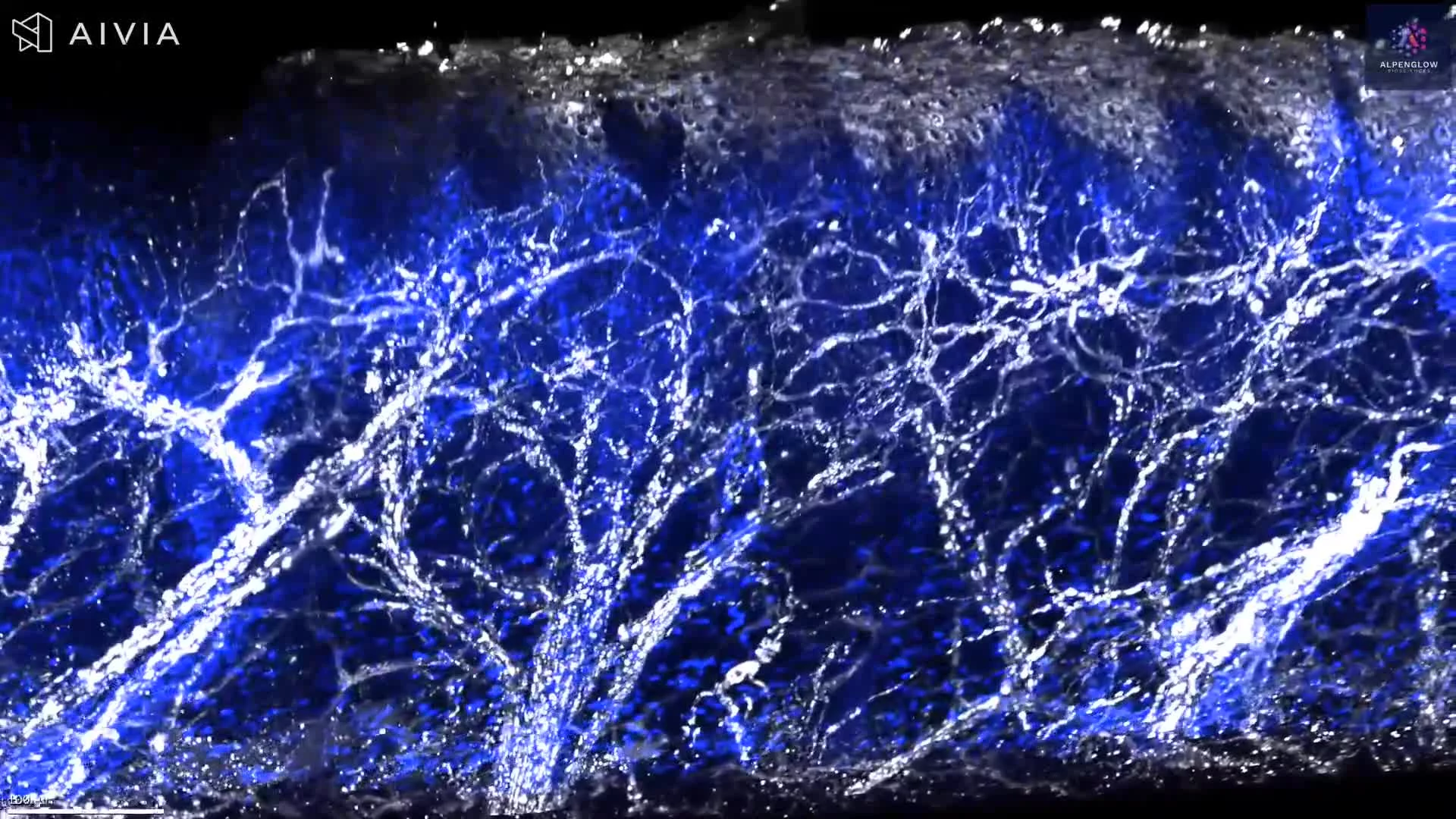
This high-resolution 3D dataset presents human skin tissue, preserving the architecture of a hair follicle, its associated gland, and the surrounding nerve network across depth.
The volumetric view allows the follicle and glandular structure to be followed through the tissue, revealing their morphology, orientation, and spatial relationships with nearby nerve fibers. This continuous view reduces the fragmentation of complex structures that can occur across individual 2D tissue sections.
Following processing with 3Dm™ data management and analysis using 3Dai™ AI-powered analysis, the dataset supports measurement of follicle dimensions, glandular morphology, nerve density, branching, and distances between neural and follicular structures. These readouts are relevant to research on alopecia, inflammatory skin conditions, hair biology, and treatment-associated changes in follicular architecture.
Explore how 3D tissue imaging supports dermatology research across skin architecture, innervation, immune organization, and spatial relationships.
The tissue was imaged using the Aurora 3D™ Spatial Biology Solution, including the 3Di™ Hybrid Open-Top Light-Sheet microscope.