3D Imaging of Collagen Architecture in Prostate Tissue
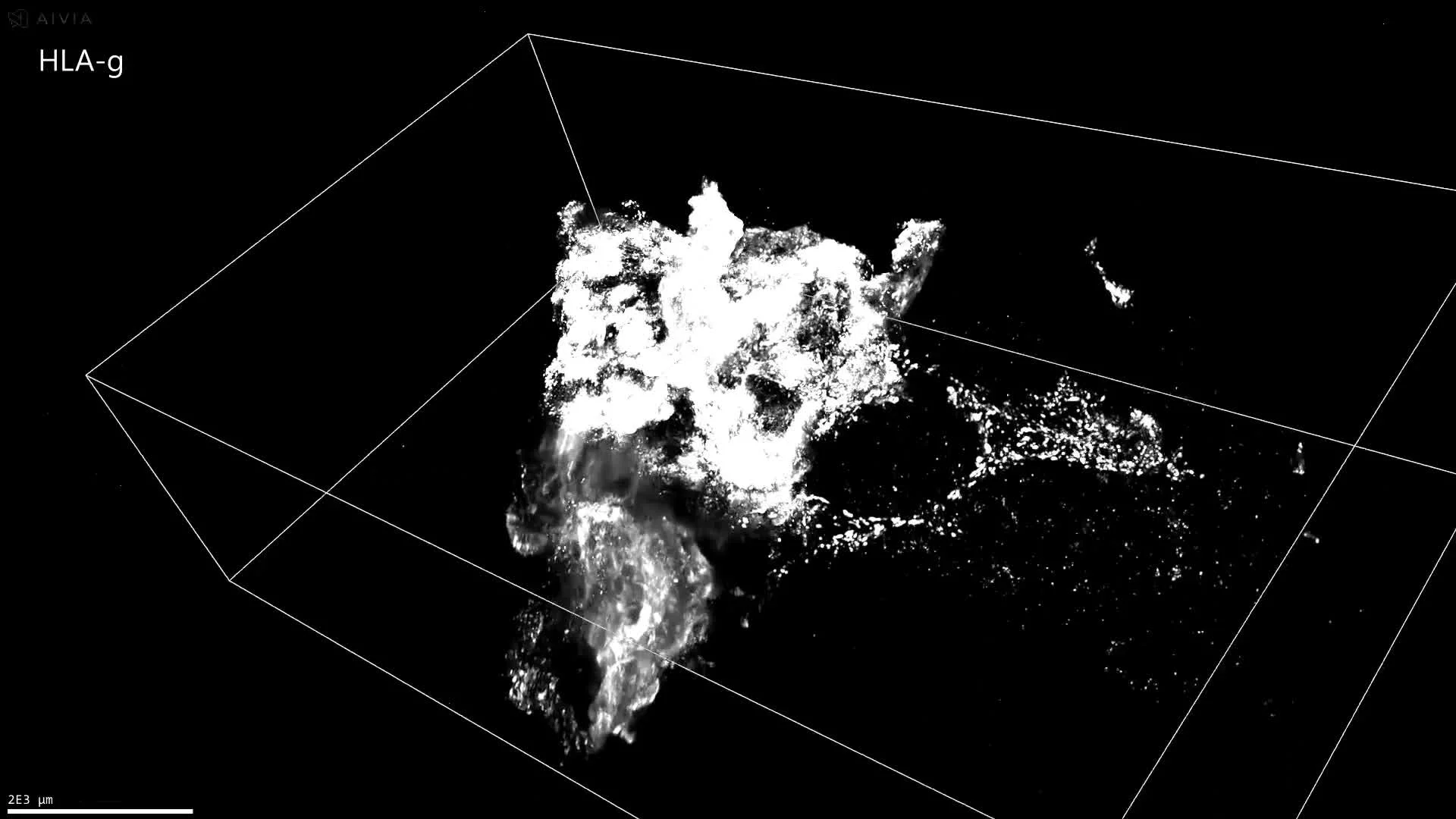
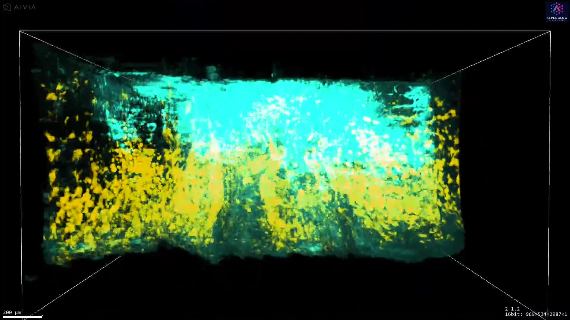
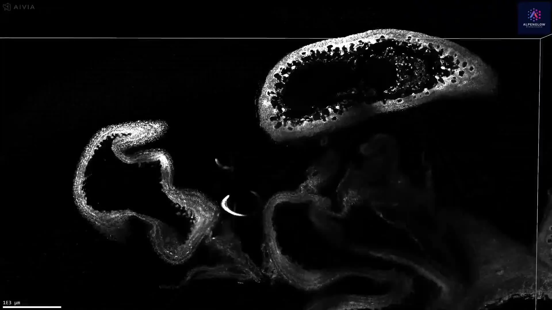
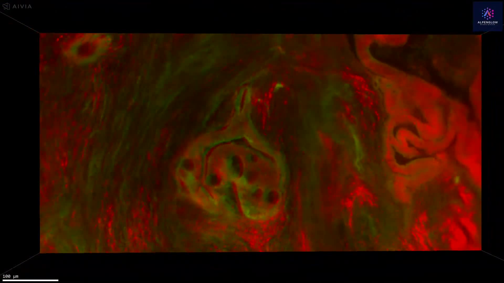
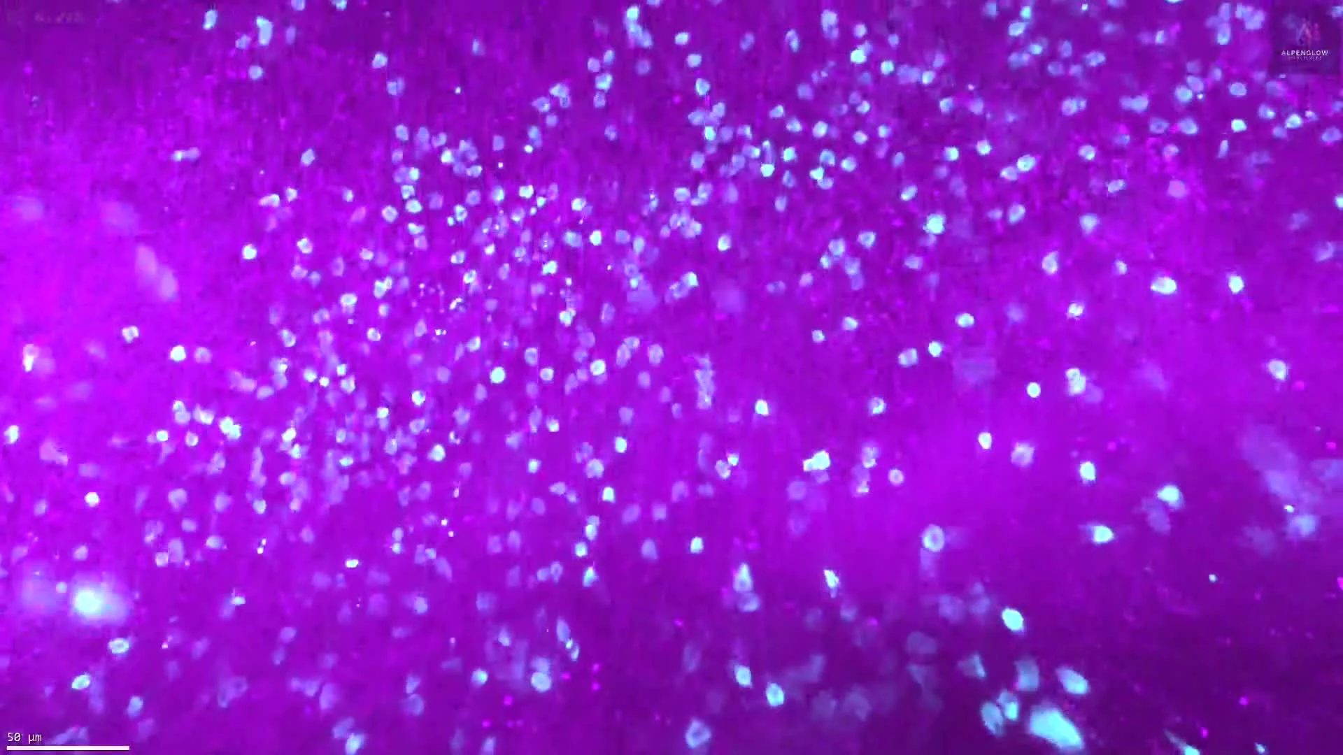
This dataset explores the structural complexity of prostate tissue using 3D fluorescence imaging on the Aurora™ 3Di Hybrid Open Top Light Sheet (HOTLS) microscope. Collagen is stained with Fast Green and rendered in vivid purple, revealing its essential role in shaping tissue architecture, influencing disease progression, and guiding therapeutic strategies.
Unlike traditional thin-sectioning, Alpenglow’s 3D digital pathology platform digitally sections intact tissue without slicing, preserving the full spatial context. This enables the visualization of collagen organization, density, and orientation in relation to surrounding cells, which are often overlooked in 2D histology.
Through integration with 3Dm data management and 3Dai AI-powered segmentation, collagen patterns can be quantified with cellular-level precision, supporting spatial profiling and high-content tissue analysis.
Applications extend to cancer research, fibrosis, and translational studies, where understanding collagen remodeling is critical for advancing biomarker development and therapeutic design.