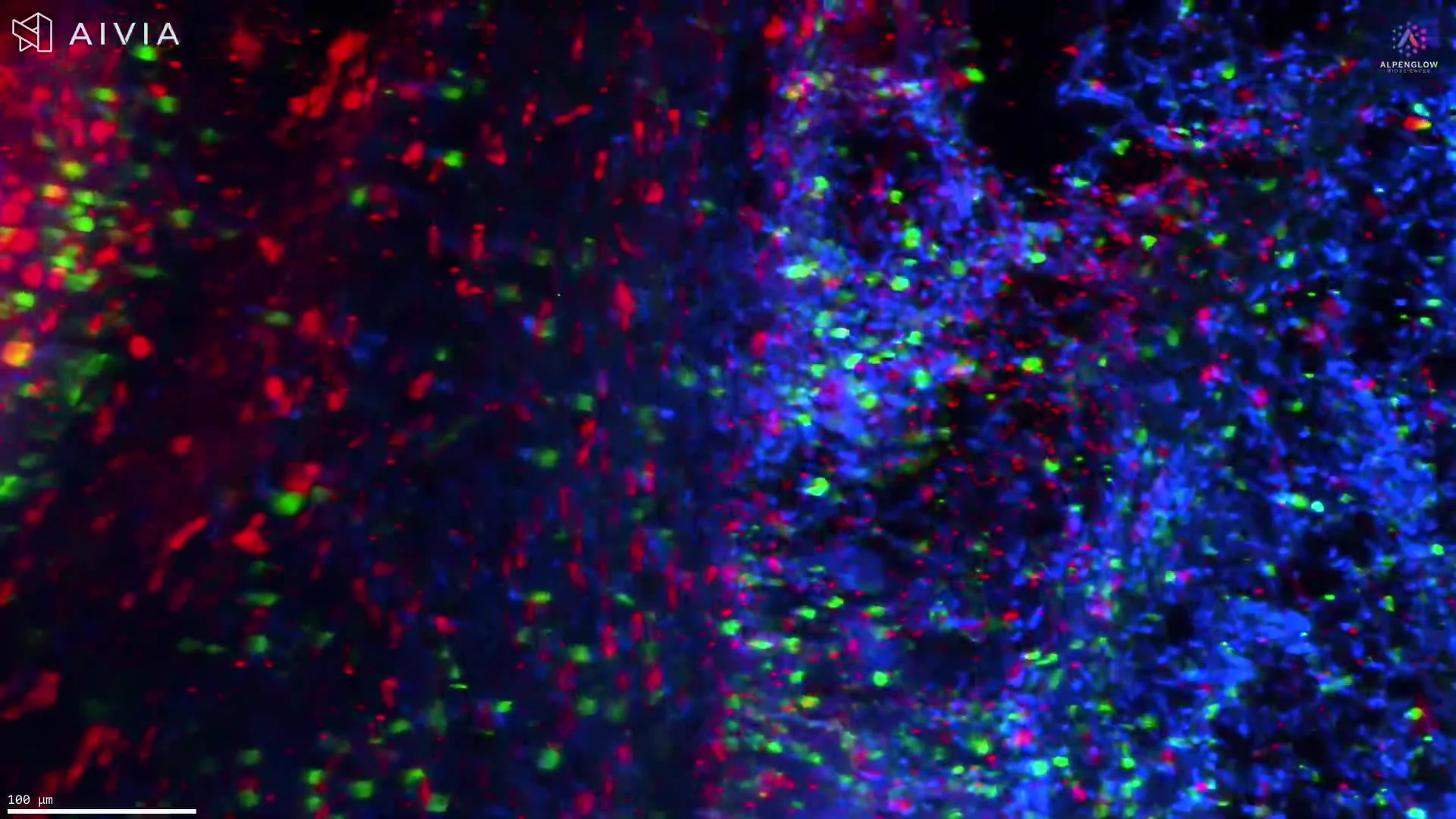
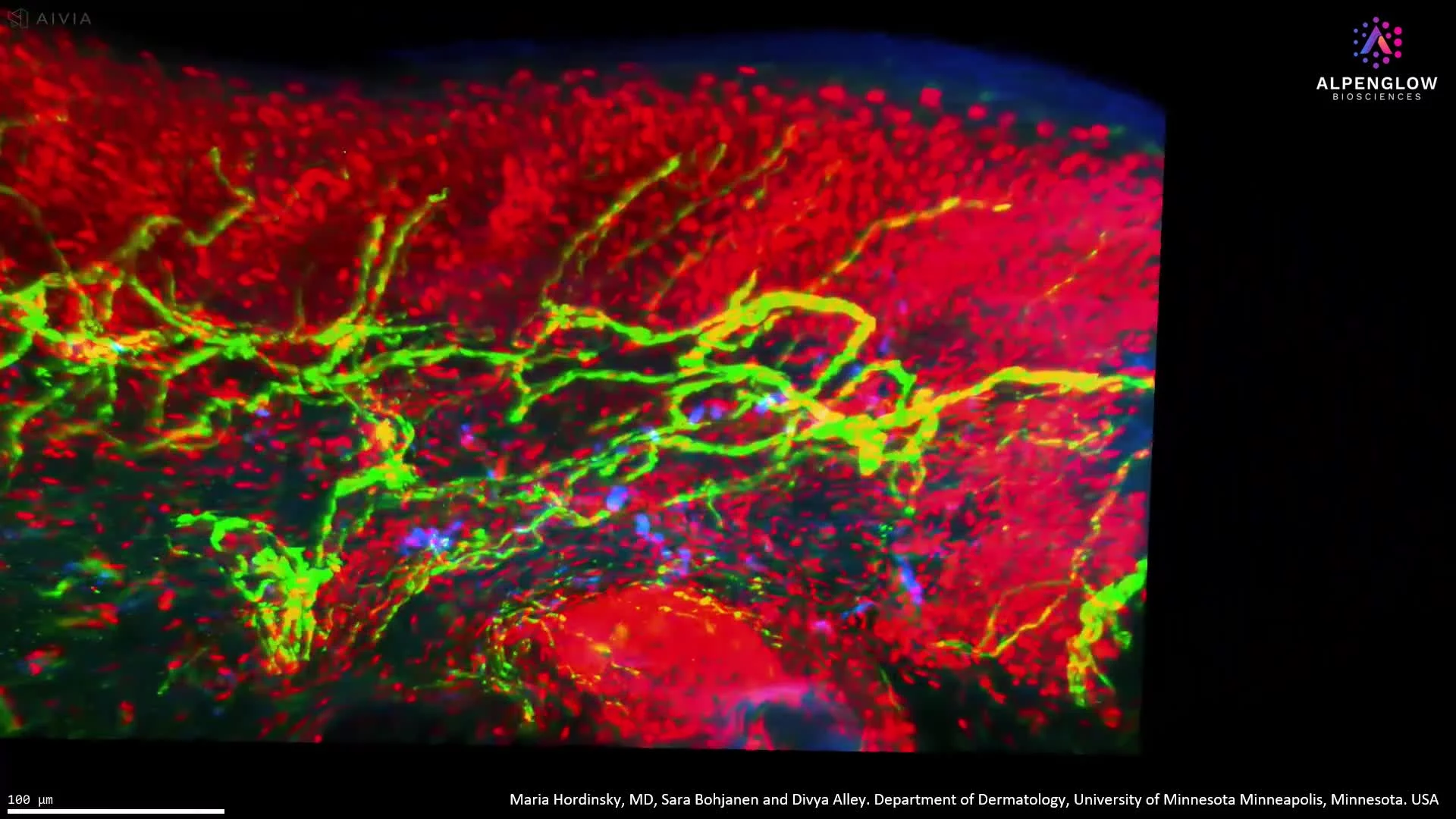
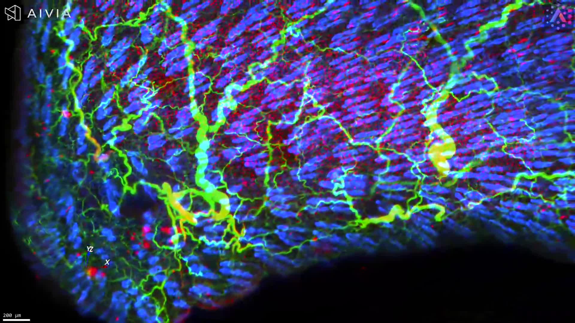
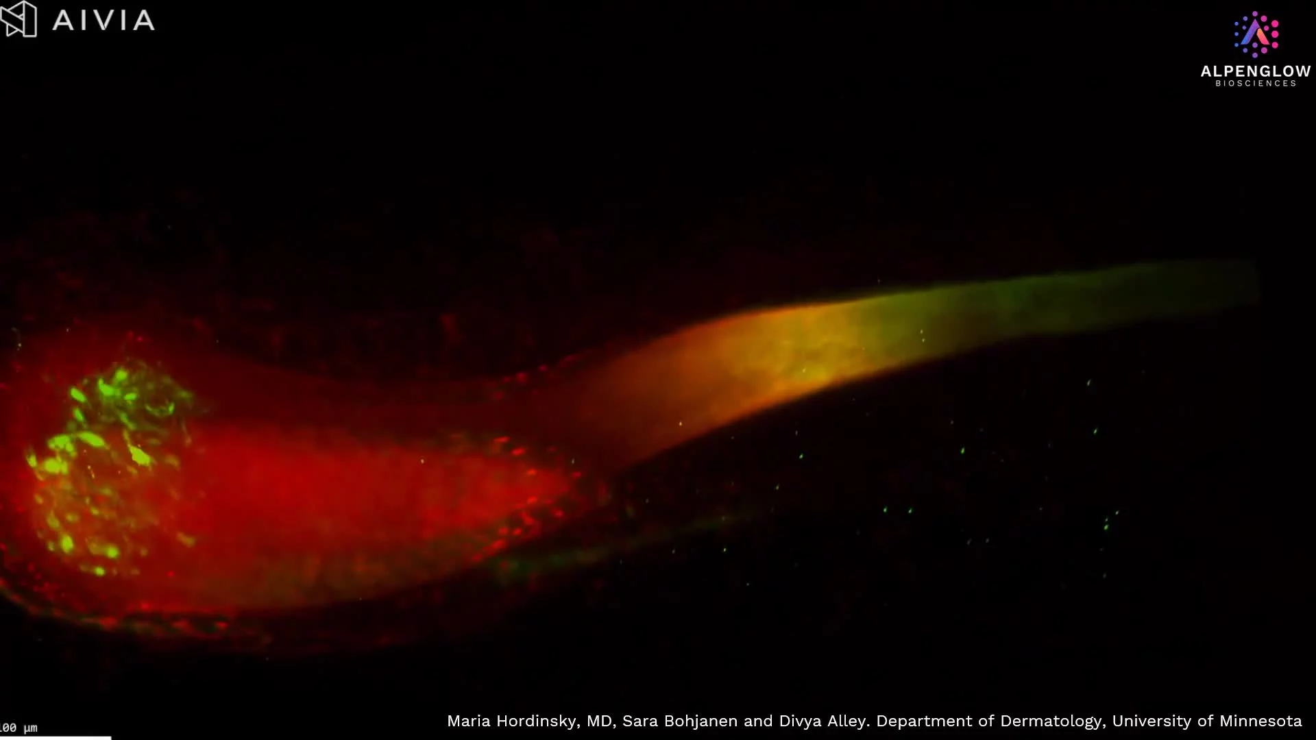
High-resolution (40X) 3D imaging of a Region of Interest in a lesional Atopic Dermatitis sample
High-resolution (40X) 3D imaging with Hybrid Open Top Light Sheet (HOTLS) microscopy of a Region of Interest in a lesional Atopic Dermatitis skin punch biopsy reveals intact tissue architecture and spatial immune organization.
The sample was stained with TO-PRO-3 for nuclei, PGP9.5 for nerve fibers, and CD3 for T cells, enabling detailed visualization of neuro-immune interactions and inflammatory pathways.
See our AD use case.
Image reproduced with permission from Incyte Corporation.
Previous
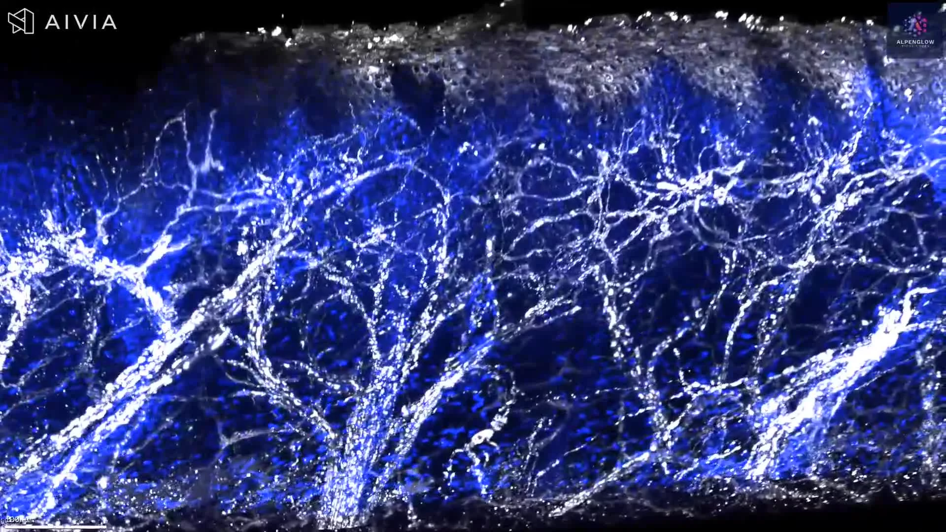
Low-resolution whole-sample 3D imaging of a lesional Atopic Dermatitis punch biopsy
Next