Why 3D tissue imaging
See tissue biology in its full 3D context.
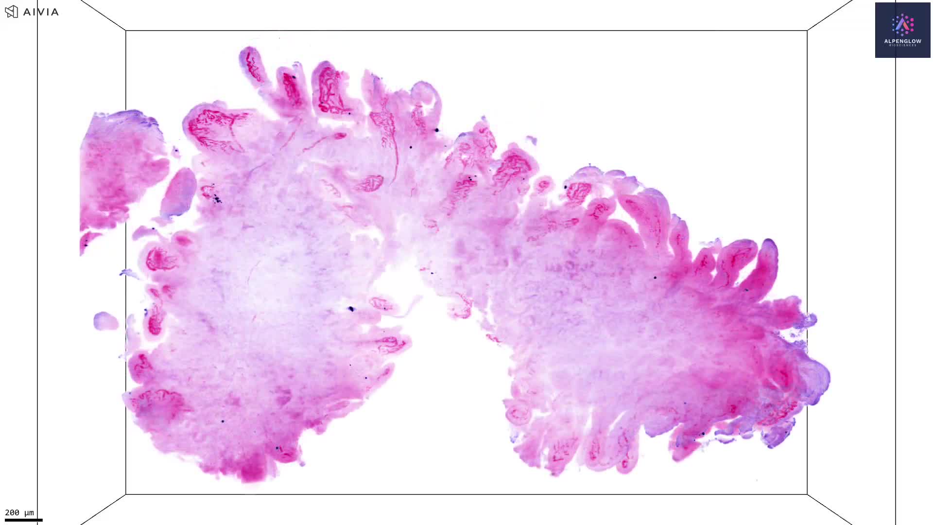
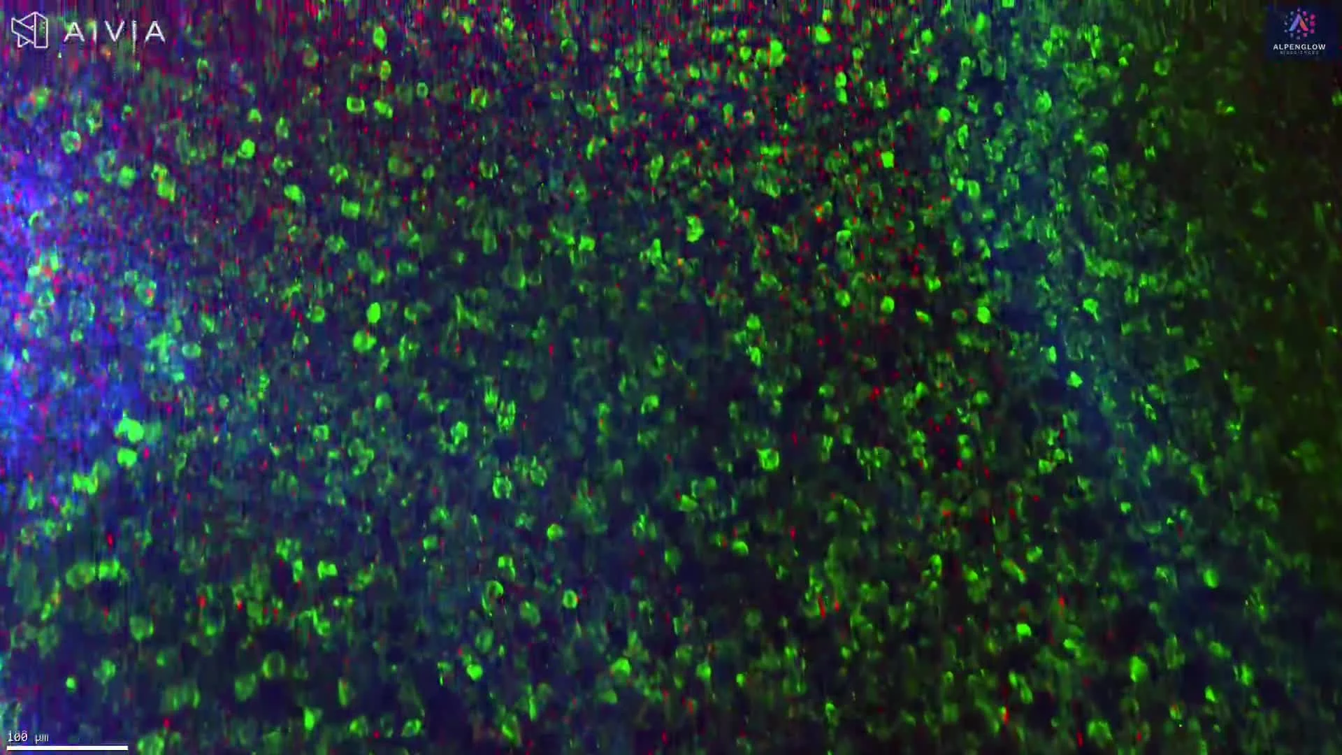
Traditional histology captures thin sections from complex tissue. 3D tissue imaging preserves depth, architecture, and spatial relationships for whole tissue imaging, spatial profiling, digital pathology, and quantitative analysis.
2D sections vs 3D context
Why can 2D tissue sections miss important spatial biology?
Thin sections capture isolated planes from a larger specimen. 3D tissue imaging expands the sampling volume, preserves depth, and retains connected tissue architecture.
Sampling coverage
Selected sections may miss sparse, localized, or unevenly distributed features.
More tissue volume can be examined across multiple regions and depths.
Spatial depth
Depth is reduced to a thin plane, limiting interpretation above and below the section.
Cells and structures retain their positions within the tissue volume.
Structural continuity
Branching structures appear as disconnected fragments across separate sections.
Vessels, nerves, glands, follicles, and immune structures remain connected.
Key biological applications
Which biological questions benefit from 3D tissue imaging?
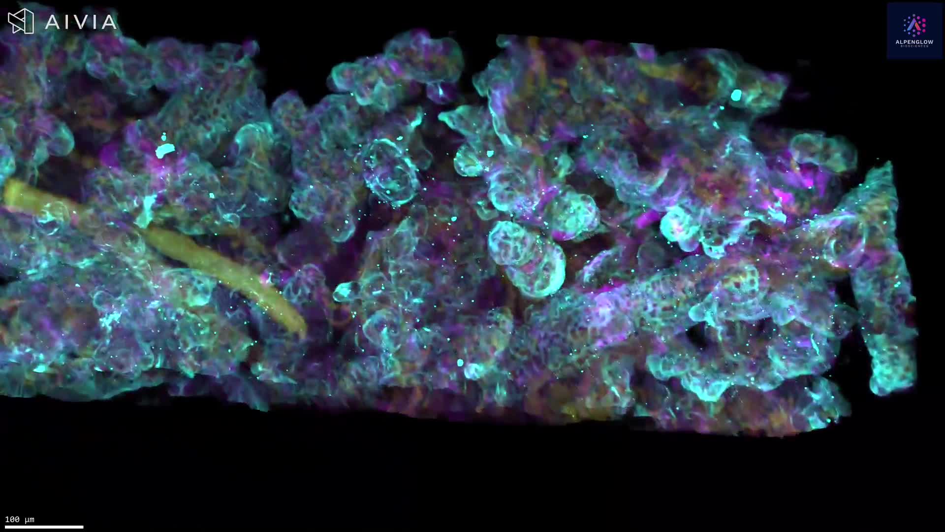
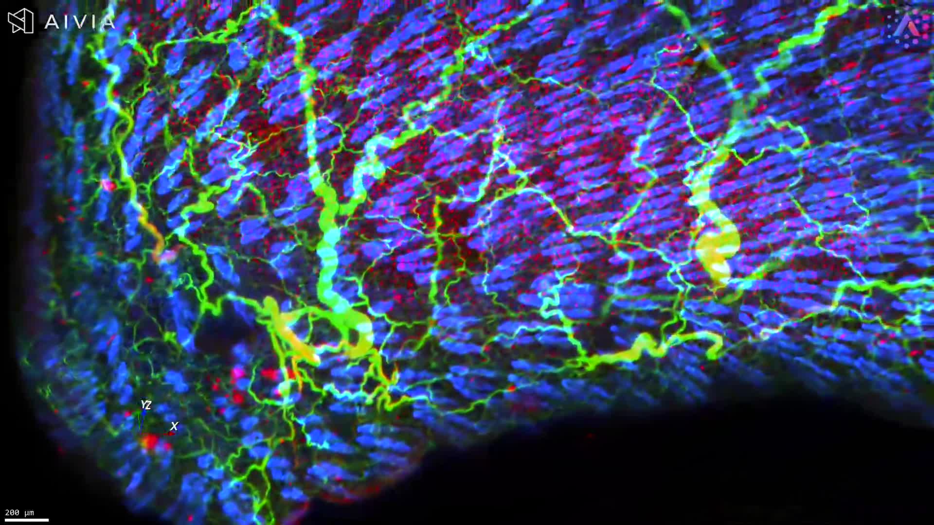
3D tissue imaging is particularly valuable when interpretation depends on complex structures, cell distributions, rare features, or tissue organization across depth.
Convoluted structures
Vessels, nerves, glands, follicles, and crypts often require 3D context to evaluate shape, branching, and continuity.
Complex cell distributions
Immune cells, tumor regions, and stromal compartments can be interpreted in relation to nearby structures across tissue depth.
Sparse biological features
Rare cells, focal lesions, tertiary lymphoid structures, and localized features can be missed when sampling is limited.
Tissue-scale architecture
Whole tissue imaging helps reveal how compartments, regions, and structures are organized across the sample.
Where this matters
These capabilities support translational research, drug development, spatial biology, and digital pathology programs where intact tissue context affects interpretation.

From tissue to data
How does Alpenglow turn intact tissue into quantitative 3D data?
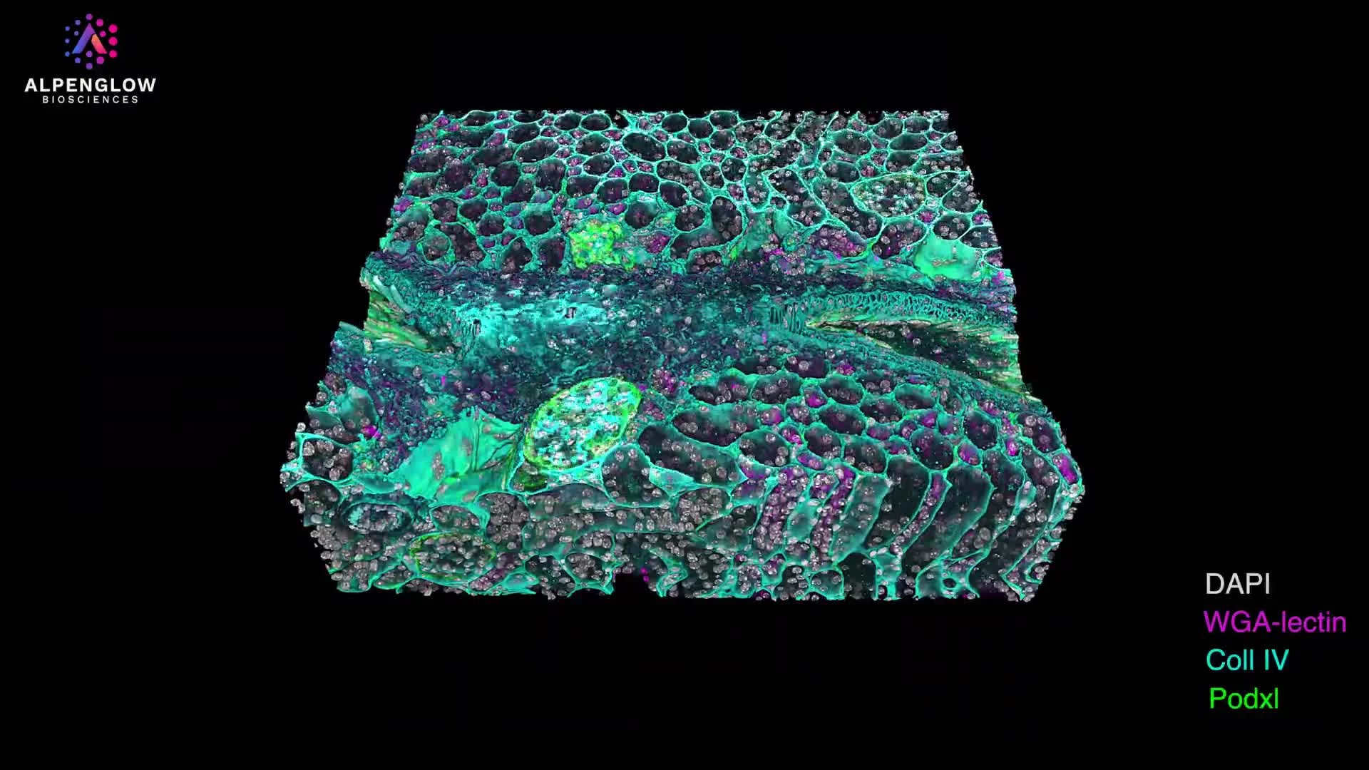
Aurora™ connects whole tissue imaging, volumetric data management, and AI-powered analysis in one end-to-end 3D spatial biology workflow.
3Di™
Image intact tissueLight-sheet microscopy captures tissue architecture as a high-resolution volumetric dataset.
3Dm™
Manage the volumeLarge 3D datasets are processed, aligned, organized, and prepared for review and analysis.
3Dai™
Quantify the biologyAI-powered analysis measures cells, structures, tissue features, and spatial relationships across the volume.
Move from sections to spatial insight
Ready to see what 3D tissue imaging can reveal in your samples?
Explore whole tissue imaging, spatial profiling, and AI-powered quantitative analysis with the Aurora 3D™ Spatial Biology Solution.