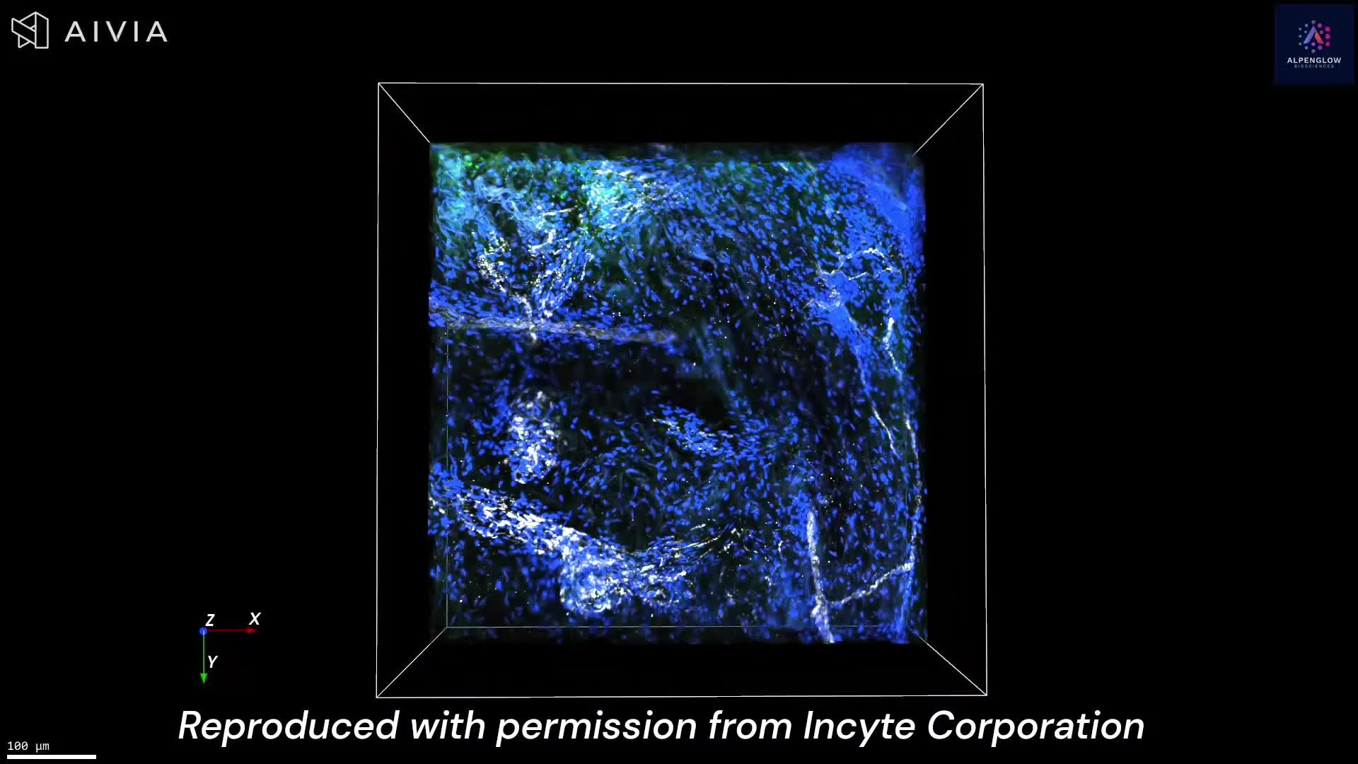
3D Tissue Imaging of Mouse Muscle Innervation
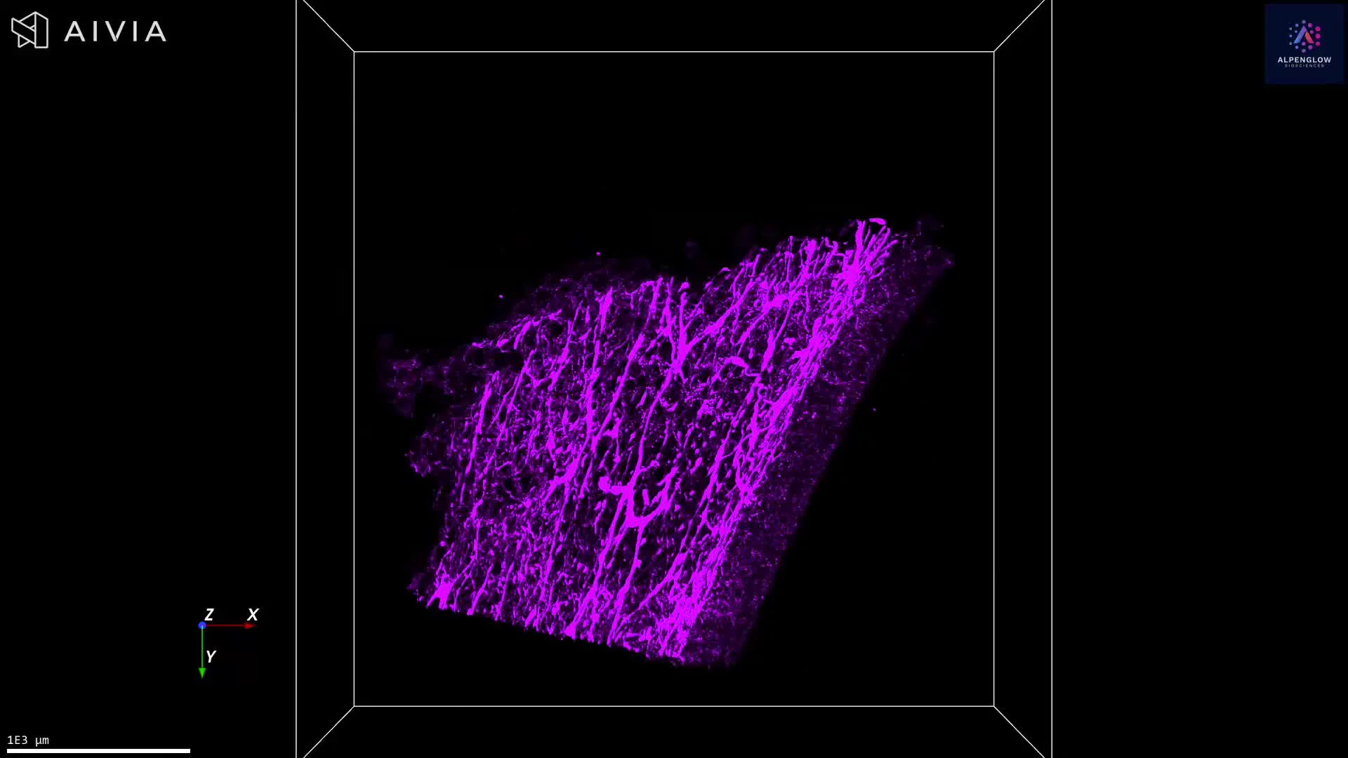
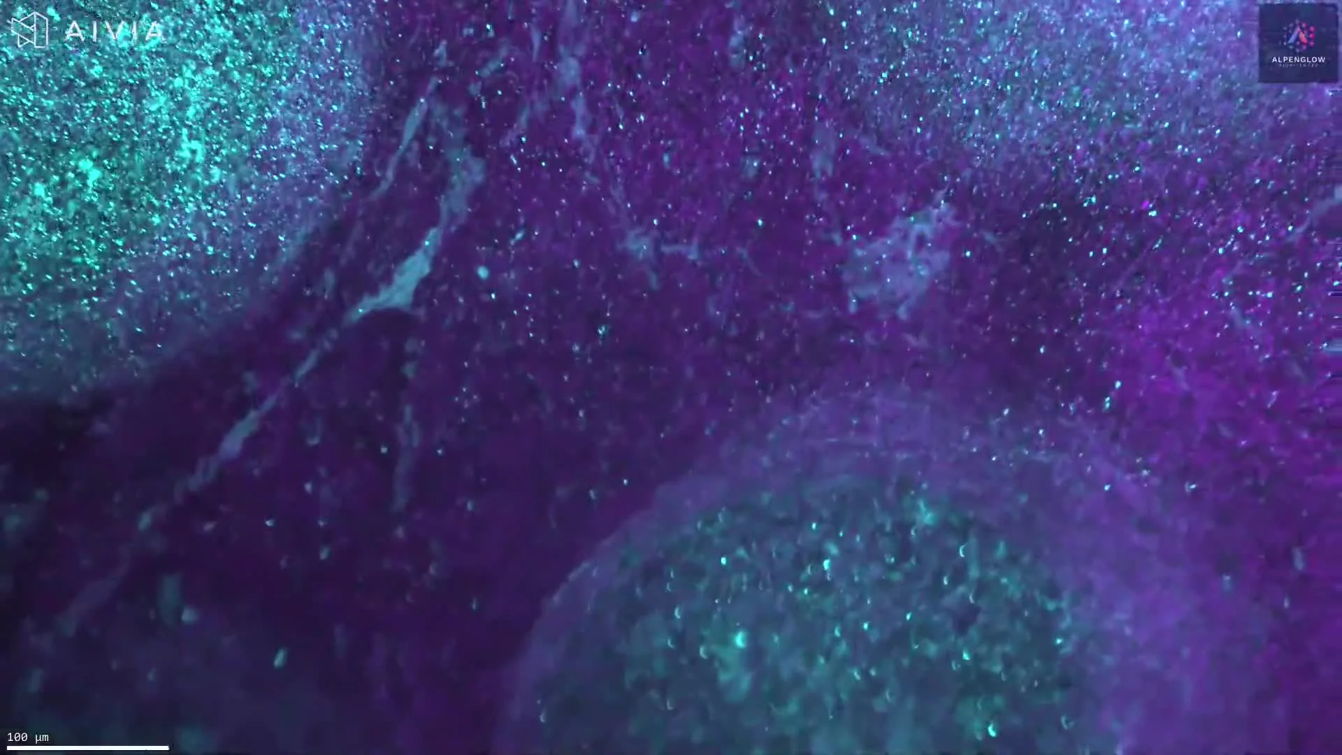
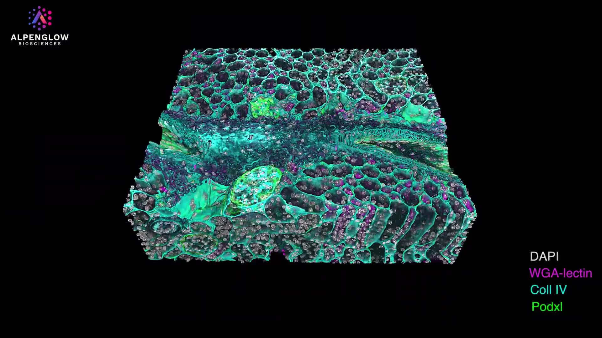
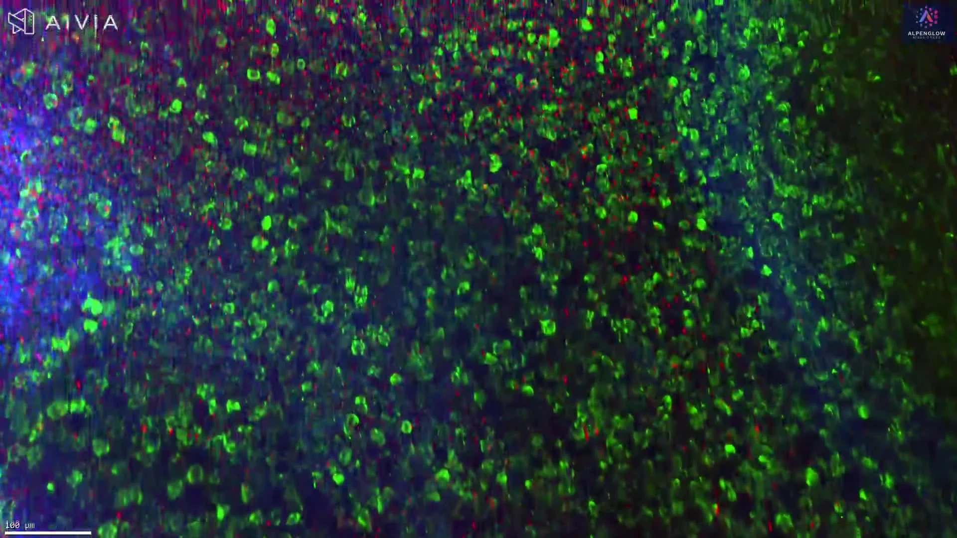
This dataset presents volumetric 3D imaging of mouse skeletal muscle stained with NF200, PGP9.5, and YO-PRO-1.
NF200, shown in green, highlights neurofilament heavy chain in larger-caliber axons and reveals major neural trajectories. PGP9.5, shown in magenta, visualizes the broader axonal network throughout the muscle. YO-PRO-1, shown in blue, labels nuclei and provides cellular context across the tissue volume.
The 3D view preserves axonal trajectories, branching patterns, and regional innervation across depth. With appropriate segmentation, the dataset can support measurements of axon length, branching complexity, regional nerve density, orientation, and spatial distribution.
Explore how 3D tissue imaging supports dermatology research across skin architecture, innervation, immune organization, and spatial relationships.
The tissue was imaged using the Aurora 3D™ Spatial Biology Solution, including the 3Di™ Hybrid Open-Top Light-Sheet microscope.