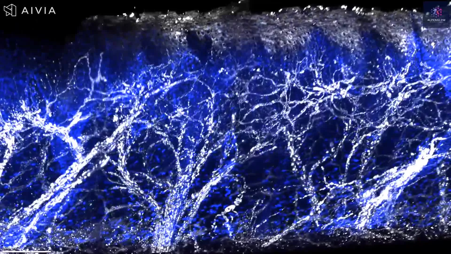
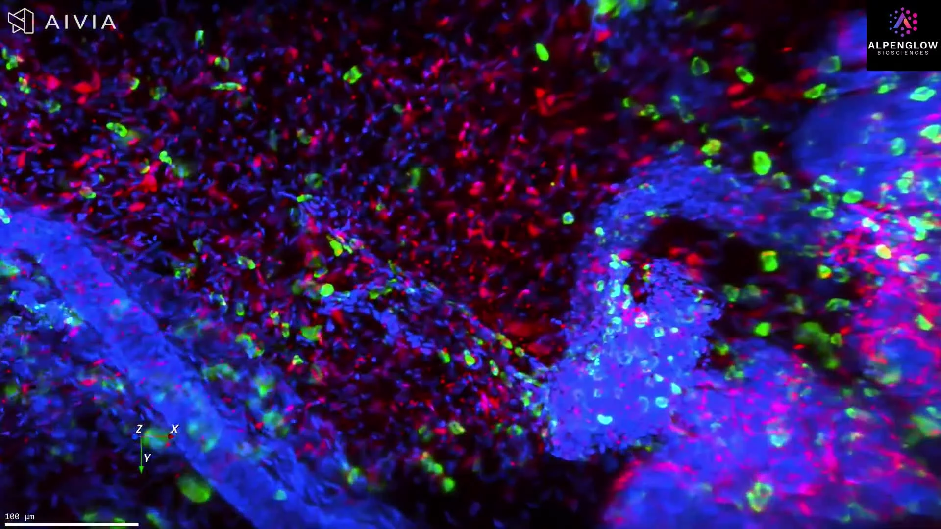
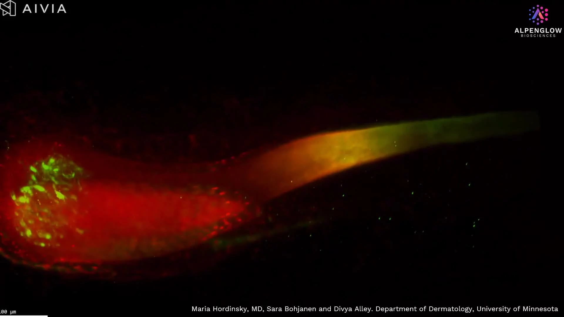
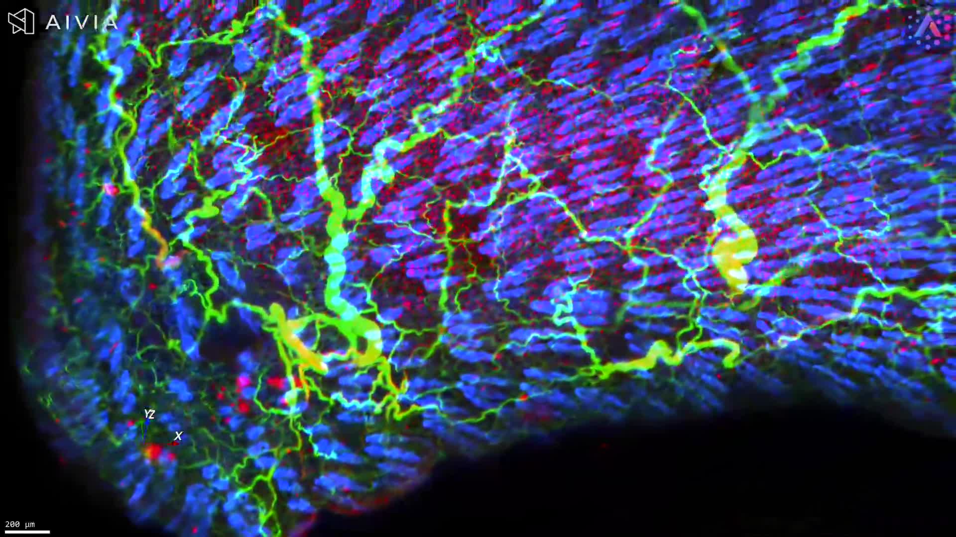
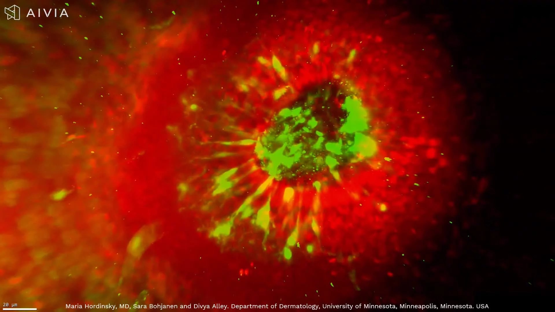
3D Imaging of Scalp Tissue for Alopecia Research
This high-resolution 3D visualization of scalp tissue provides unprecedented clarity into structures relevant to hair biology and disease. The dataset captures the epidermal layer, peripheral nerve distribution, and piliferous bulb architecture in measurable detail, features critical to understanding alopecia and related conditions.
Stains used:
TO-PRO-3 (Red): Highlights nuclei in the epidermis
CD45 (Blue): Marks immune cell distribution
PGP9.5 (Green): Maps peripheral nerve innervation
By comparing volumetric 3D analysis with conventional 2D slices, this dataset demonstrates the significant loss of information in single-plane histology, underscoring the value of whole-tissue imaging with HOTLS microscopy. Leveraging advanced data management and AI-powered segmentation, immune and neural structures are quantified with precision.
The insights extend beyond visualization, contributing to dermatology research in alopecia areata, prurigo nodularis, hidradenitis suppurativa, atopic dermatitis (see our AD use case), and other inflammatory skin diseases.
These breakthroughs highlight the transformative potential of 3D spatial biology and digital pathology in uncovering the mechanisms underlying hair loss and skin disorders.