3D Imaging of a Rabbit Retina: High-Resolution Tissue Mapping
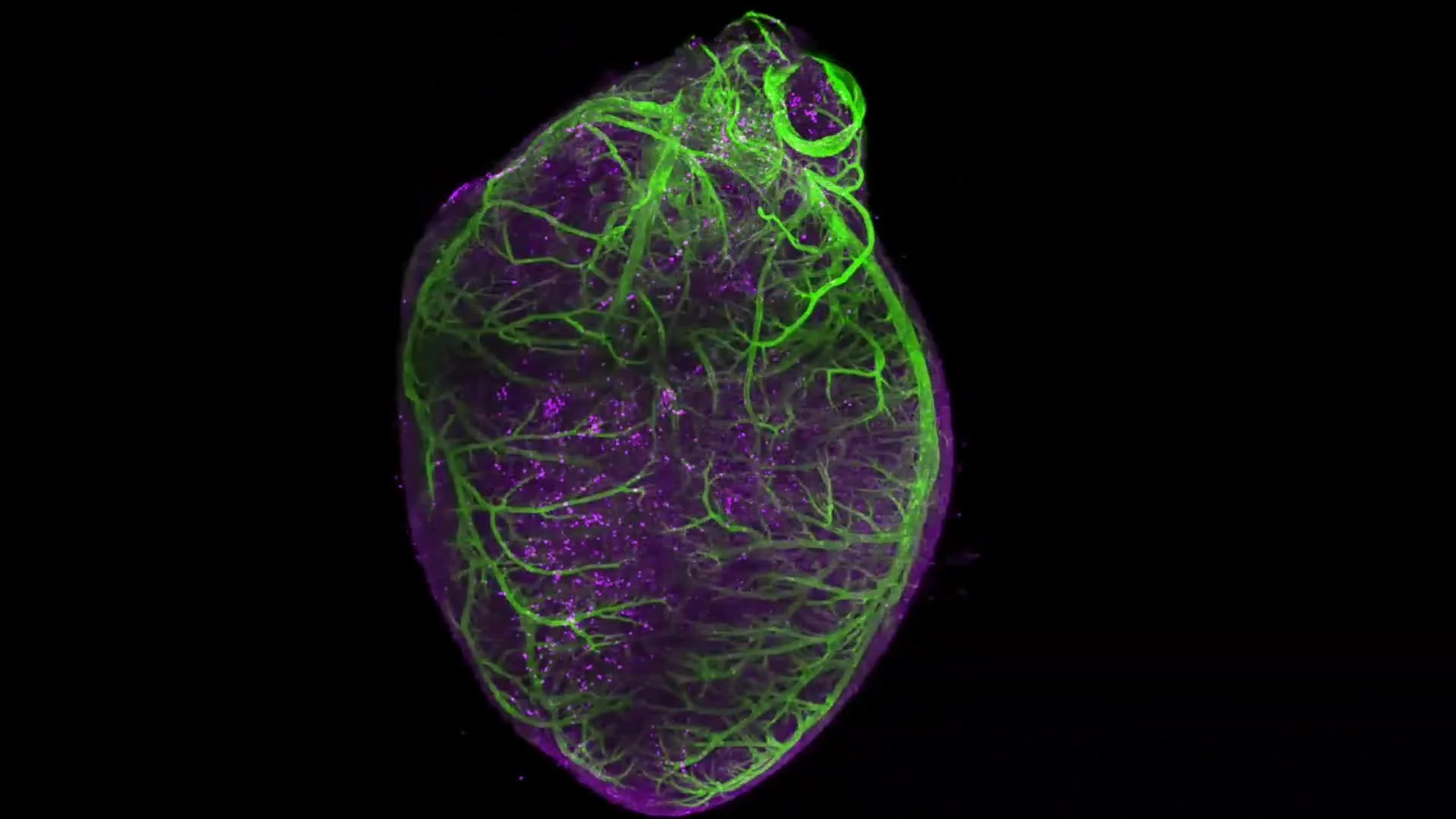
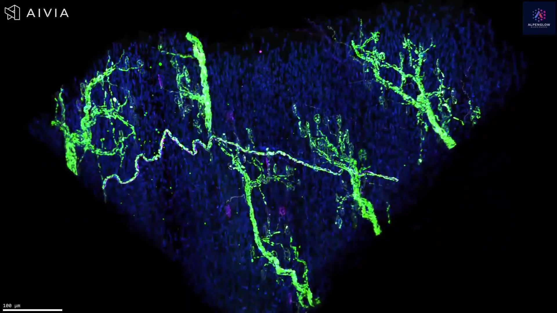
This dataset presents a high-resolution 3D visualization of a cleared rabbit retina, captured intact on the Aurora™ 3Di Hybrid Open Top Light Sheet (HOTLS) microscope. By avoiding physical sectioning, the imaging preserves the true spatial architecture of the tissue.
Stains used:
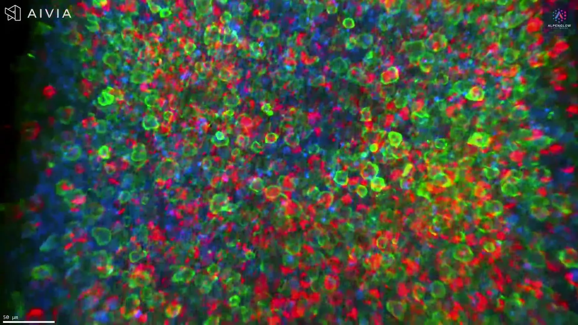
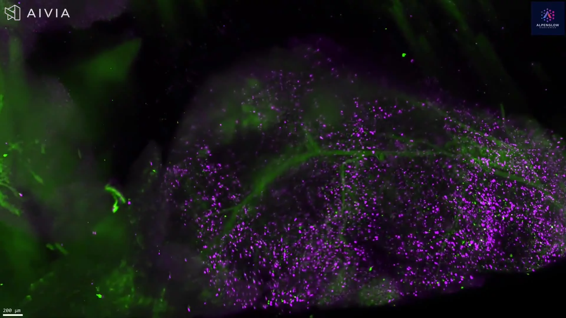
TO-PRO-3 (Magenta, pseudocolor): Labels nuclei
Eosin (Green, pseudocolor): Highlights cytoplasmic structures
The fly-through reveals the full thickness of the sample (≈1 × 1 × 4 mm), capturing the retina, choroid, and sclera in their native spatial organization. This volumetric dataset enables accurate morphological and contextual analysis at cellular resolution, overcoming the limitations of 2D histology.
When combined with 3Dm data management and 3Dai AI-powered segmentation, the dataset can be further analyzed for nuclear density, layer-specific organization, and cellular distribution. Applications include ophthalmology research, retinal disease modeling, and translational studies focused on vision science.