Deep Tissue Staining with Anti-CD3 to Identify T Cells
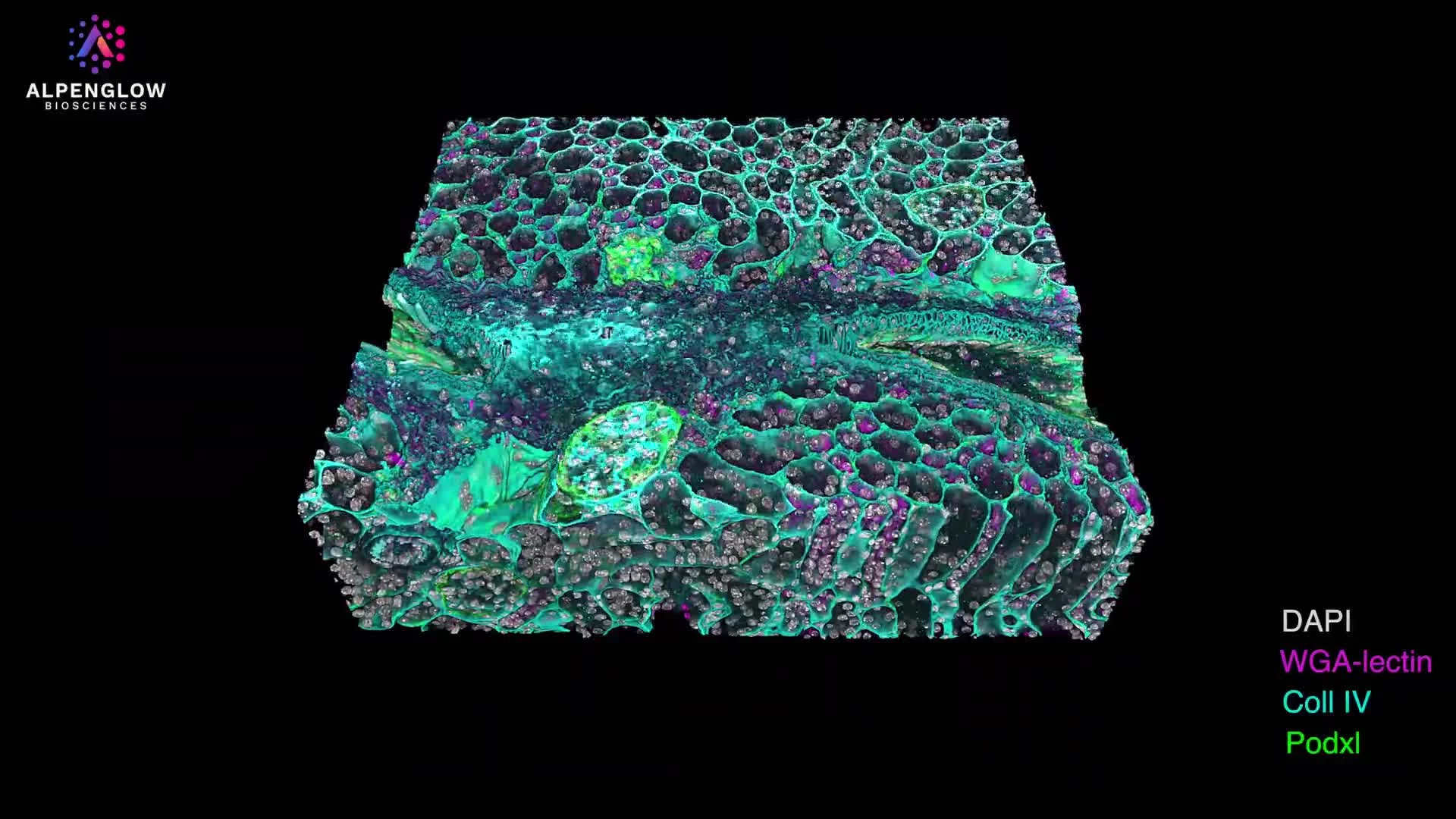
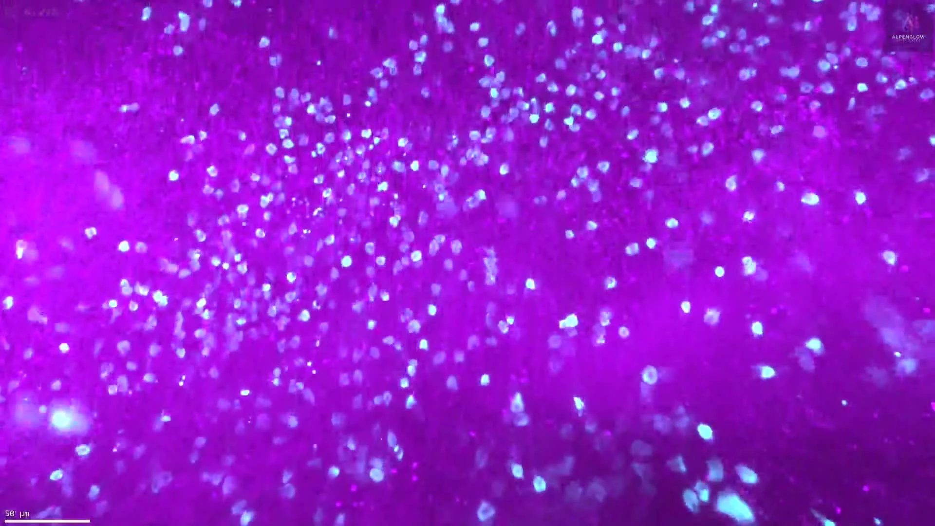
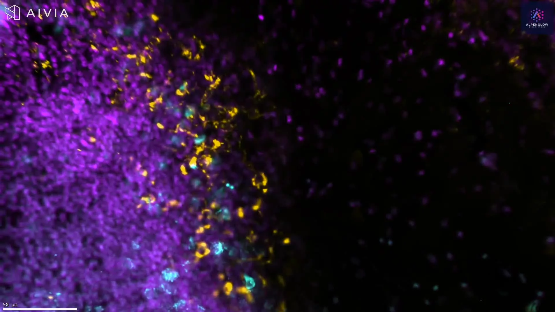
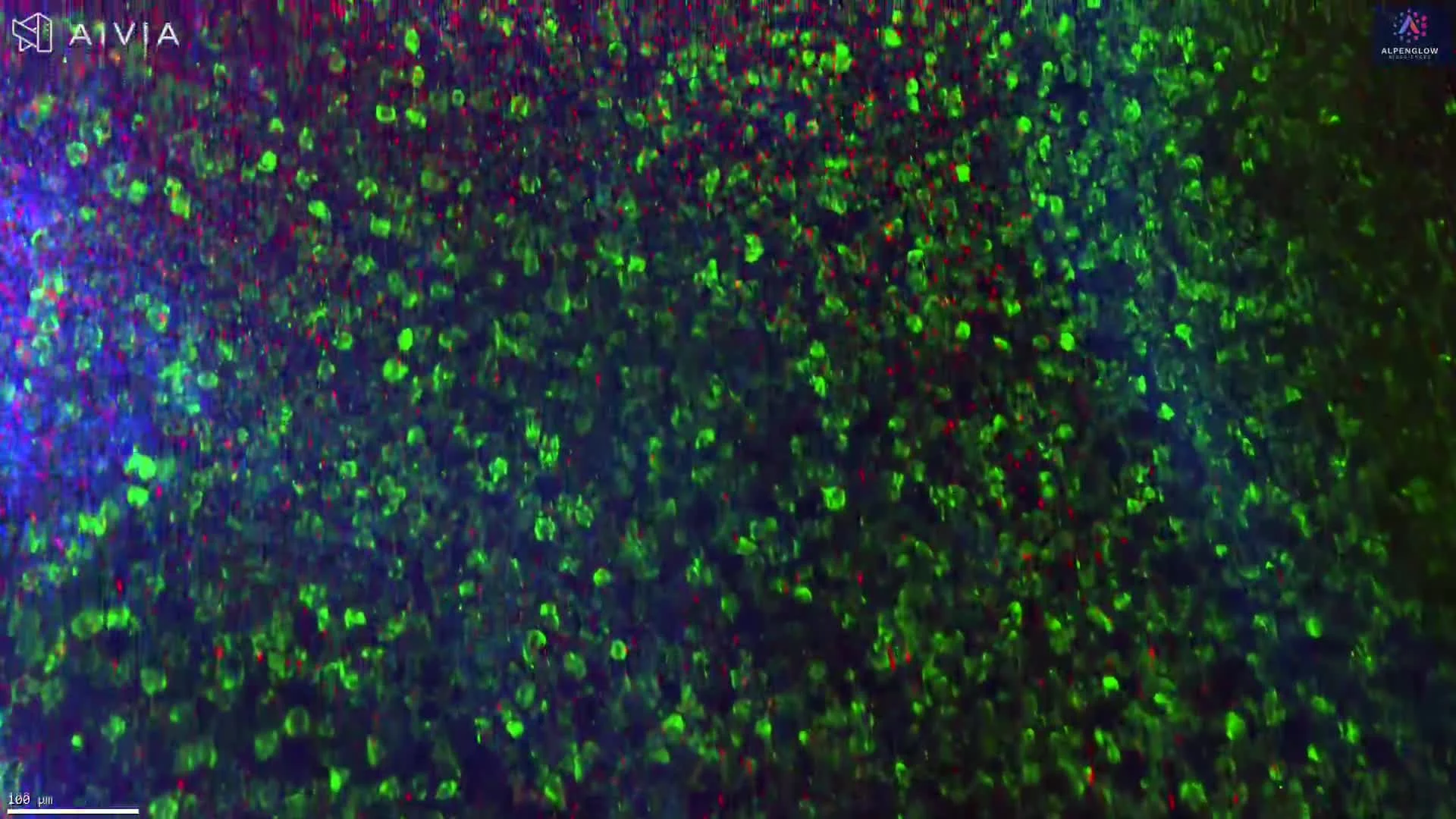
This dataset demonstrates the use of optimized staining and clearing protocols to visualize T cells in formalin-fixed, paraffin-embedded (FFPE) human tonsil tissue. Samples up to 3 mm were de-waxed, pretreated, and subject to antigen retrieval using an EDTA/Methanol buffer system before staining with Biocare Medical anti-CD3 (clone BC33, 1:100) and the nuclear marker TO-PRO-3 (Thermo Fisher).
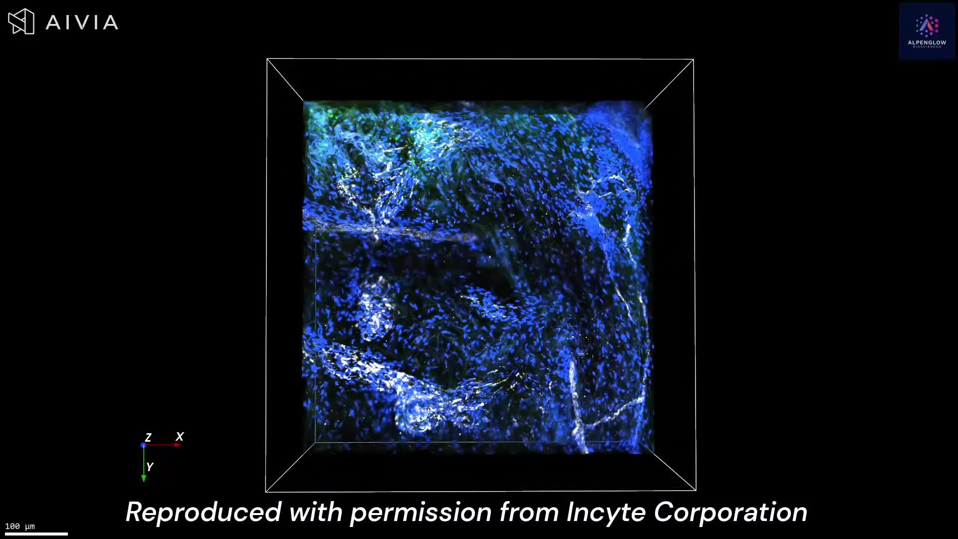
The tissue was cleared using a hybrid iDISCO protocol to enable antibody penetration throughout the full sample, then imaged on the Aurora™ 3Di Hybrid Open Top Light Sheet (HOTLS) fluorescent microscope. This workflow preserves tissue integrity while providing whole-sample 3D histology that reveals T cell distribution in its complete spatial context.
Combining validated antibodies with advanced digital pathology methods allows researchers to quantify immune architecture with precision. Data integration through 3Dm data managementand automated 3Dai AI-powered segmentationfurther supports reproducible immune cell mapping and analysis.
For more information and a list of validated antibodies compatible with deep-tissue imaging, see our brochure: Staining with Immunomarkers within Deep Tissues.