Mapping Epidermal Innervation Patterns in the Scalp with 3D Imaging
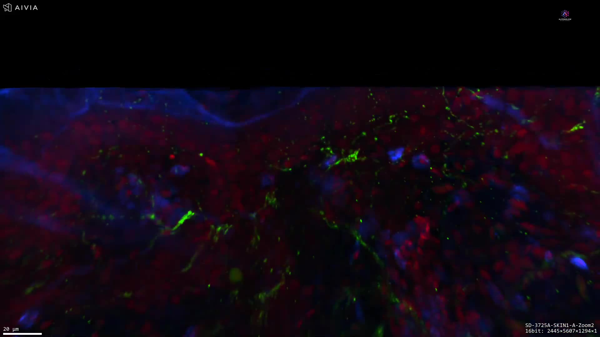
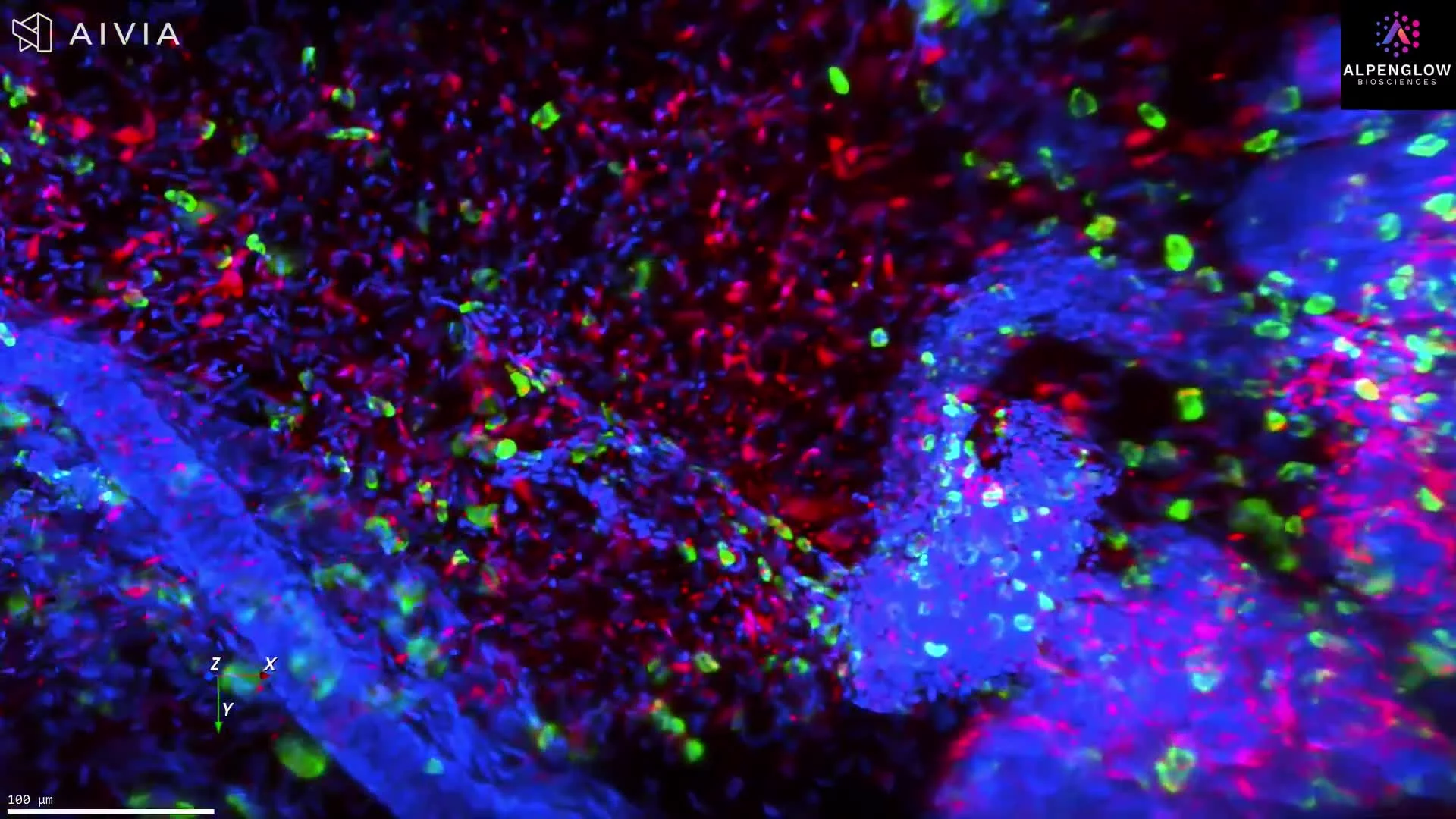
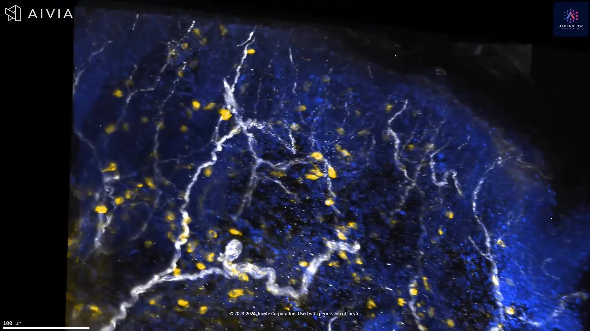
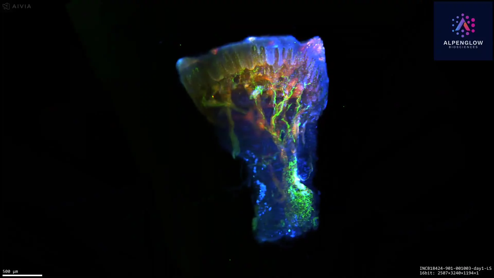
This stunning 3D visualization of the human scalp highlights the intricate patterns of epidermal innervation. Using HOTLS microscopy on the Aurora platform, the dataset captures how large peripheral nerves branch upward into fine structures that extend through the upper epidermis, creating a detailed map of nerve distribution.
Stains used:
TO-PRO-3 (Red): Labels cell nuclei in the epidermis
PGP9.5 (Green): Maps peripheral nerve fibers and branching
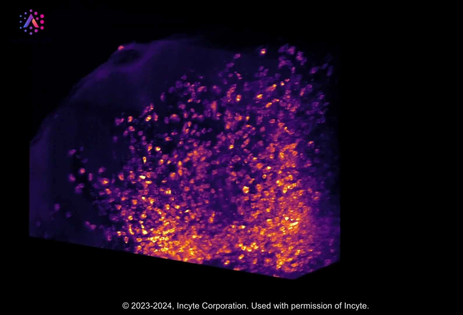
The ability to map nerves in their true three-dimensional context provides insights not possible with 2D slices. Through 3Dm data management and 3Dai AI-powered segmentation, this visualization supports quantitative analysis of neural density and distribution in scalp tissue.
These insights are especially valuable for alopecia research, where nerve–epidermis interactions may play a role in hair follicle health, and more broadly in dermatology research focused on sensory innervation and inflammatory conditions.
By connecting 3D histology with digital pathology, this work demonstrates how advanced imaging can transform understanding of scalp biology and disease.