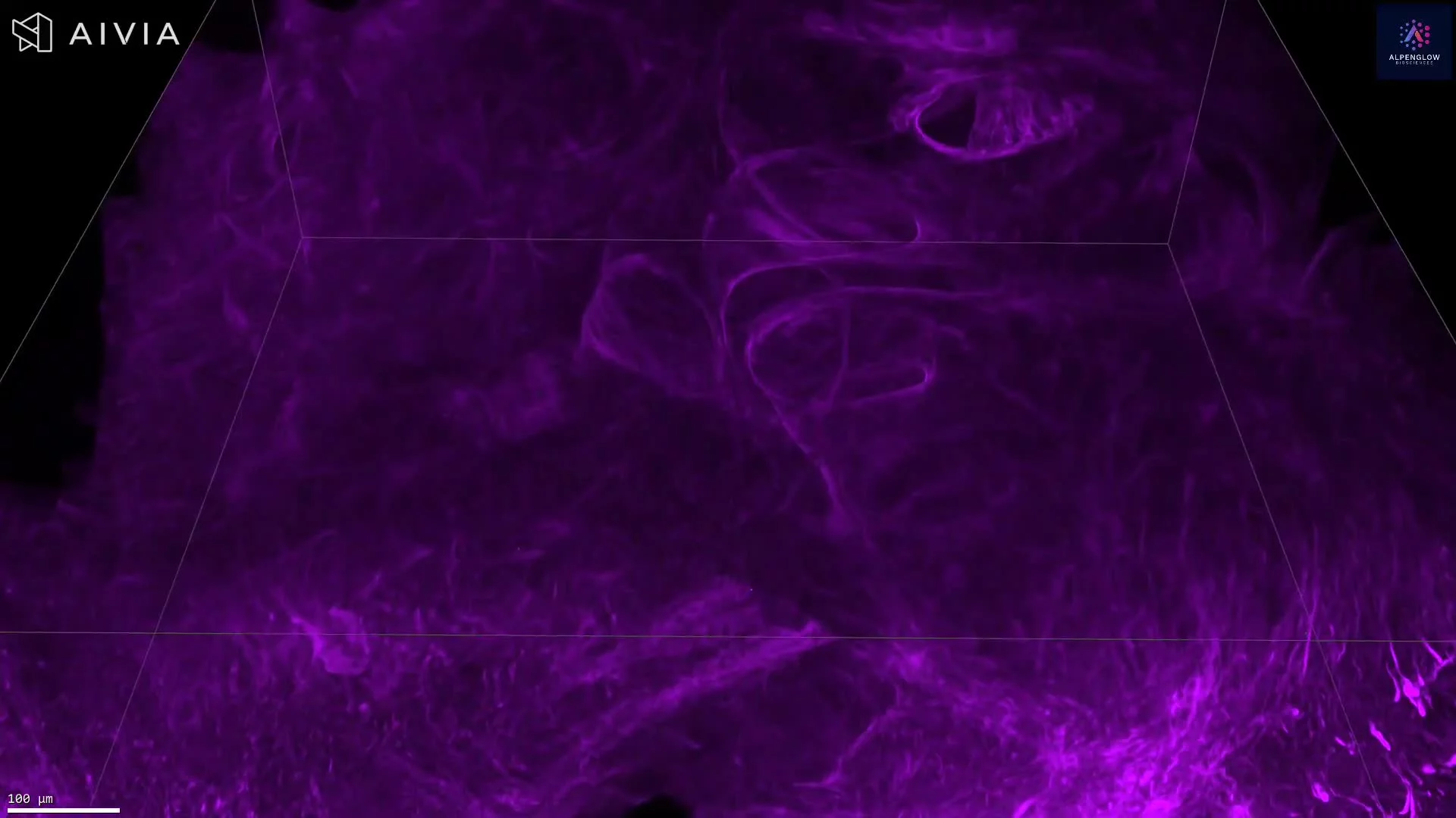
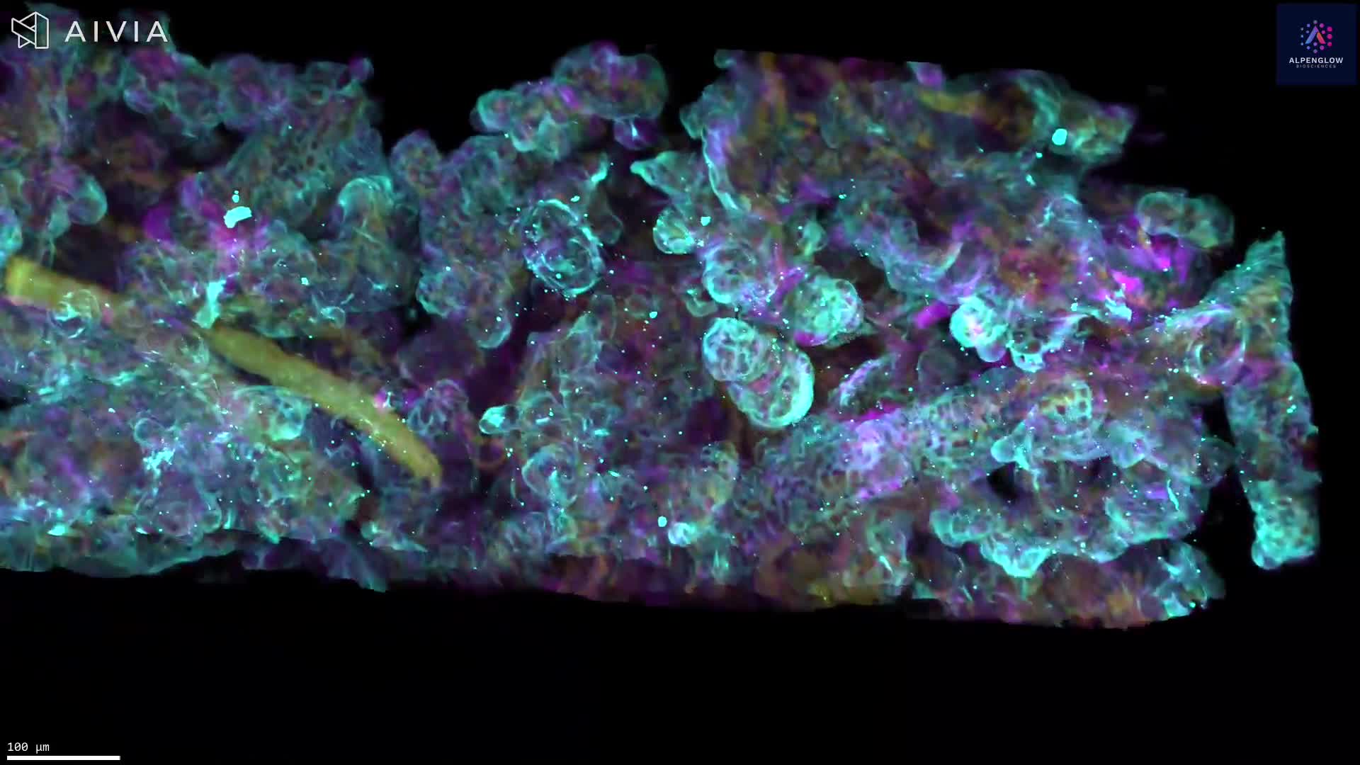
Segmentation Analysis of Liver Fibrosis and Steatosis in 3D
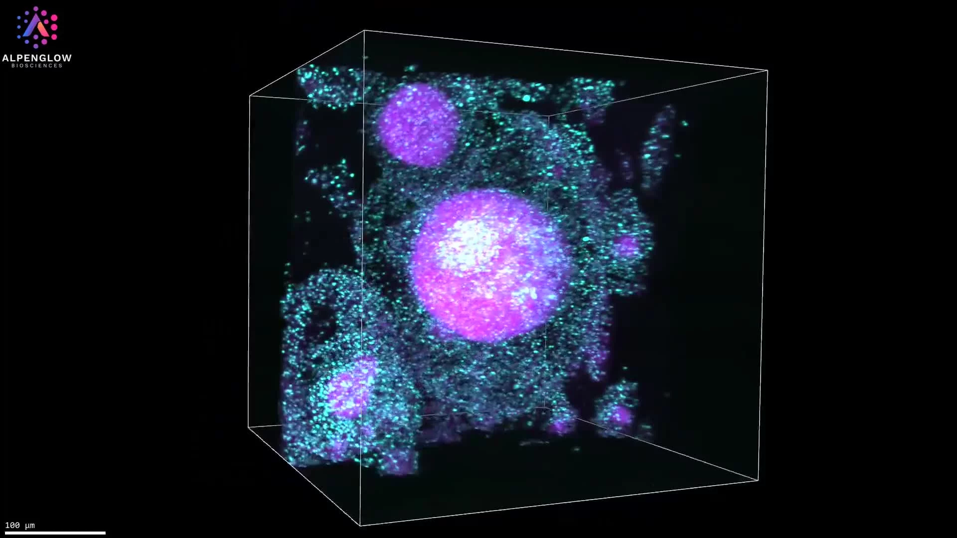
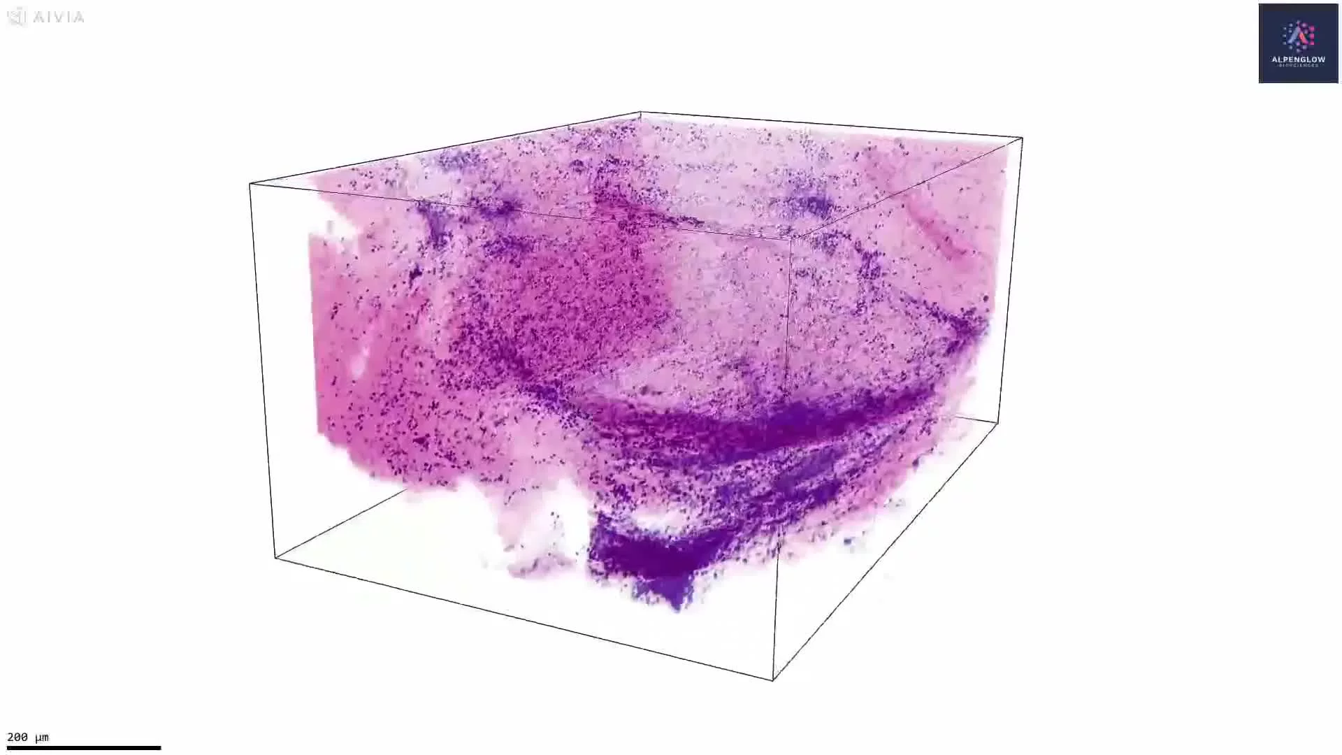
This 3D spatial biology visualization of a human liver biopsy demonstrates advanced image segmentation to separate pathological features of fibrosis and steatosis. Using a pixel classifier, fibrosis is segmented in cyan and steatosis in yellow, enabling precise tissue-scale quantification.
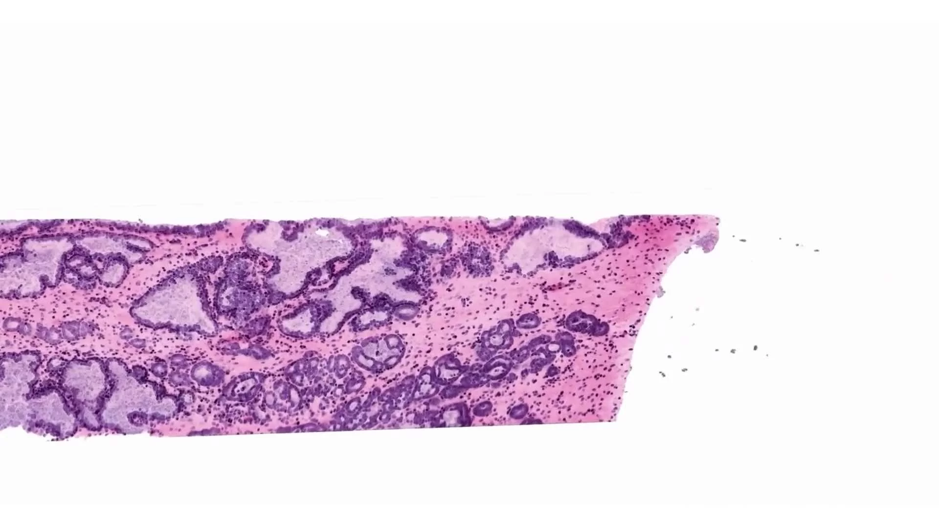
Captured on the Aurora™ 3Di Hybrid Open Top Light Sheet (HOTLS) microscope, the dataset integrates with 3Dm data management and automated 3Dai AI-powered segmentation to perform reproducible classification across intact tissue volumes. This workflow transforms qualitative histological assessment into measurable, quantitative metrics, revealing the extent and distribution of liver pathology in three dimensions.
Such capabilities are critical for digital pathology, 3D histology, and spatial profiling in hepatology, supporting drug development and translational research in liver diseases.
By combining whole-tissue imaging with automated segmentation, this approach delivers the ground truth needed to advance studies of fibrosis, steatosis, and other metabolic and inflammatory conditions.
Contact us to learn how Alpenglow’s 3D spatial biology platform can advance your research with large-scale, quantitative imaging datasets.