
From vasculature to single cells, 3D light-sheet imaging reveals the intricate architecture of a whole mouse brain in unparalleled detail.
High-resolution 3D imaging of a cleared rabbit retina stained with TO-PRO-3 and eosin, preserving architecture across retinal, choroid, and scleral layers.
See the full Aurora Workflow in action with 3D imaging of murine heart vasculature, delivering vessel measurements, branching analysis, and cell counts.
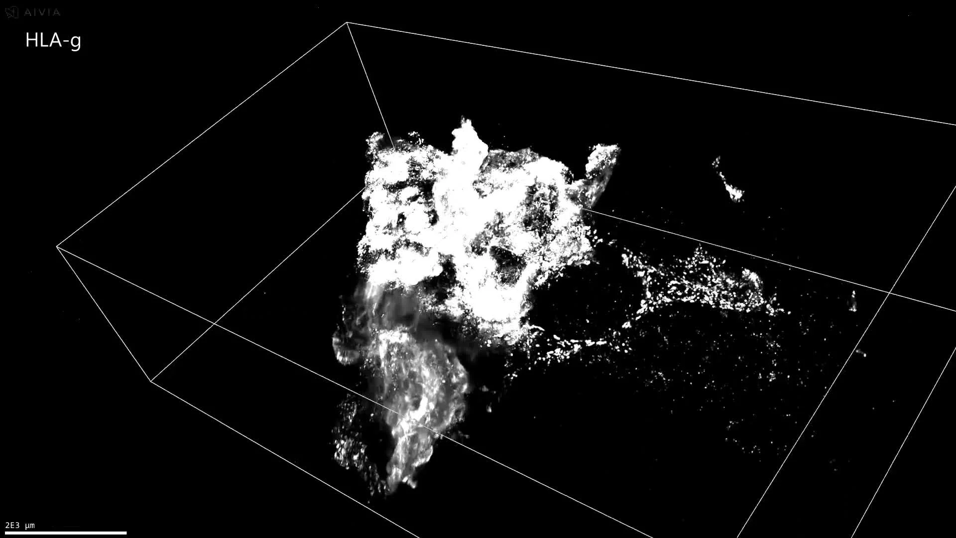
3D imaging of human placenta tissue stained with SMA, HLA-G, and CD31. From whole-tissue scans to single-cell resolution, uncover tissue complexity in detail.
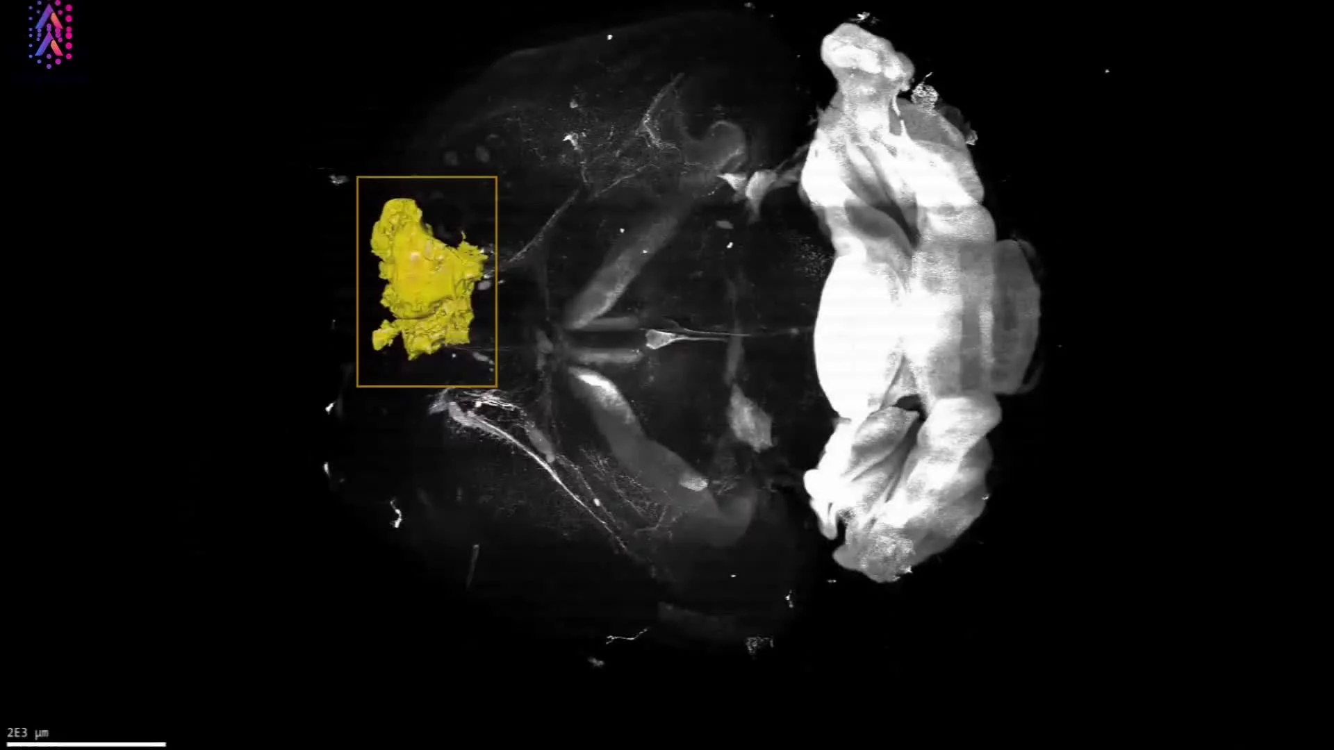
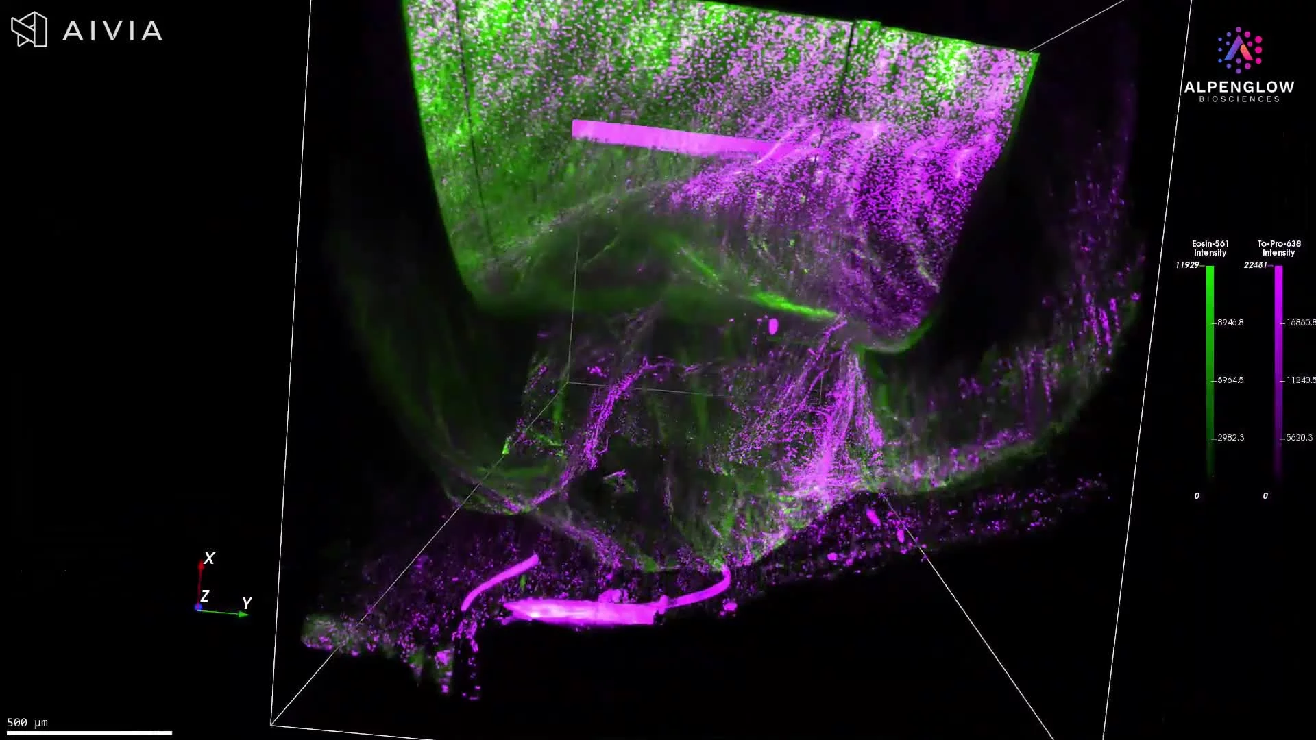
Whole mouse brain imaged in 3D with TO-PRO-3 and eosin. Scout and Zoom workflows capture glioblastoma ROI, segmentation, and high-resolution morphology.
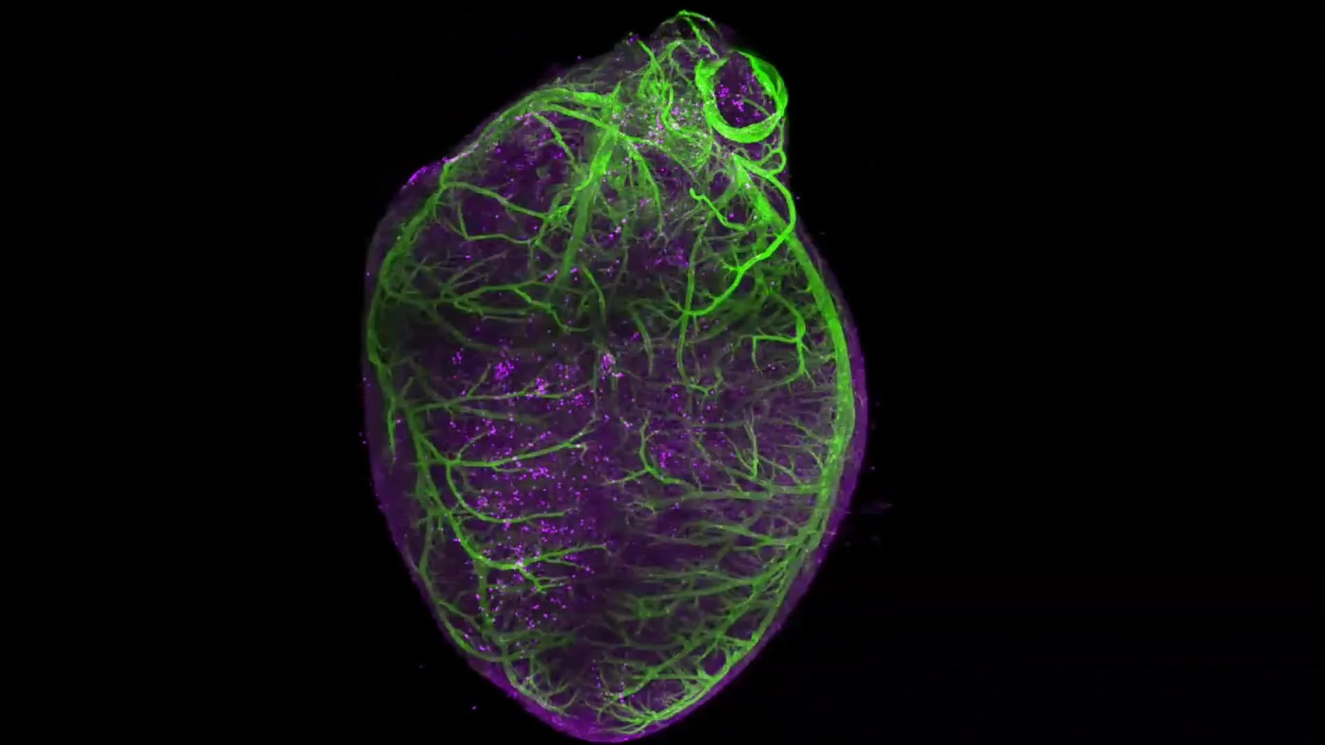
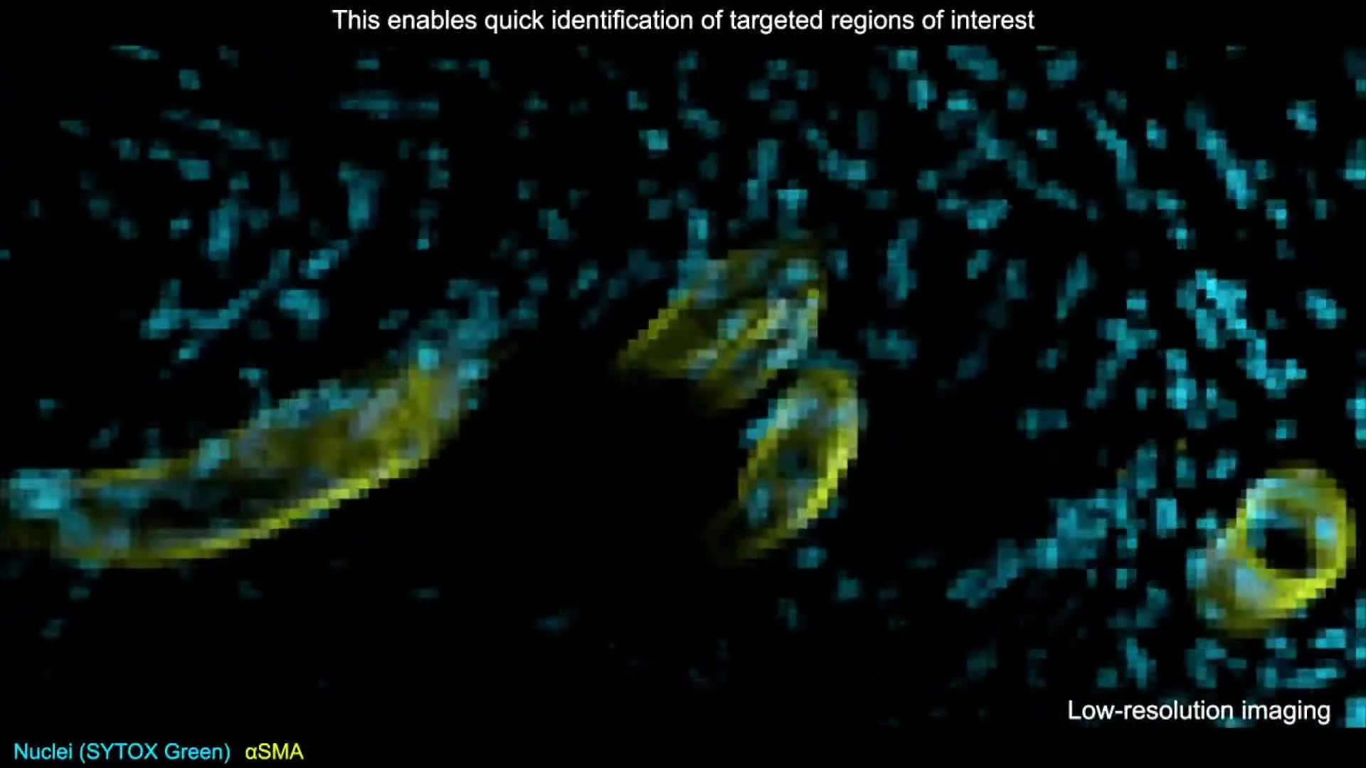
3D imaging of a whole murine heart stained with SMA reveals vascular structures and mural cells, from large vessels down to pericytes.
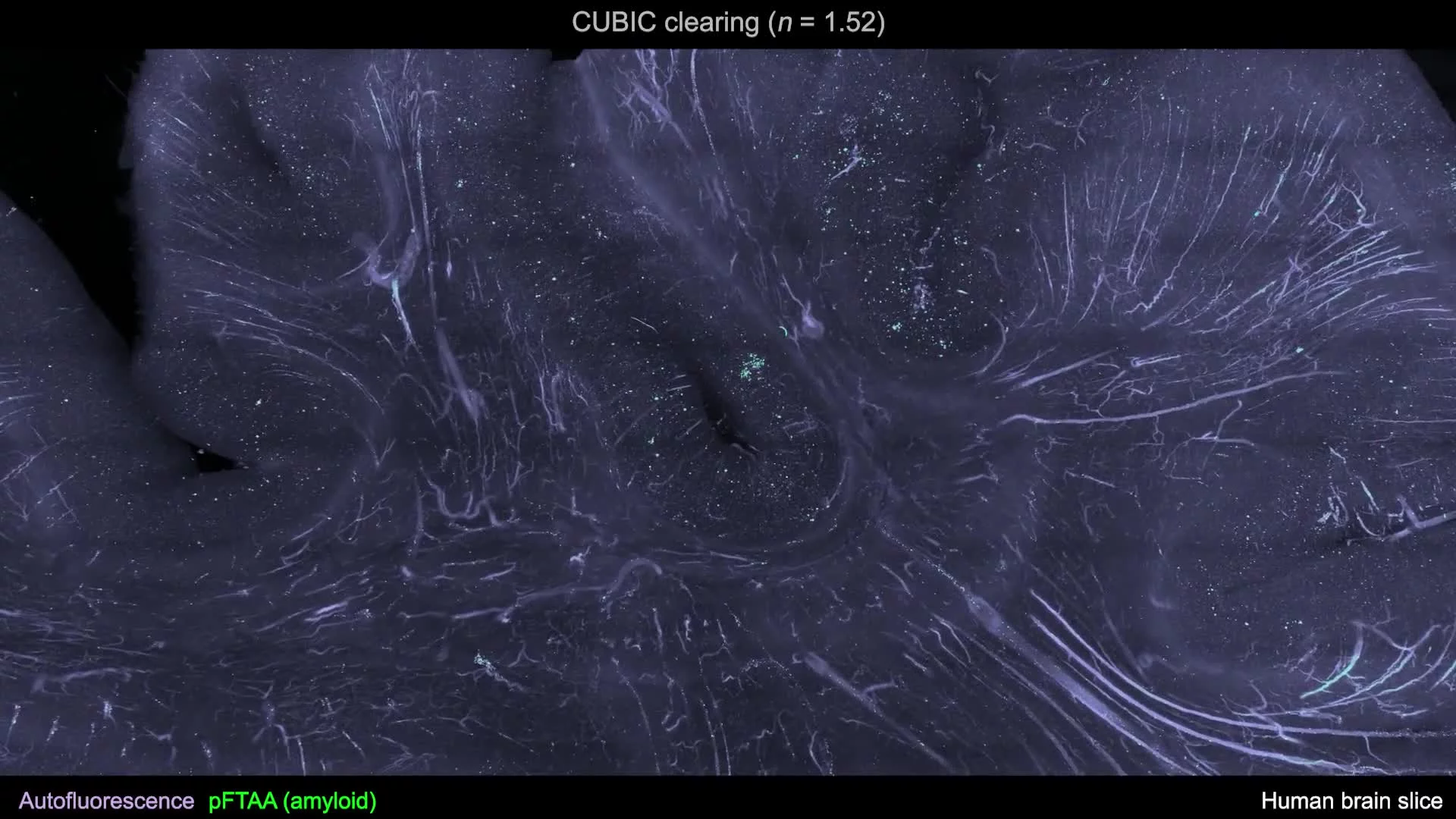
Large cleared human brain slice (10 × 7 × 0.3 cm) imaged at 0.17 microns per pixel using the CUBIC protocol for high-resolution volumetric analysis.