Ground-truth 3D data for robust AI models
Alpenglow Biosciences generates rigorously annotated, multi-modal 3D datasets at scale so your models, biomarkers, and decisions align with biological reality.
What is Ground Truth 3D Tissue Data
Ground truth 3D tissue data combines whole tissue 3D imaging, digital pathology, and expert annotations into a single volumetric dataset. Instead of inferring biology from a handful of 2D slides, AI and diagnostics teams train on intact 3D histology volumes that capture real tissue architecture, spatial organization, and rare events.
These AI-ready datasets come from Aurora™ 3D Spatial Biology workflows, including 3Di™ HOTLS light sheet microscopy, 3Dm™ data management, and 3Dai™ image analysis, as well as 3D histology imaging services for teams that want ground truth data without installing hardware.
What we do. Ground truth 3D tissue data and annotations
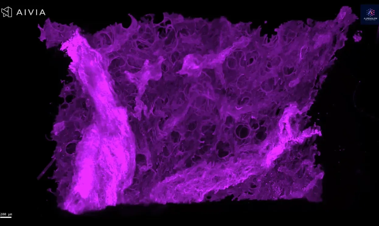
Alpenglow’s imaging platform produces high-resolution 3D microscope images that capture architectural context, cellular detail, and rare features not visible with current clinical imaging modalities. Alpenglow produces 10X the data at 1/10 the cost of conventional digital imaging technologies.
Establishing a new Gold Standard in Ground Truth for AI
Whole-specimen coverage captures complete cross-sections and intact volumes, reducing the uncertainty of traditional digital pathology.
Non-destructive, volumetric imaging preserves authentic tissue architecture for follow-up molecular assays and orthogonal analysis.
Cell-level fidelity enables precise definition of boundaries, phenotypes, and spatial relationships across the tissue microenvironment.
Rare feature visibility surfaces subtle events, rare cells, and edge cases that 2D slides and small fields of view can miss.
Multi-modal alignment enables labels and analyses to co-register in the same volume.
Auditable datasets with versioned volumes and annotations.
Whole rat heart imaged at Scout resolution 2 um/pixel
Establishing a reliable standard in ground truth for AI
Ground truth 3D tissue data anchors AI models to the real biology in intact tissue instead of partial views.
With Alpenglow datasets, you can:
Train and validate models on whole-tissue 3D histology, not data inferred from a few slices.
Improve robustness across diverse specimens, staining conditions, and rare edge cases.
Create benchmark datasets that support clinical validation and regulatory evaluation in digital pathology and radiology pathology correlation.
Who uses ground truth 3D tissue data
AI and ML teams building digital pathology and spatial biology models
Diagnostics and device teams validating new readouts and workflows
Radiology and pathology groups aligning non invasive imaging with tissue ground truth
Multi-modal spatial biology annotation framework
Our ground truth comprises comprehensive spatial biology annotations spanning multiple data modalities and biological scales. Cell segmentation forms the foundation, with precise boundary delineation of individual cells and subcellular compartments.
Marker co-localization
Mapping across fluorescent channels to identify protein interactions and cellular states.
Spatial neighborhood analysis
Understanding tissue architecture and cellular communication patterns in 3D.
Molecular expression profiling
integrates gene and protein expression data with spatial context, linking molecular signatures to tissue structure.
This multimodal approach helps AI models learn from complete tissue biology instead of flattened or inferred signals.
Our Systematic Ground Truth Generation Workflow
01
Sample Preparation & Controls
Standardized protocols ensure consistent tissue preservation and staining quality. Positive and negative controls validate every batch, while systematic documentation tracks sample provenance and processing conditions.
02
High-Resolution 3D Tissue Imaging
Gold-standard imaging protocols capture tissue architecture at sub-cellular resolution. Multi-channel fluorescence imaging preserves spatial relationships while maintaining quantitative accuracy across imaging sessions.
03
Expert Manual Annotation
Board-certified pathologists and imaging scientists perform detailed annotations using specialized software. Each annotation undergoes peer review to ensure accuracy and consistency with established biological knowledge.
04
Quality Control & Validation
Rigorous QC protocols include inter-annotator agreement metrics, systematic bias detection, and continuous refinement processes. Version control systems track all changes and maintain annotation provenance.
Ground truth 3D tissue data from Alpenglow Biosciences means standardized preparation, high-resolution 3D tissue imaging, expert annotation, and rigorous QC in one workflow.


