3D Imaging of Glioblastoma in Whole Mouse Brain
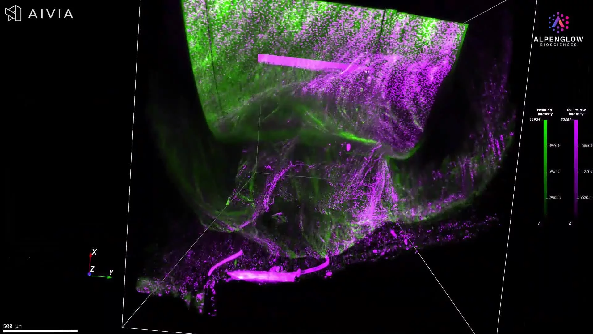
This dataset presents a full 3D visualization of a whole mouse brain stained with TO-PRO-3 and eosin, revealing nuclei and protein structures. The workflow begins with low-resolution Scout imaging (2 μm/pixel) to capture the entire organ, followed by stepwise refinement to focus on the glioblastoma region.
Workflow highlights:
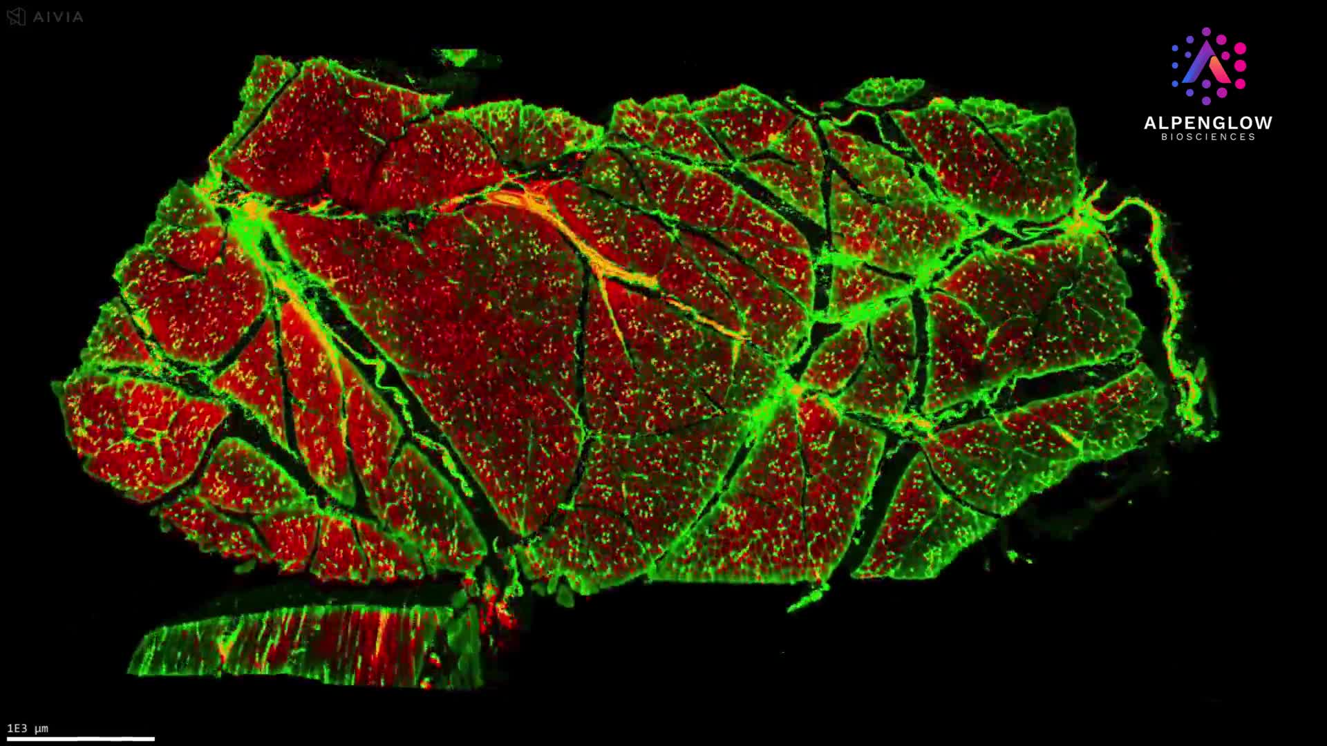
Whole-organ imaging (Scout): Broad view at 2 μm/pixel resolution
Tumor identification: Selecting the glioblastoma region of interest (ROI)
Segmentation and mesh construction: ROI highlighted in yellow, mesh built around glioblastoma pixels to calculate tumor volume and capture morphology
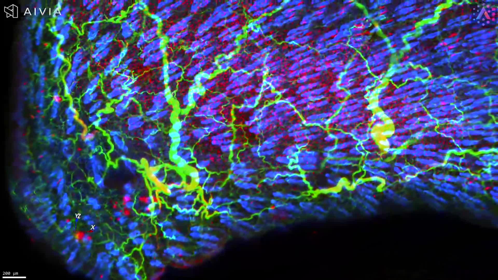
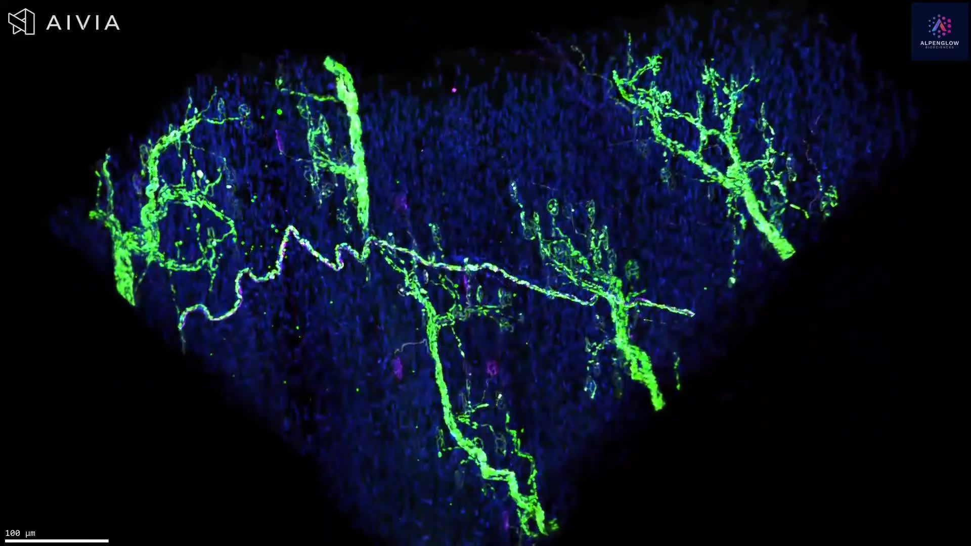
High-resolution imaging (Zoom): ROI imaged at 0.34 μm/pixel, showing TO-PRO-3 in purple and eosin in pink
Captured using the Aurora 3Di Hybrid Open Top Light Sheet (HOTLS) microscope, this dataset showcases the dual-path Scout + Zoom workflow, allowing researchers to transition seamlessly from organ-level context to subcellular resolution. Integration with 3Dm data management and 3Dai AI-powered segmentation supports accurate volume calculations, morphology analysis, and reproducible quantification.
This powerful approach provides actionable data for oncology research, supporting drug discovery and therapeutic development in glioblastoma and other brain tumors. By combining whole-brain imaging with quantitative segmentation, Alpenglow’s 3D spatial biology platform provides insights that are not possible with conventional 2D histology.