Whole-Tissue 3D Imaging of Mast Cell–Nerve Crosstalk in Skin
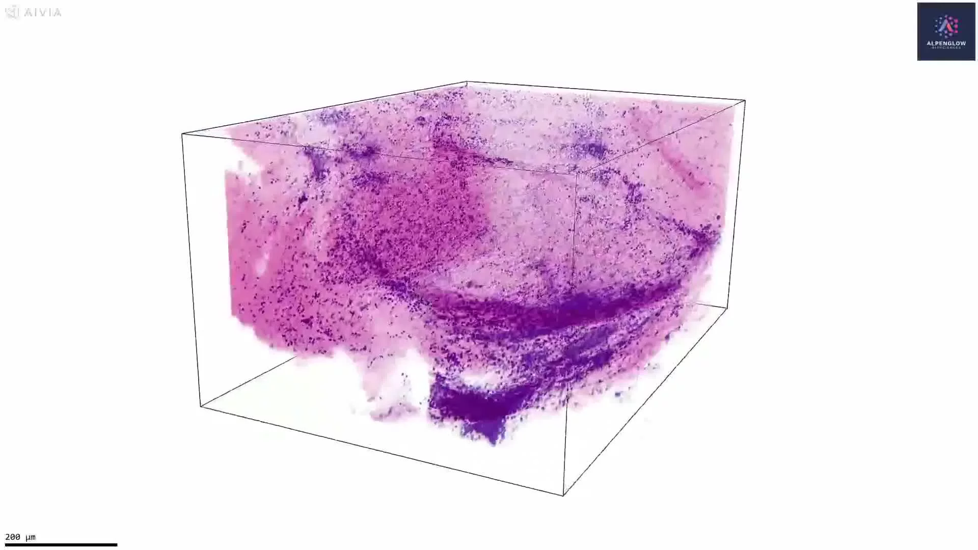
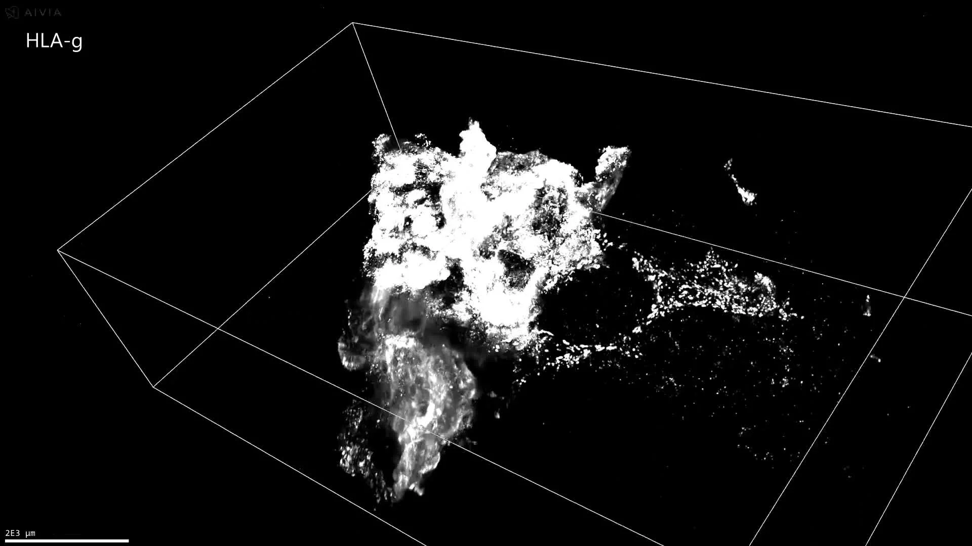
This dataset presents an intact, fluorescence-labeled human skin biopsy imaged in true 3D with the Aurora™ 3Di Hybrid Open Top Light Sheet (HOTLS) microscope. The volumetric view reveals the native architecture of neuroimmune interactions that conventional 2D slices cannot capture.
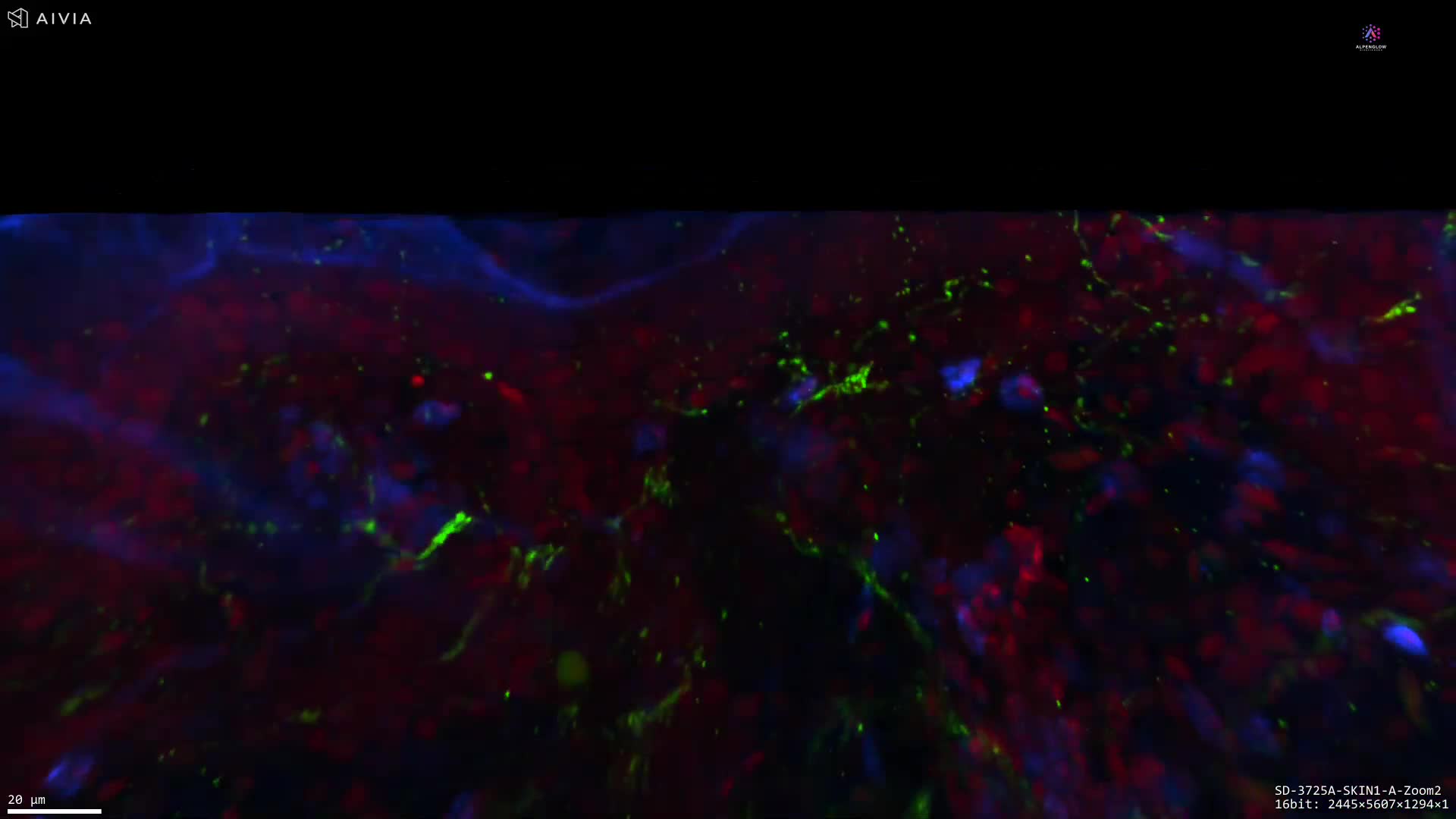
Stains used:
Tryptase (Green): Labels mast cells
TO-PRO-3 (Blue): Marks nuclei
PGP9.5 (Red): Traces nerves
By preserving full cell morphology and spatial relationships, this dataset provides the ground truth for quantifying mast cell density, mapping nerve proximity, and investigating mechanisms underlying chronic itch, fibrosis, and inflammatory skin disorders.
With integration of 3Dm data management and 3Dai AI-powered segmentation, researchers can perform reproducible, high-content quantification of neuroimmune interactions. This 3D approach eliminates slice bias and ensures accurate analysis at scale.
Applications span translational dermatology, where mast cells play a role in inflammatory skin disease, and immuno-oncology, where mast cell–nerve dynamics may influence tumor microenvironments. This example illustrates how 3D histology and digital pathology offer actionable insights that extend beyond visualization to measurable data.