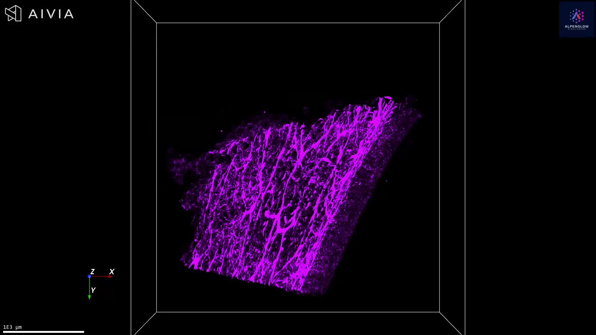
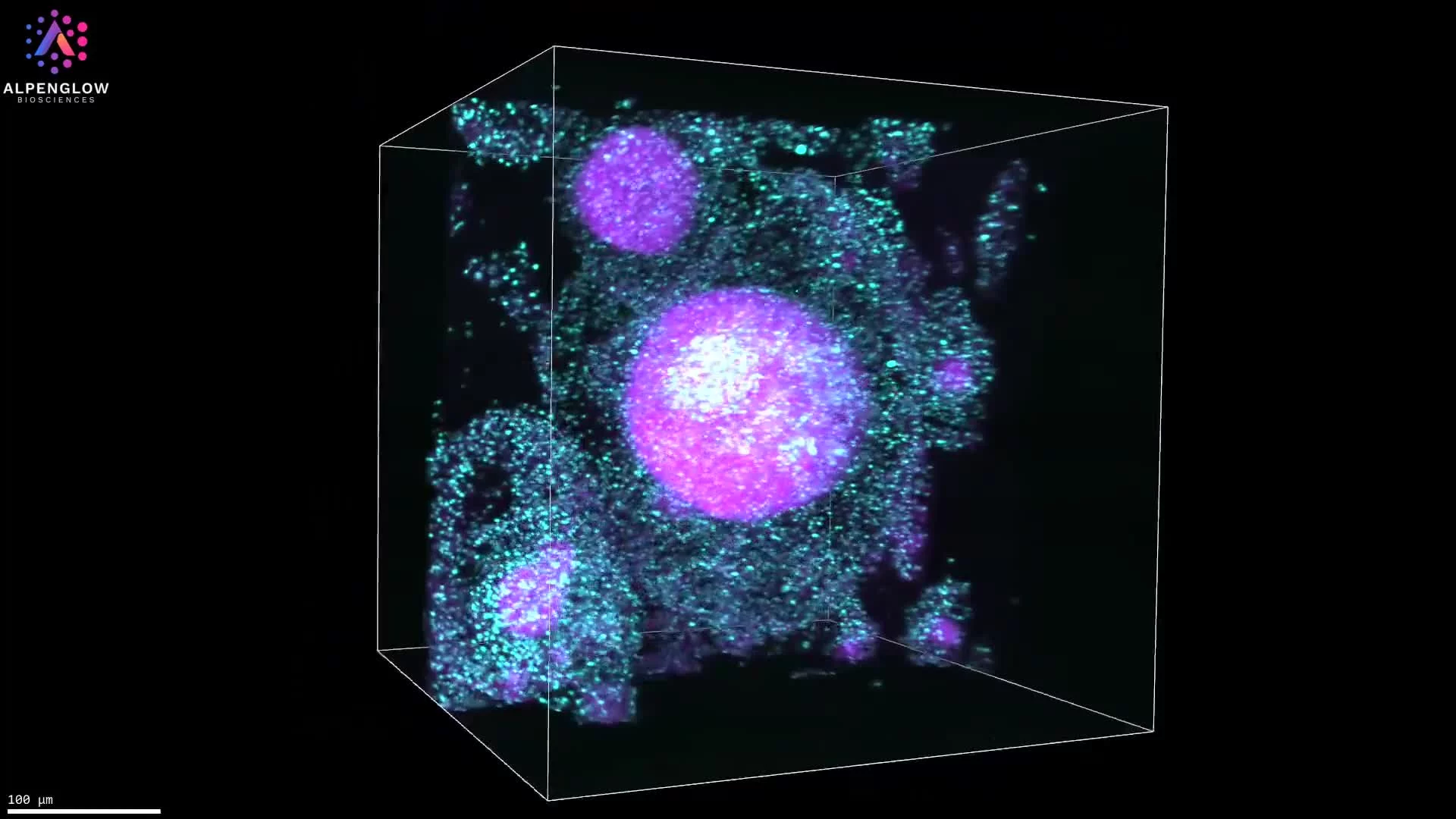
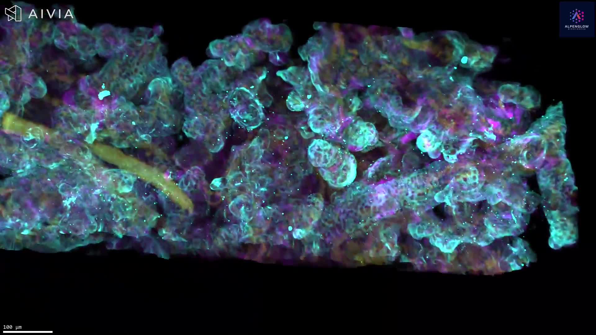
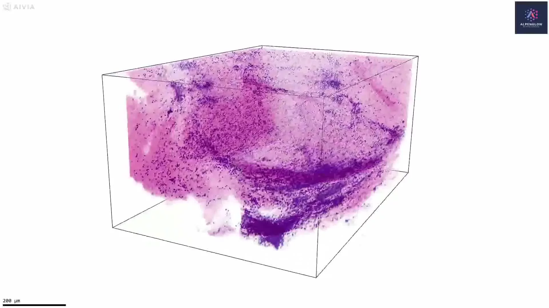
3D Imaging of a Human Ileocecal Sample
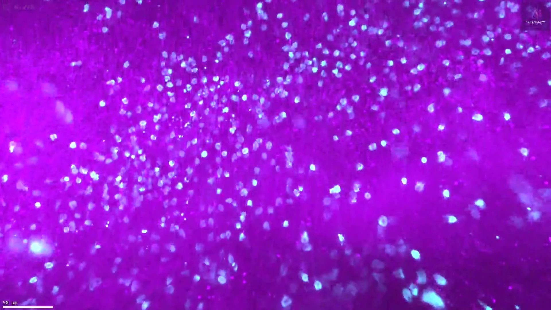
This remarkable dataset showcases a human ileocecal sample imaged in collaboration with Professor Raj Kapur, MD, PhD (UW Medicine & Seattle Children’s Hospital). Captured on the Aurora™ 3Di Hybrid Open Top Light Sheet (HOTLS) microscope, the scan encompasses a total volume of 2,350 cubic millimeters and contains more than 840 billion pixels — more data points than there are stars in the Milky Way.
The sheer scale of this dataset demonstrates the unparalleled capacity of Alpenglow’s 3D tissue imaging technology to preserve intact morphology and capture entire tissue regions in volumetric detail. By combining high-throughput imaging with advanced 3Dm data management and 3Dai AI-powered analysis, researchers can generate quantitative spatial statistics across whole-tissue volumes.
Applications extend to gastrointestinal and developmental disorders such as Hirschsprung disease, where mapping the enteric nervous system structure is essential for understanding pathology. This 3D histology approach is also transforming digital pathology, enabling translational research and providing the ground truth necessary for drug development and disease discovery.
Contact us to learn how Alpenglow’s 3D spatial biology platform can advance your research with large-scale, quantitative imaging datasets.