3D Imaging and TLS Quantification in NSCLC
Tertiary lymphoid structures (TLS) are critical immune aggregates that influence prognosis and response to immunotherapy in non-small cell lung cancer (NSCLC). Traditional single cross-section imaging often misrepresents TLS size, cellular composition, and maturity classification due to sampling bias.
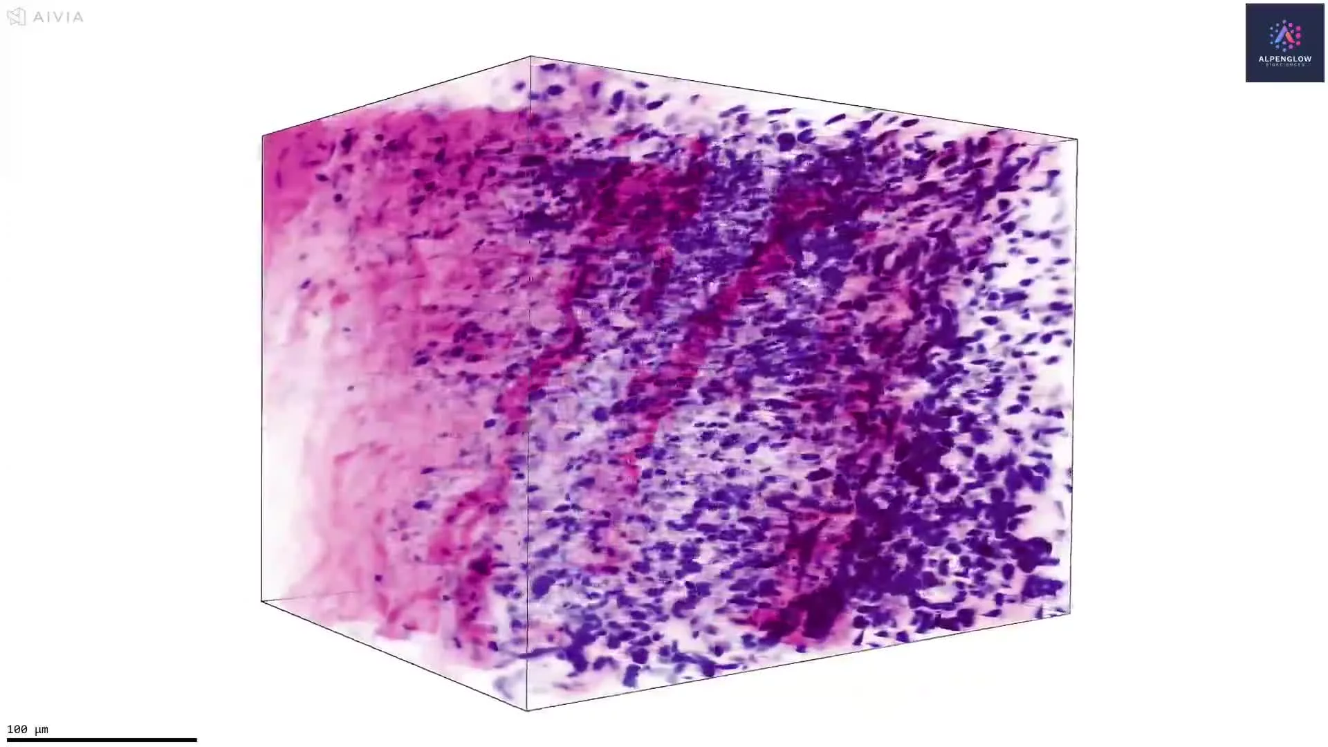
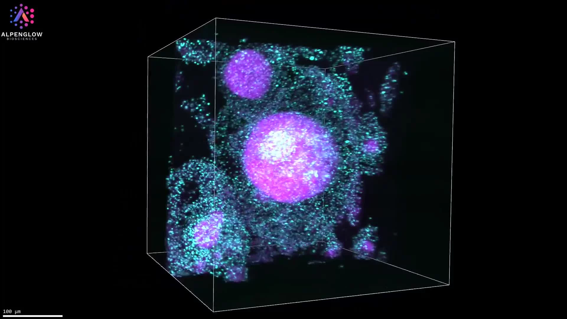
Using Alpenglow’s 3Di spatial imaging platform and computational H&E staining, TLS can be visualized and quantified across entire tumor biopsies in 3D, non-destructively.
Computational H&E staining replicates the contrast of standard histology while preserving the intact tissue. In this NSCLC study, samples were stained with:
TO-PRO-3: nuclear marker for hematoxylin contrast
Eosin to highlight cytoplasm and extracellular matrix
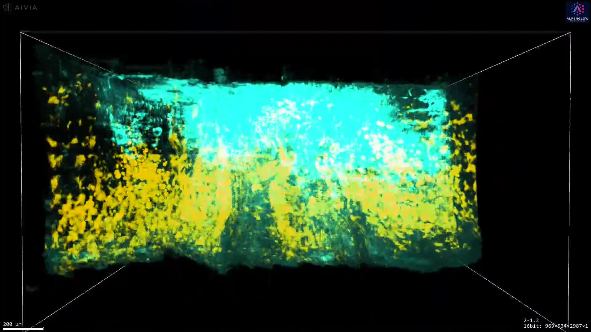
After staining, the tissues were optically cleared and imaged at whole-block scale using Scout mode, followed by imaging at subcellular resolution in Zoom mode. This workflow maintains architectural context while enabling single-cell segmentation in 3D.
Key advantages:
Accurate detection and measurement of TLS in whole tissue samples
3D quantification of TLS volume, surface area, and cellular density
Improved classification of TLS maturity and organization
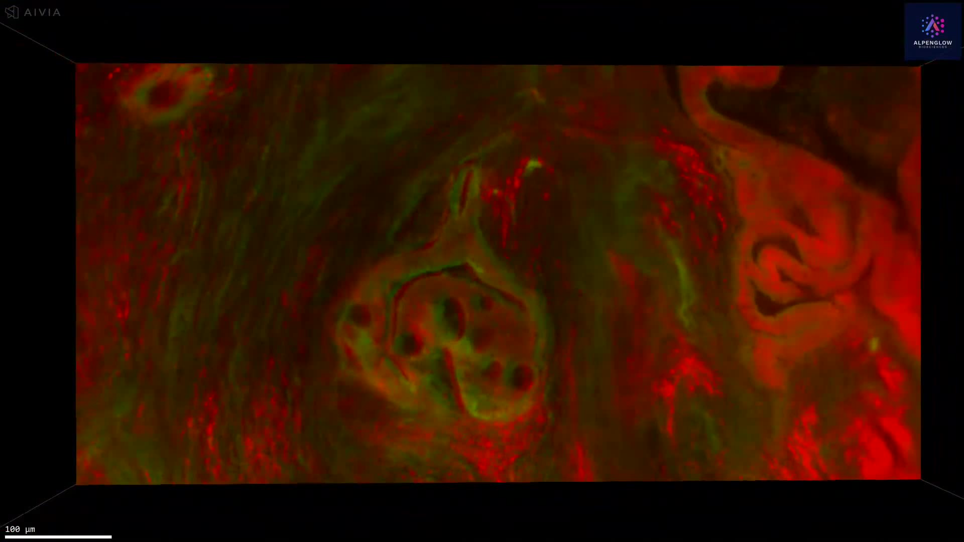
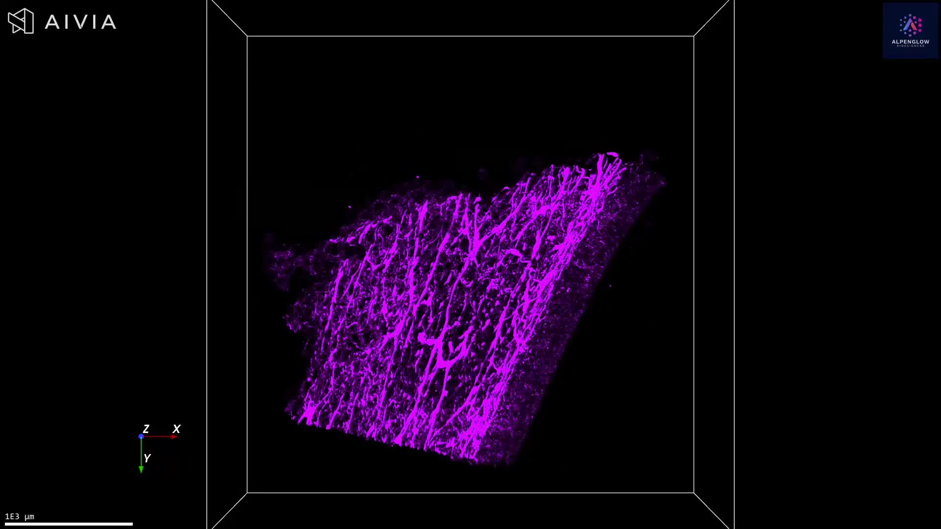
Enhanced biological insight through visualization of spatial context with surrounding tumor, stroma, and vasculature
In one NSCLC sample, what appeared as a single TLS in 2D was revealed in 3D to be two distinct structures with unique volumes and surface areas. Such insights demonstrate the power of combining computational H&E with 3D imaging to reveal structures invisible to conventional pathology.
By moving beyond 2D, researchers gain a more accurate and comprehensive understanding of TLS in oncology samples—opening new opportunities for translational research, biomarker development, and therapeutic discovery.