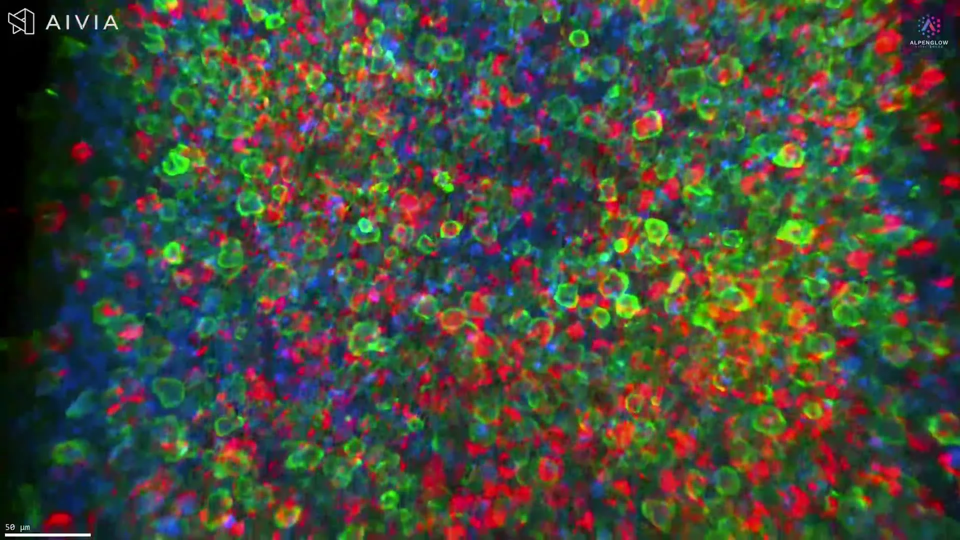
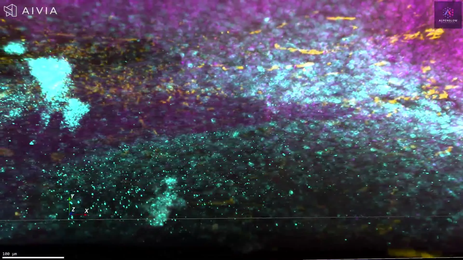
Identification of B Cells and Follicular Dendritic Cells through CD21 Staining
This dataset demonstrates the identification of B cells and follicular dendritic cells in formalin-fixed, paraffin-embedded (FFPE) human tonsil tissue using optimized staining and clearing protocols. Samples up to 3 mm were de-waxed, pretreated, and subject to antigen retrieval with an EDTA/Methanol buffer system. They were then stained with BioLegend anti-CD21 (clone Bu32) and the nuclear dye TO-PRO-3 (Thermo Fisher).
The tissue was cleared using a hybrid iDISCO protocol, ensuring deep antibody penetration, and imaged with the Aurora™ 3Di Hybrid Open Top Light Sheet (HOTLS) fluorescent microscope. This workflow maintains tissue architecture and enables intact 3D histology of immune cell networks in lymphoid tissue.
By combining validated antibody staining with 3Dm data management and 3Dai AI-powered segmentation, researchers can achieve quantitative mapping of B cell and dendritic cell organization.
These high-content datasets provide valuable insights for immunology research, vaccine studies, and immuno-oncology, where B cell activation and follicular dendritic cell function play critical roles in immune response.