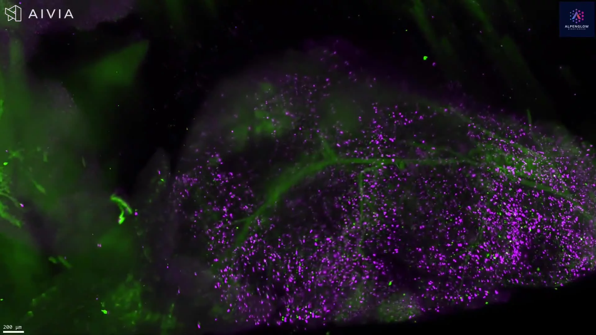
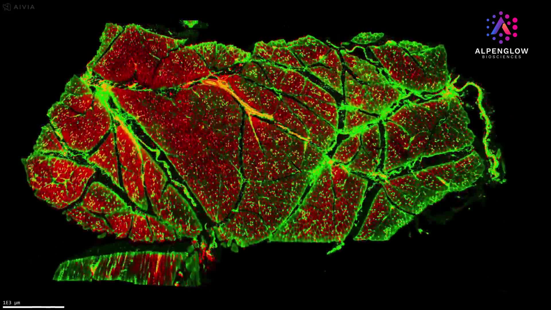
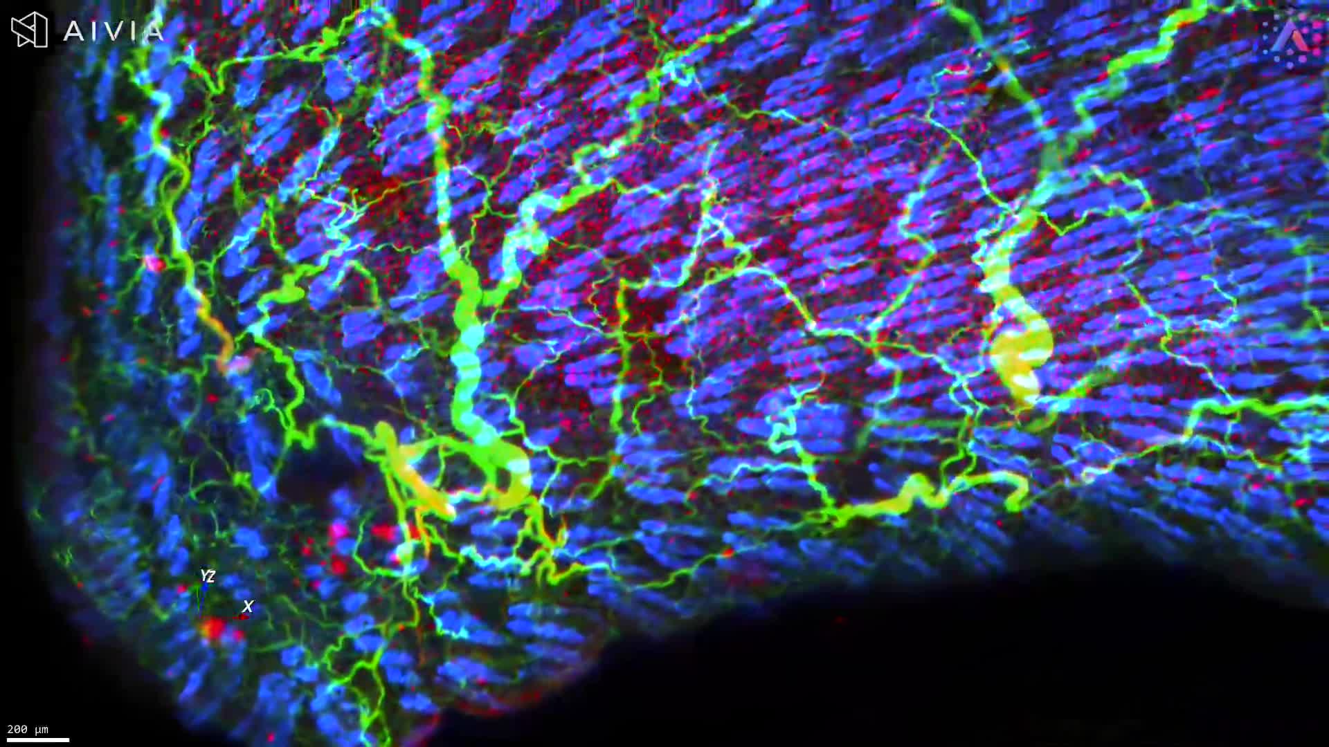
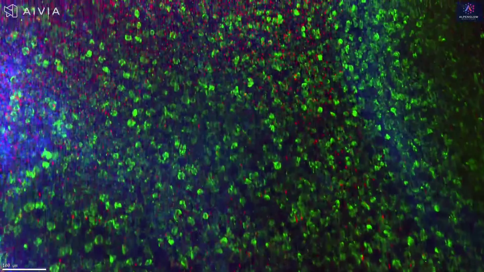
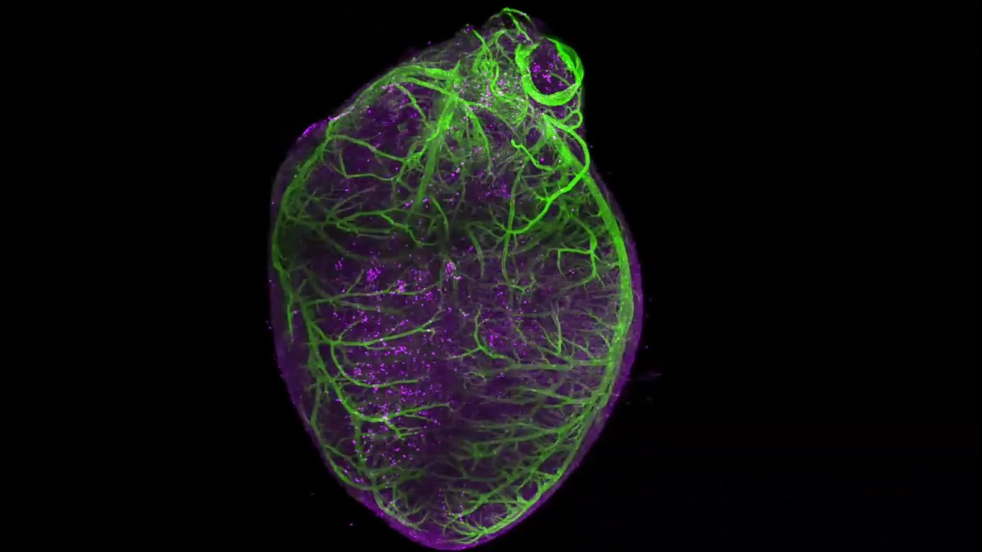
3D Light-Sheet Imaging of a Whole Mouse Brain
This dataset showcases 3D light-sheet imaging of a whole mouse brain, captured with the Aurora™ 3Di Hybrid Open Top Light Sheet (HOTLS) microscope. From organ-level vasculature down to single-cell resolution, the imaging reveals the full intricacy of brain architecture across every layer.
The dataset highlights how volumetric imaging preserves intact morphology, making it possible to study neuronal networks, vascular pathways, and cellular organization without the distortions of traditional 2D histology.
Complete visualization of neuronal and vascular networks across the brain
Cellular context preserved in full volumetric detail
Accurate mapping of tissue complexity that 2D slices cannot replicate
This dataset connects directly to the Nature Methods paper presenting our light-sheet microscope: Read the paper.
By combining 3Dm data management with 3Dai AI-powered segmentation, researchers can extend these visuals into actionable analyses, from neuronal density measurements to vascular branching and spatial profiling.
Applications include neuroscience research, brain mapping, and studies of neurodegenerative diseases, where understanding tissue complexity in situ is critical.