Scout-to-Zoom 3D Computational H&E Imaging of TLS in NSCLC
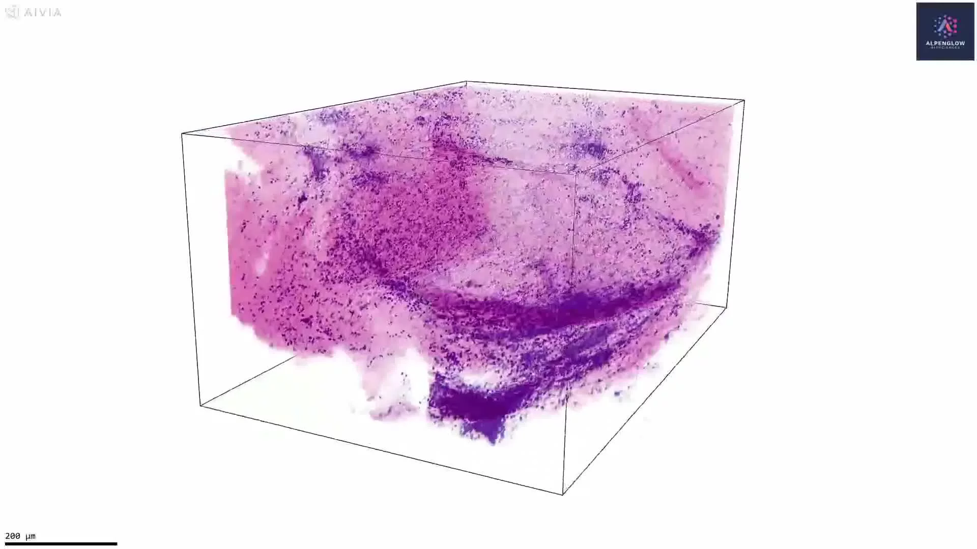
This dataset presents 3D computational H&E imaging of tertiary lymphoid structures in a human non-small cell lung cancer sample.
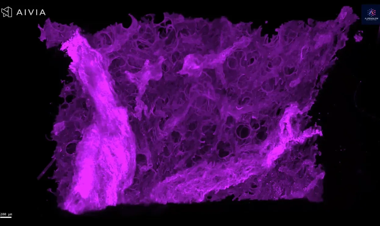
TO-PRO-3 labels nuclei, while eosin highlights protein-rich tissue architecture. Together, the fluorescent signals are computationally rendered to create an H&E-like view of the intact tissue volume.
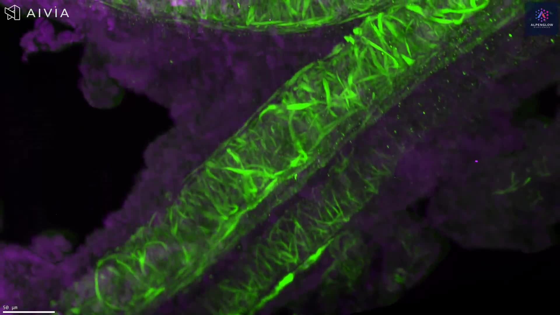
The Scout-to-Zoom workflow begins with rapid volumetric imaging at 2 µm per pixel to identify nuclear aggregates and other regions of interest across the tissue. Selected regions are then acquired at high resolution at 0.167 µm per pixel, revealing cellular morphology and TLS organization in greater detail.
In this NSCLC sample, Scout imaging identified dense nuclear aggregates that were examined using Zoom imaging and classified as two distinct tertiary lymphoid structures approximately 200 µm below the tissue surface. Their volumes and surface areas were quantified in 3D.
The dataset supports measurement of TLS number, volume, surface area, cellular density, distribution, and spatial relationships with surrounding tumor and stromal regions. Preserving each structure across depth reduces the sampling and orientation effects associated with individual 2D sections.
Explore how 3D tissue imaging supports immuno-oncology research across tumor architecture, immune-cell organization, and spatial relationships.
The tissue was imaged using the Aurora 3D™ Spatial Biology Solution, including the 3Di™ Hybrid Open-Top Light-Sheet microscope.