Identifying Cytotoxic T Cells in Cleared Tissue with CD8 Staining
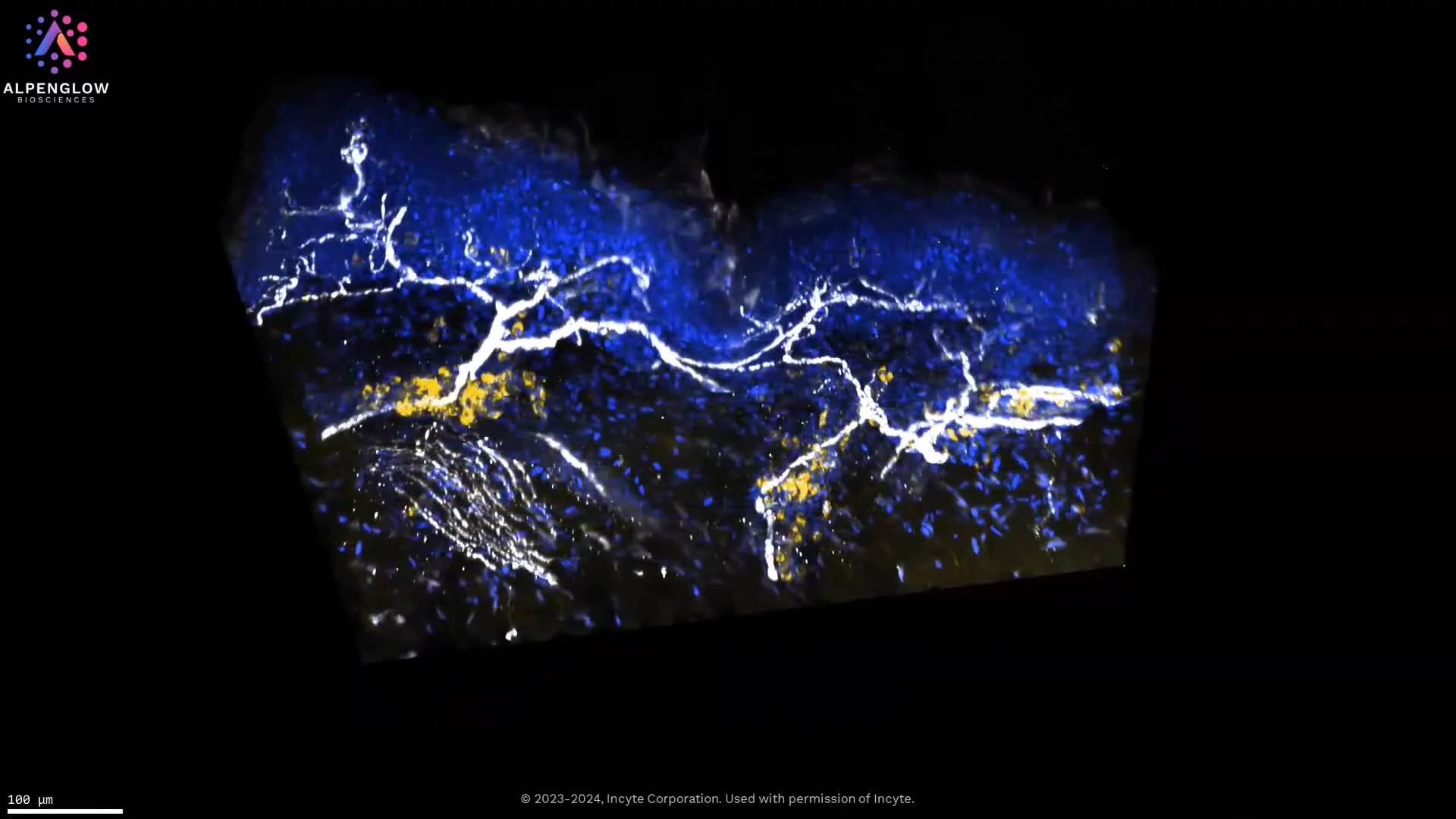
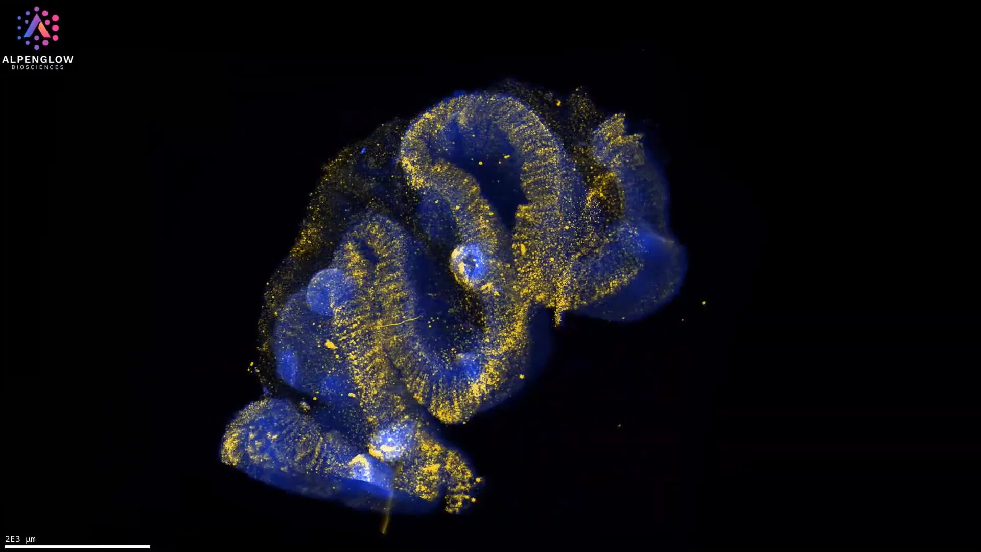
This dataset demonstrates the identification of cytotoxic T cells in formalin-fixed, paraffin-embedded (FFPE) human tonsil tissue using optimized deep-tissue staining and clearing protocols. Samples up to 3 mm were de-waxed, pretreated, and subject to antigen retrieval with an EDTA/Methanol system, then stained with Biocare Medical anti-CD8 (SP6, 1:100) and the nuclear marker TO-PRO-3 (Thermo Fisher).
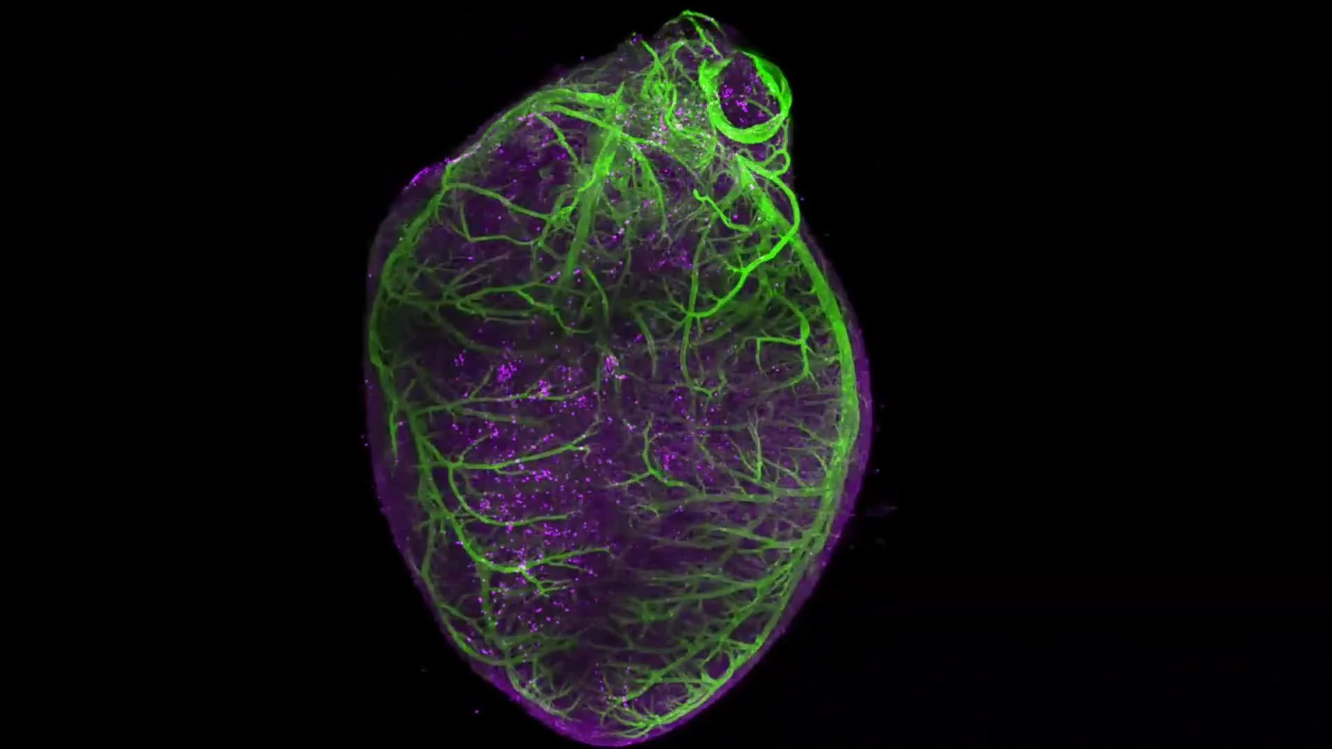
The tissue was cleared using a hybrid iDISCO protocol, ensuring antibody penetration throughout the sample, and imaged with the Aurora™ 3Di Hybrid Open Top Light Sheet (HOTLS) microscope.
This approach maintains tissue integrity, enabling visualization of cytotoxic T cells in their full spatial context.
By combining antibody optimization with 3D histology, digital pathology, and advanced spatial profiling, researchers can quantify the distribution of CD8-positive cells within lymphoid tissue.
For more information and a list of validated antibodies compatible with deep-tissue imaging, see our brochure: Staining with Immunomarkers within Deep Tissues.