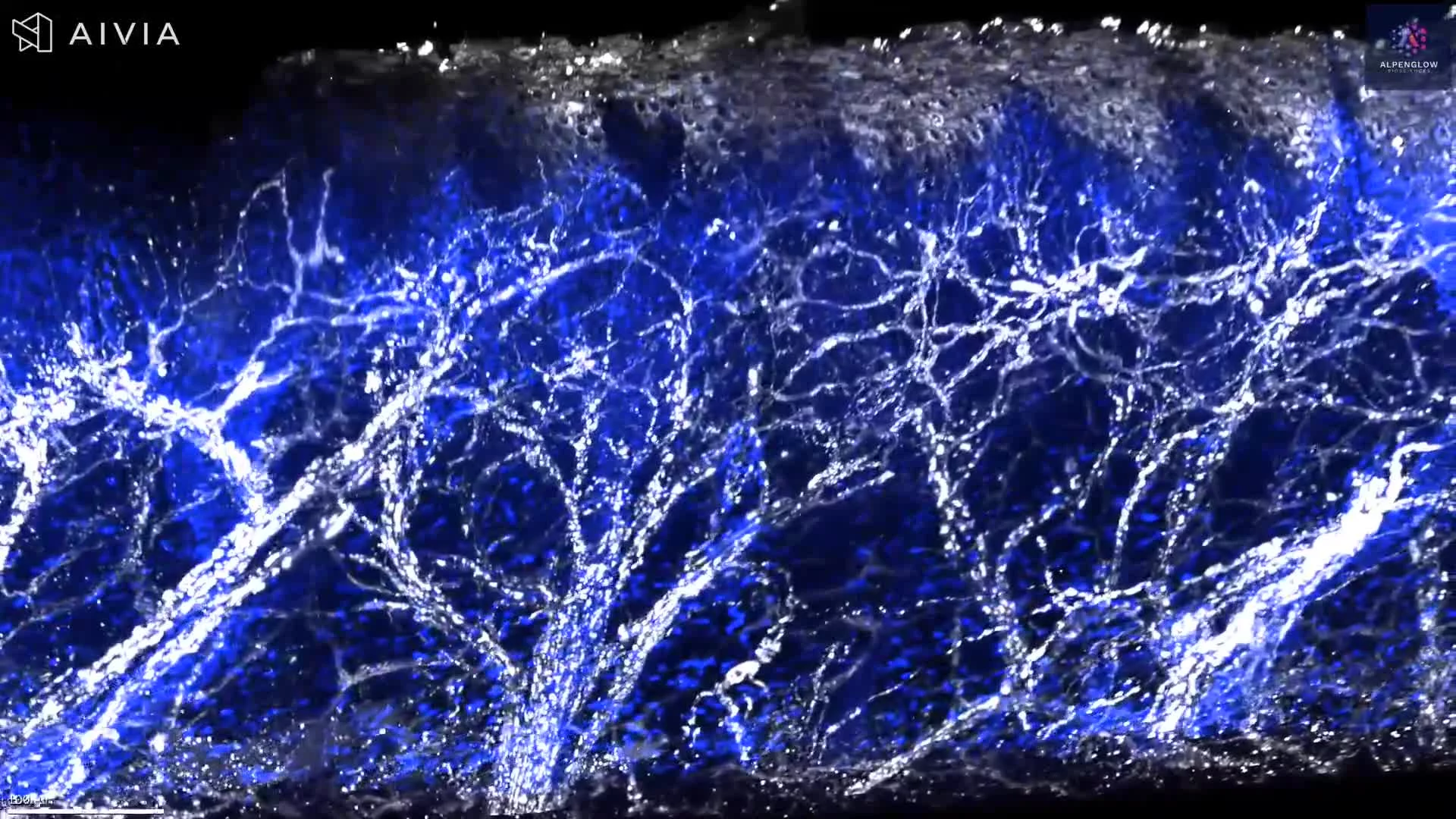
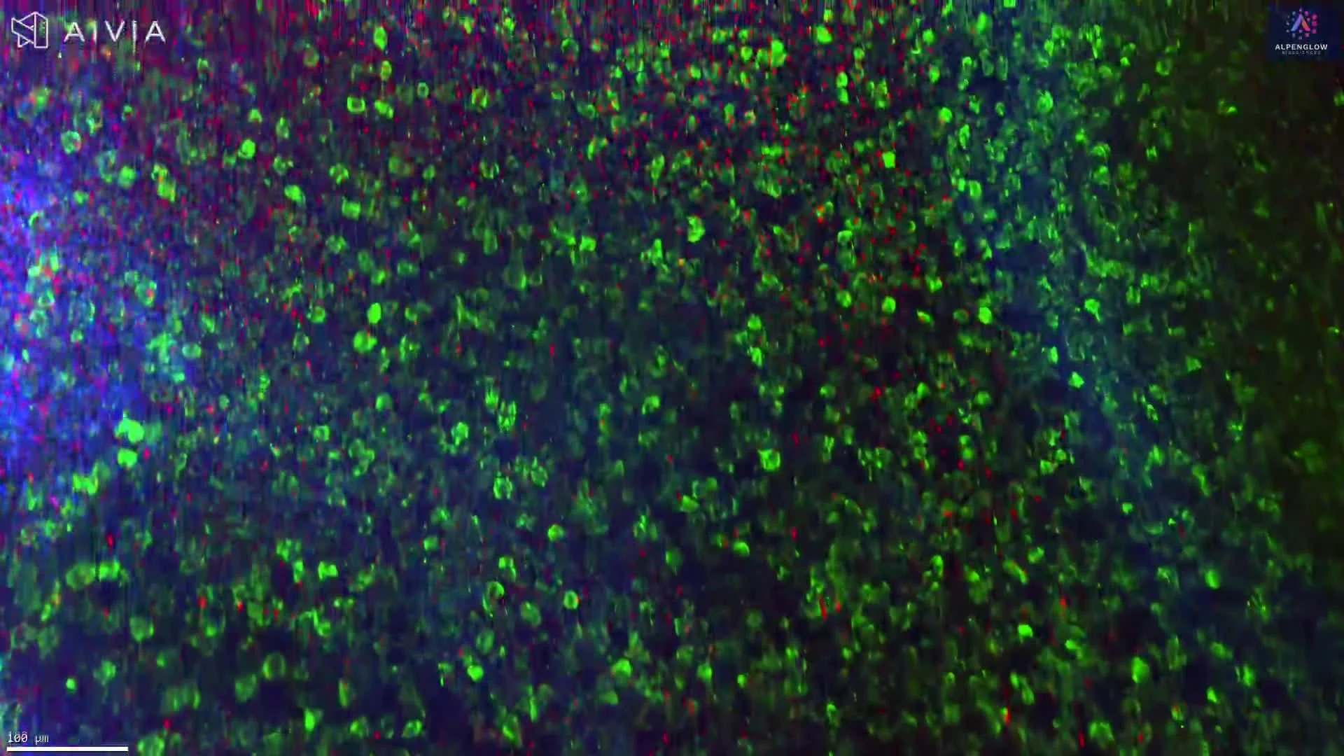
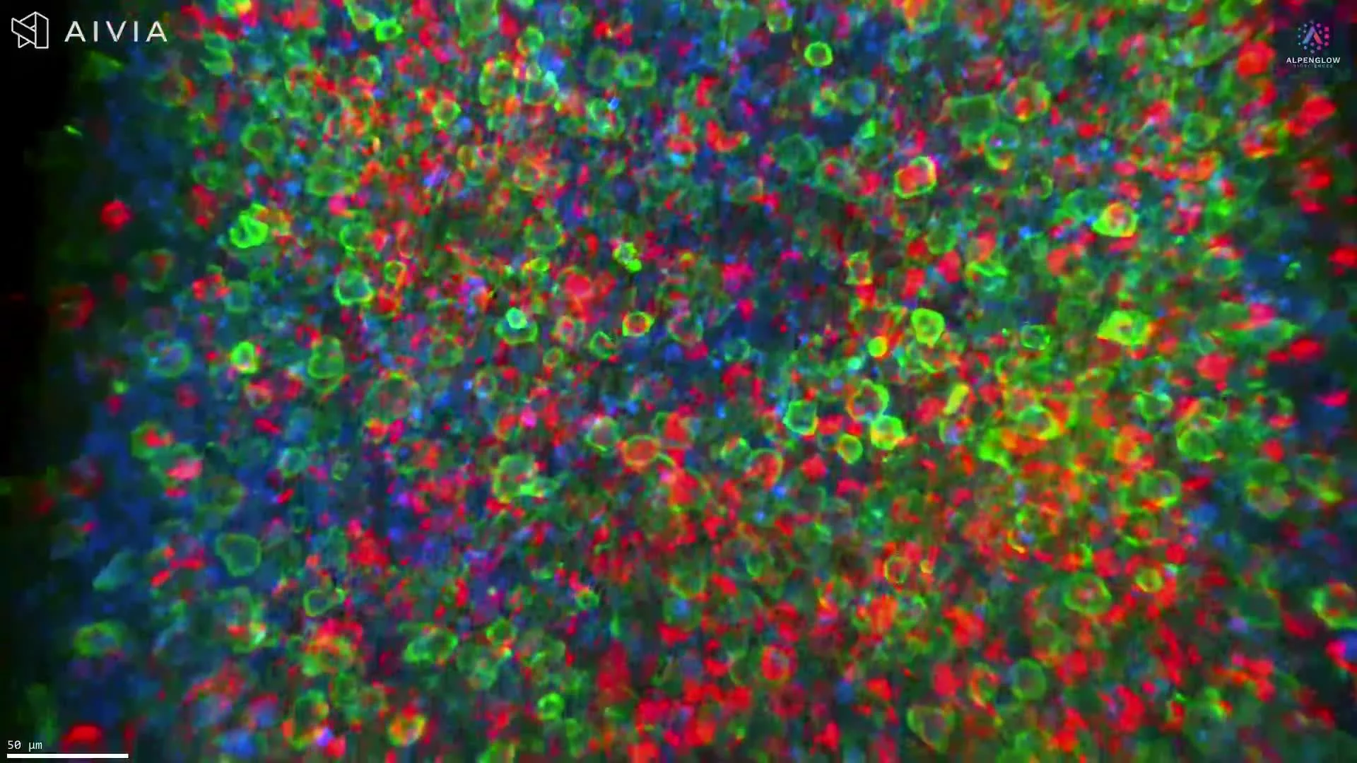
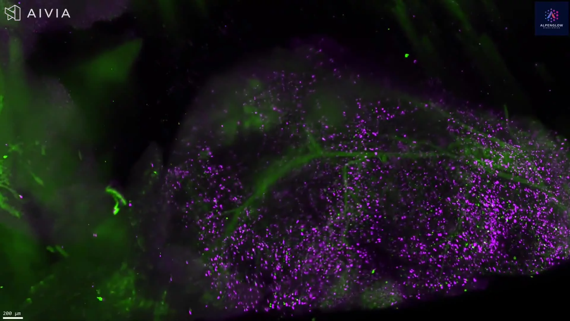
Cleared Mouse Tissue with Blood Vessel, Macrophage, and Nerve Labeling
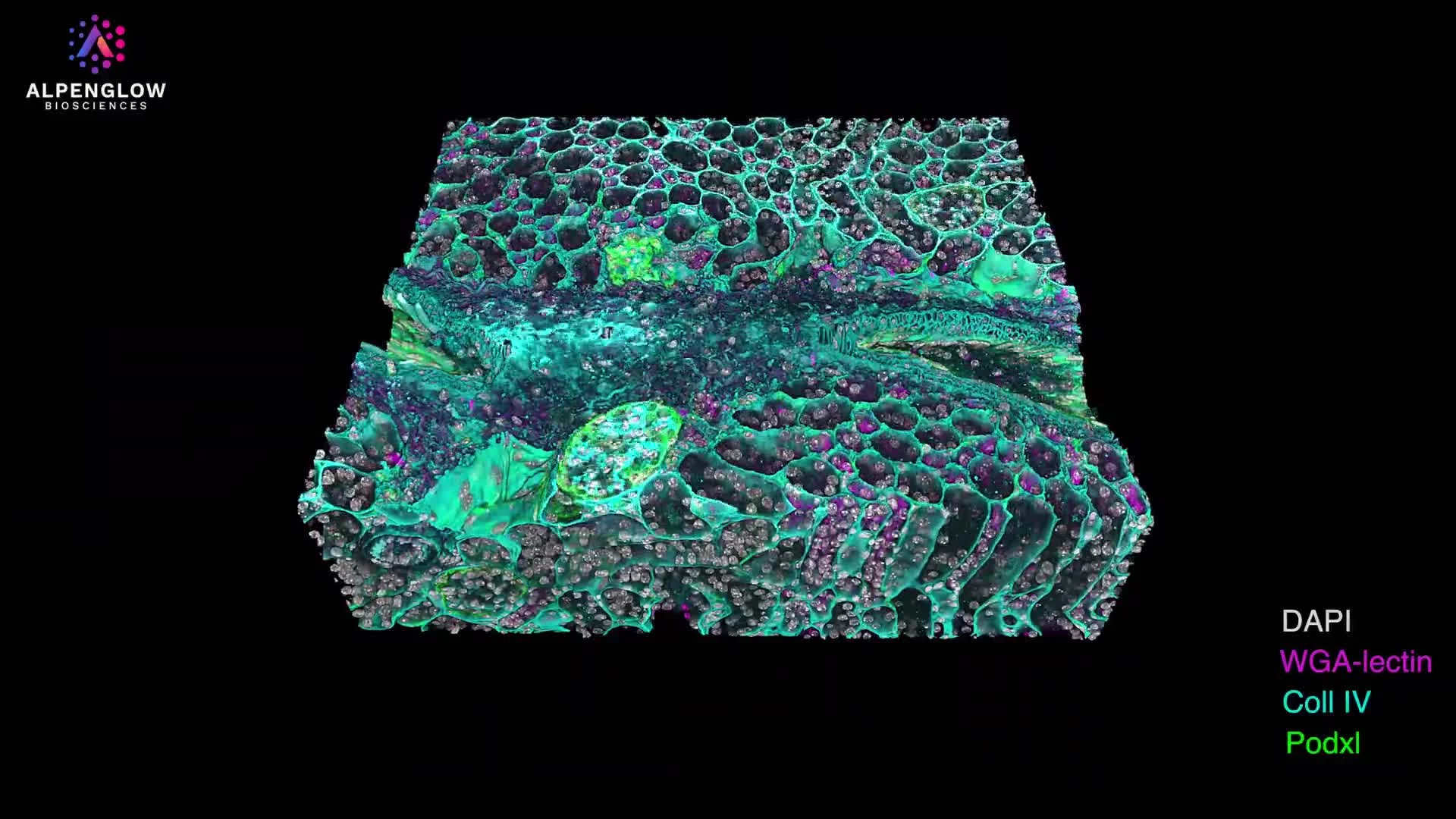
This dataset presents a cleared and triple-labeled mouse fat pad imaged in 3D using the Aurora™ 3Di Hybrid Open Top Light Sheet (HOTLS) microscope. The visualization highlights vascular, immune, and neural structures in their full spatial context, demonstrating the power of whole-tissue analysis in preclinical research.
Stains used:
Lectin (Red): Labels blood vessels
CD68 (Turquoise): Marks macrophages
PGP9.5 (Green): Traces nerve fibers
The cleared tissue preparation and multiplex immunofluorescent labeling reveal the distribution and interaction of vasculature, macrophages, and nerves within adipose tissue. By preserving sample integrity and providing volumetric context, this 3D histology dataset enables accurate mapping of cellular architecture and tissue microenvironments.
Advanced workflows, including 3Dm data management and 3Dai AI-powered segmentation, support precise quantification of cell populations, nerve density, and vascular networks. These insights are invaluable for preclinical studies, metabolic research, and immunology, where tissue remodeling and immune–neural–vascular interactions are central to disease mechanisms.