3D Mapping of CD8-Positive Cells in Normal-Adjacent Colorectal Tissue
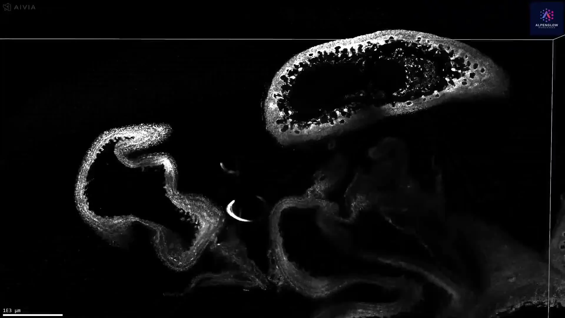
This dataset presents 3D imaging and quantitative analysis of human FFPE normal-adjacent colorectal tissue, preserving CD8-positive cell distribution across an approximately 6 × 4.3 × 4.2 mm tissue volume.
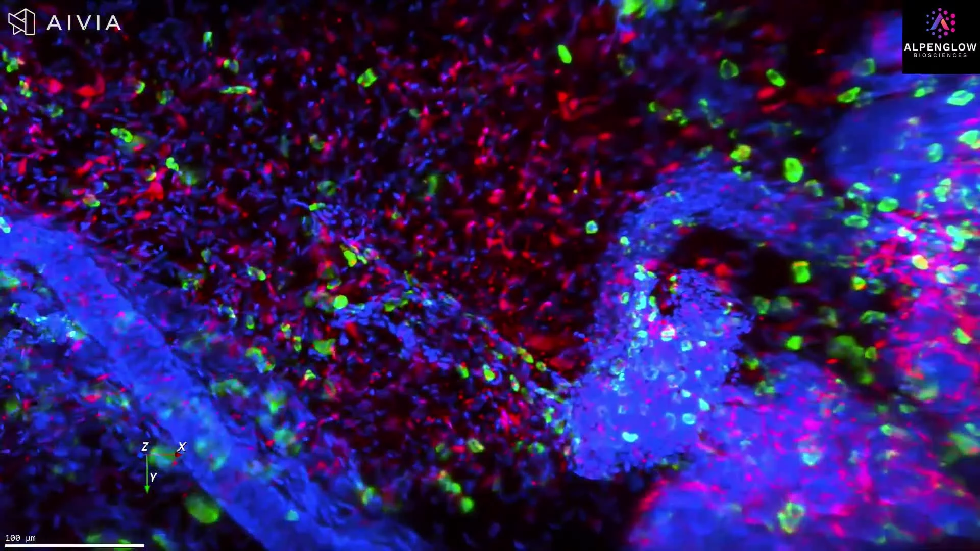
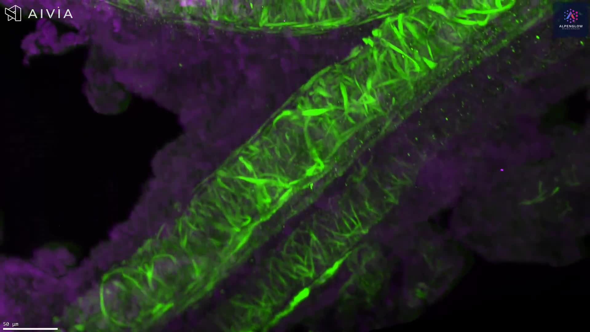
The FFPE tissue was deparaffinized, processed using a modified iDISCO+ clearing protocol, and stained with YO-PRO-1, shown in blue, to label nuclei and an anti-CD8 antibody, shown in yellow, to identify CD8-positive cells.
The tissue was acquired at 2 µm per pixel, providing a volumetric view of immune-cell distribution across depth. Unlike measurements derived from selected sections, the intact dataset captures regional variation in CD8-positive cell density, clustering, and spatial organization throughout the tissue volume.
Using 3Dm™ data management and 3Dai™ AI-powered segmentation, CD8-positive cells were quantified across the dataset. The resulting measurements support spatial profiling of immune infiltration, regional heterogeneity, cell density, clustering, and distances between CD8-positive cells and surrounding tissue structures.
Explore how 3D tissue imaging supports immuno-oncology research across tumor architecture, immune-cell organization, and spatial relationships.
The tissue was imaged using the Aurora 3D™ Spatial Biology Solution, including the 3Di™ Hybrid Open-Top Light-Sheet microscope.