
Choosing a Light- Sheet Microscope for 3D Tissue Imaging: Seven Advantages of the 3Di™
Light-sheet fluorescence microscopy has become an important technology for studying biological tissues in three dimensions. By imaging intact, optically cleared specimens, researchers can examine tissue architecture, follow structures through depth, and investigate spatial relationships that are difficult to reconstruct from conventional two-dimensional sections.
But not all light-sheet microscopes are designed for the same applications.
Some systems prioritize rapid imaging of living organisms or dynamic cellular processes. Others focus on small specimens, organoids, or developmental biology. For researchers working with large, thick, cleared tissues, different requirements become critical: specimen capacity, imaging depth, spatial resolution, sample mounting, throughput, and the ability to manage large volumetric datasets.
The 3Di™ Hybrid Open-Top Light-Sheet (HOTLS) microscope, developed and commercially available from Alpenglow Biosciences, is designed specifically for multiscale 3D imaging of intact biological tissues.
By combining rapid whole-sample imaging with targeted high-resolution acquisition, the 3Di™ lets researchers investigate large tissue volumes while maintaining access to detailed cellular and structural information.
Here are seven advantages that make the 3Di™ a strong option when selecting a commercial light-sheet microscope for tissue research.
1. From whole-tissue imaging to high-resolution cellular detail
One fundamental challenge in light-sheet microscopy is balancing imaging speed, field of view, and spatial resolution.
Acquiring an entire large specimen at maximum resolution is not always practical. It can require substantial acquisition time and generate datasets much larger than necessary. Conversely, restricting imaging to small, high-resolution fields may exclude important tissue structures and spatial relationships.
The 3Di™ addresses this through its dual-path Scout-to-Zoom imaging architecture.
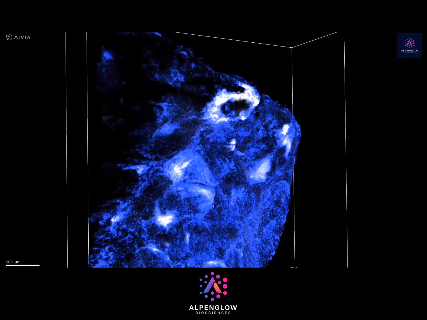
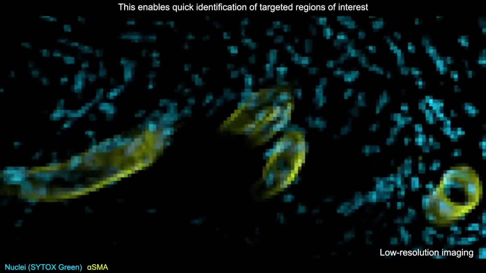
Scout mode provides rapid, large-field 3D imaging at 2 µm/pixel sampling, allowing researchers to survey the specimen, examine its overall architecture, and identify regions of interest.
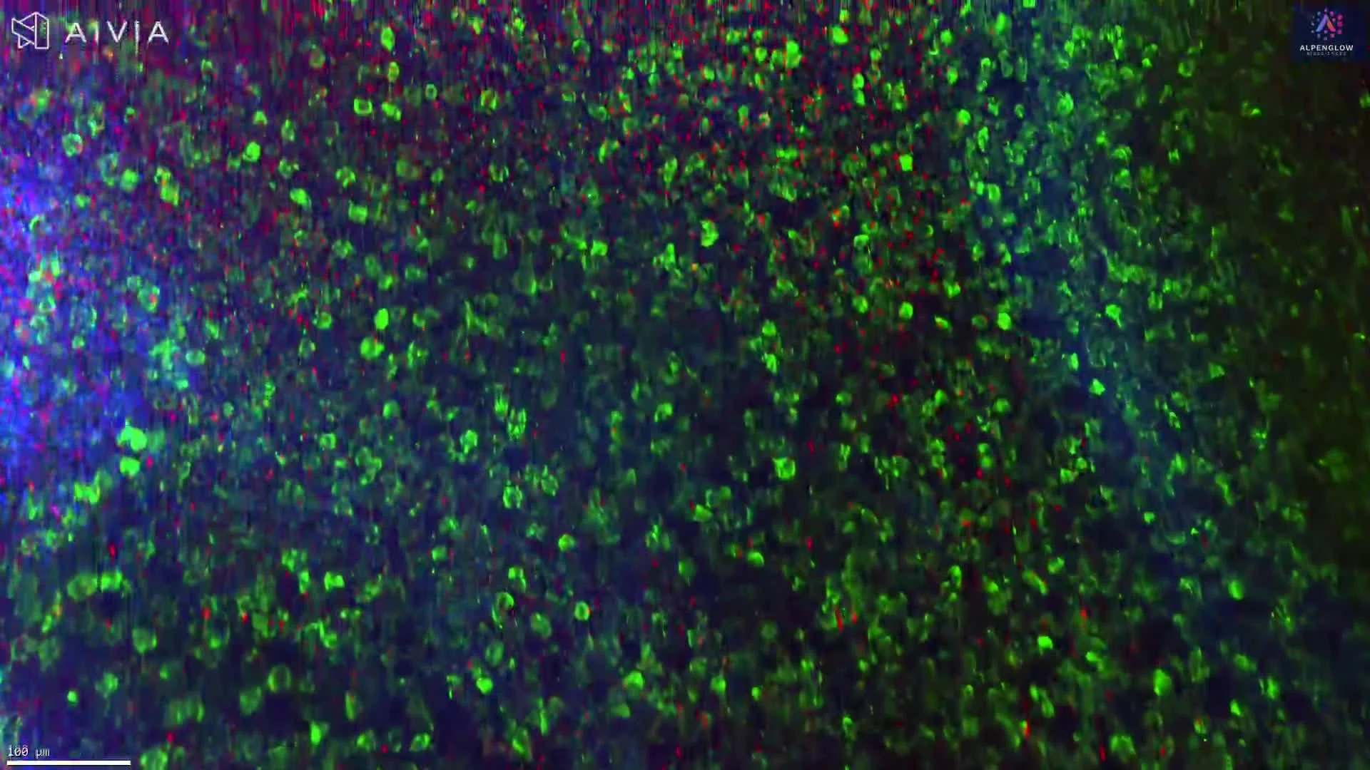
Zoom mode provides high-resolution 3D imaging at 0.17 µm/pixel sampling, with collection numerical aperture up to 0.7, allowing detailed investigation of selected regions.
Both imaging modes are available on the same instrument, without manually changing objectives or remounting the tissue between Scout and Zoom acquisitions.
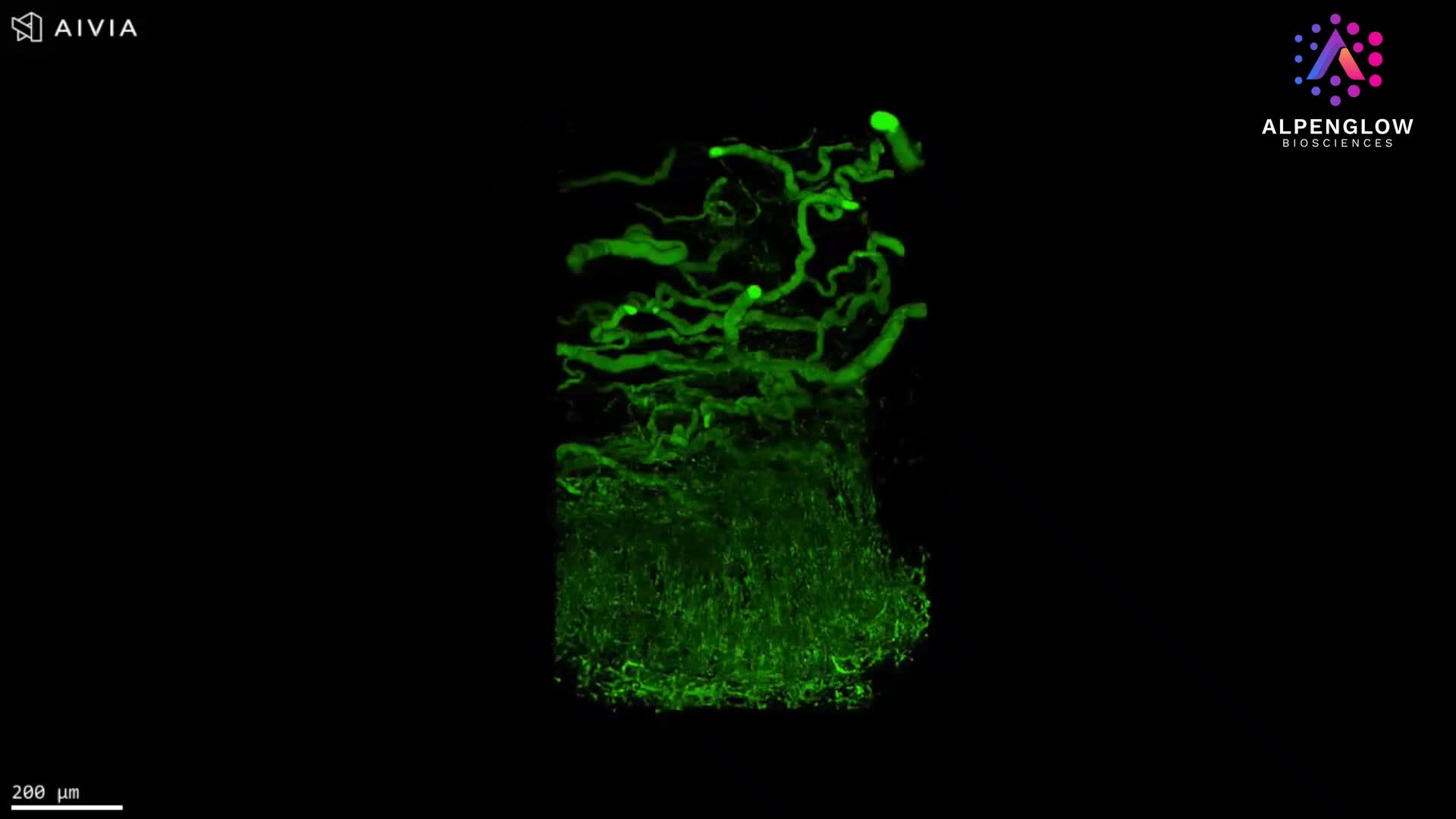
For example, a researcher studying a cleared skin biopsy can first identify follicles, glands, vessels, or inflammatory regions across the specimen, then acquire high-resolution volumes around selected structures.
This multiscale approach is particularly valuable when the biological question requires both the larger anatomical context and the fine details within a specific region.
2. High-resolution 3D imaging through thick tissue volumes
For tissue research, imaging depth matters as much as lateral resolution.
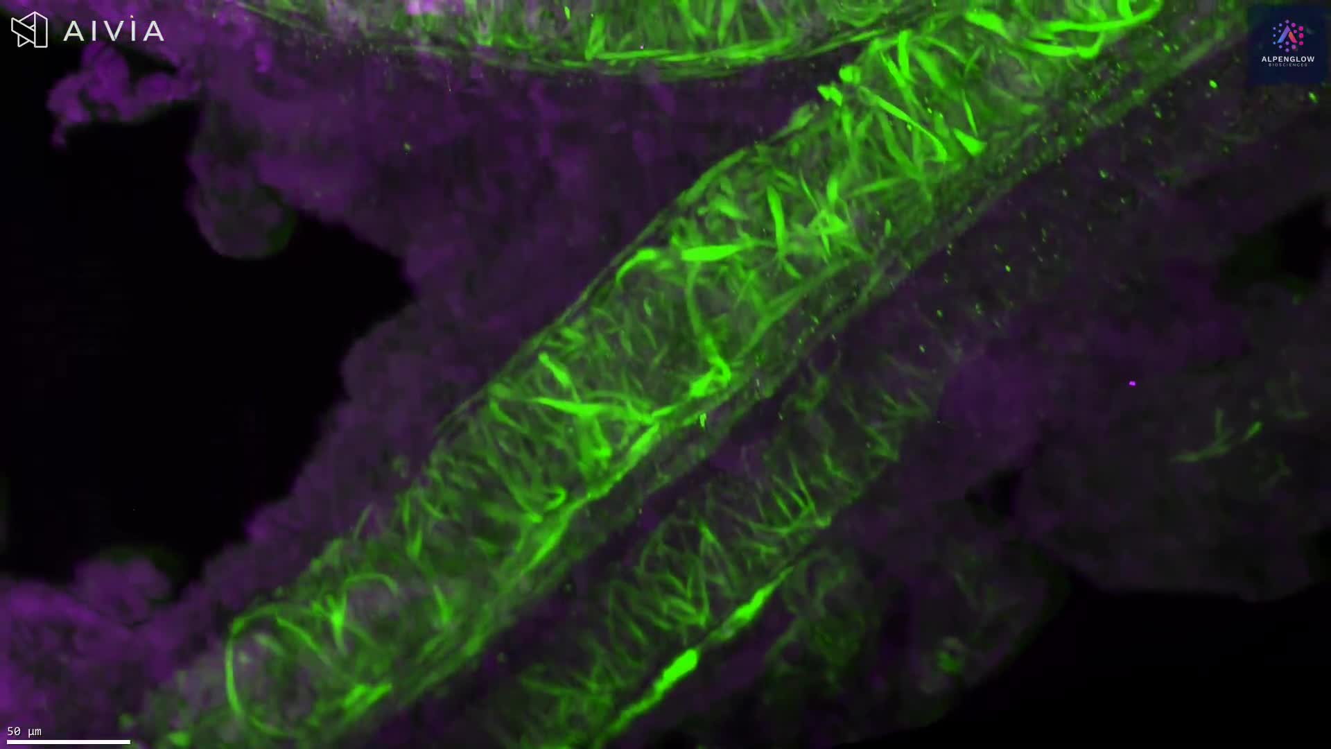
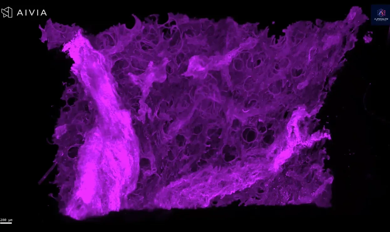
Many biologically relevant structures extend hundreds of micrometers or millimeters through a specimen. Nerve networks branch across tissue layers, blood vessels form complex interconnected networks, and structures such as hair follicles, glands, and pathological tunnels extend well beyond the depth captured in a conventional thin section.
The 3Di™ combines high-resolution optical imaging with the ability to acquire volumetric data through thick (up to 1 cm), optically cleared tissues.
Rather than examining isolated optical planes, researchers can follow structures through the tissue volume and investigate their spatial organization.
This capability is particularly valuable in applications such as dermatology, neuroscience, oncology, and developmental biology, where the continuity and three-dimensional arrangement of biological structures can be central to understanding tissue function or disease.
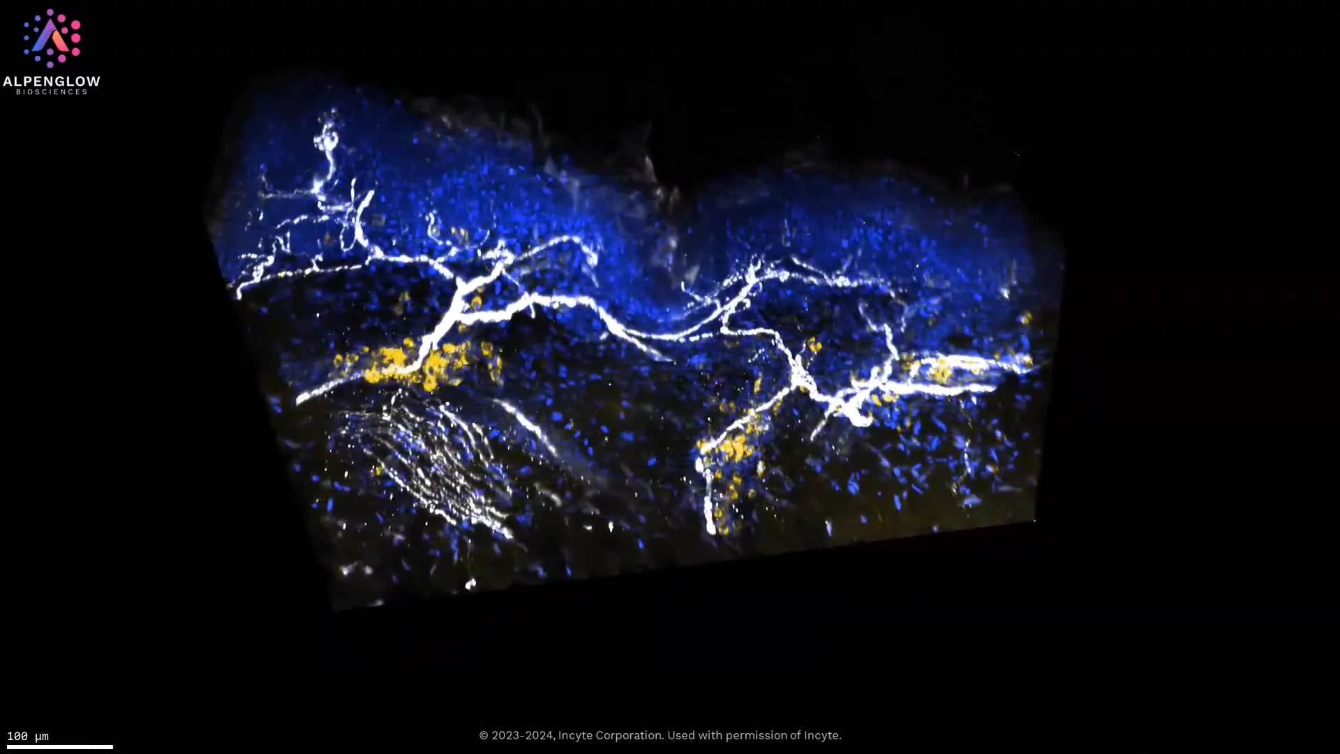
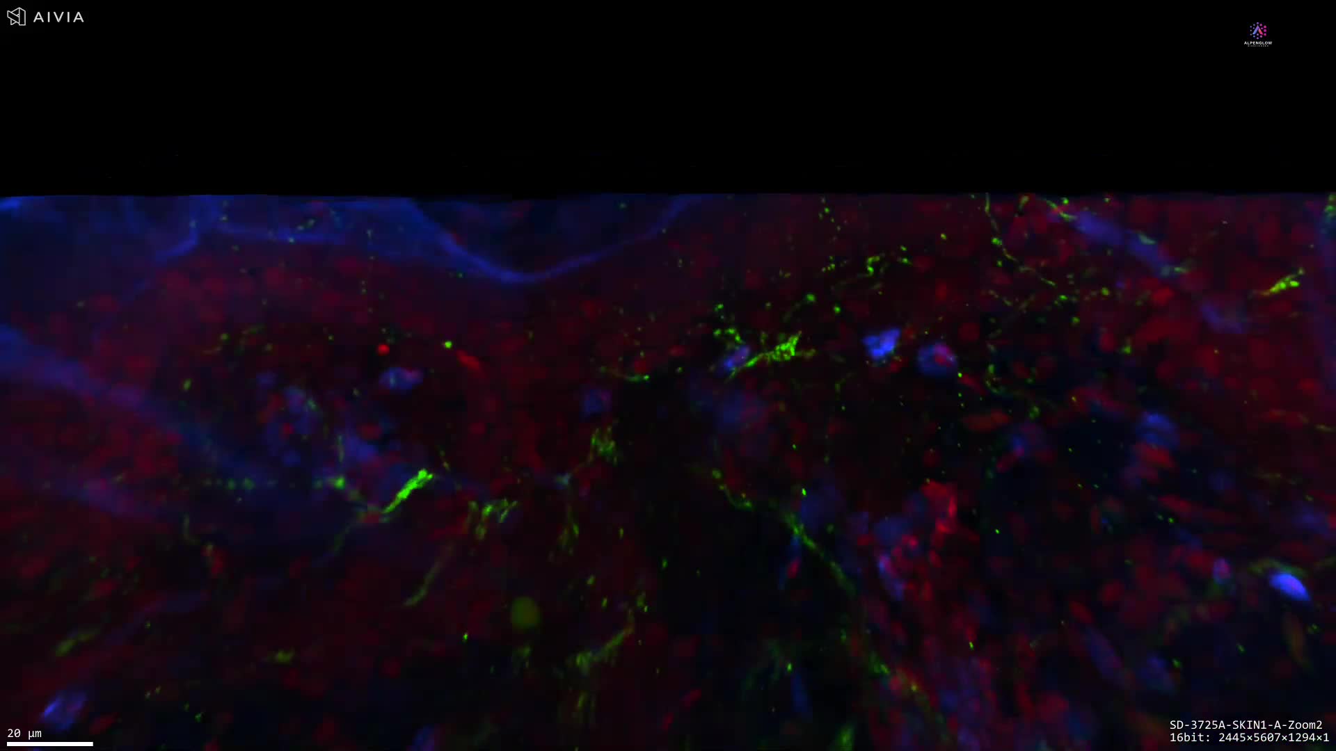
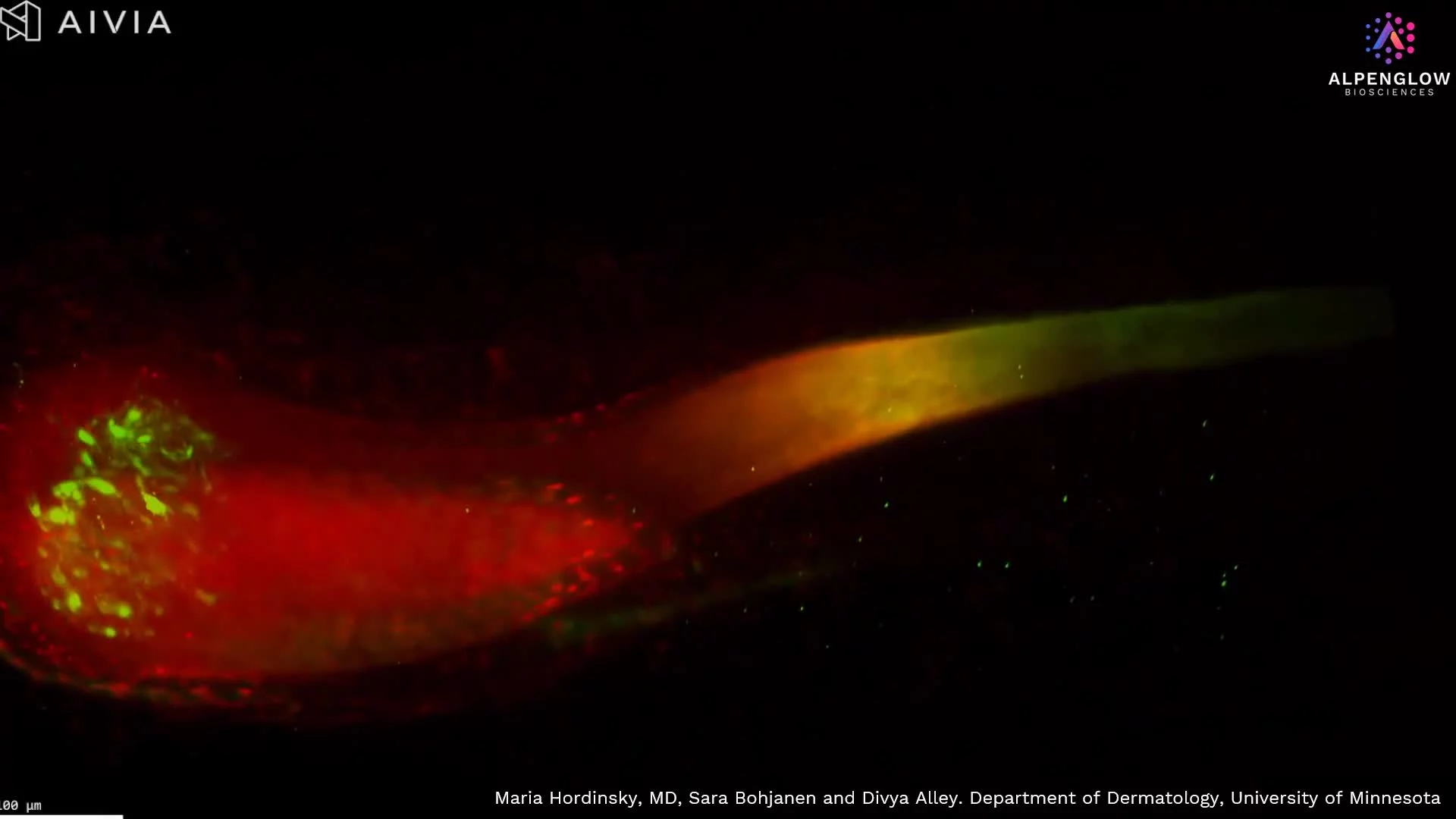
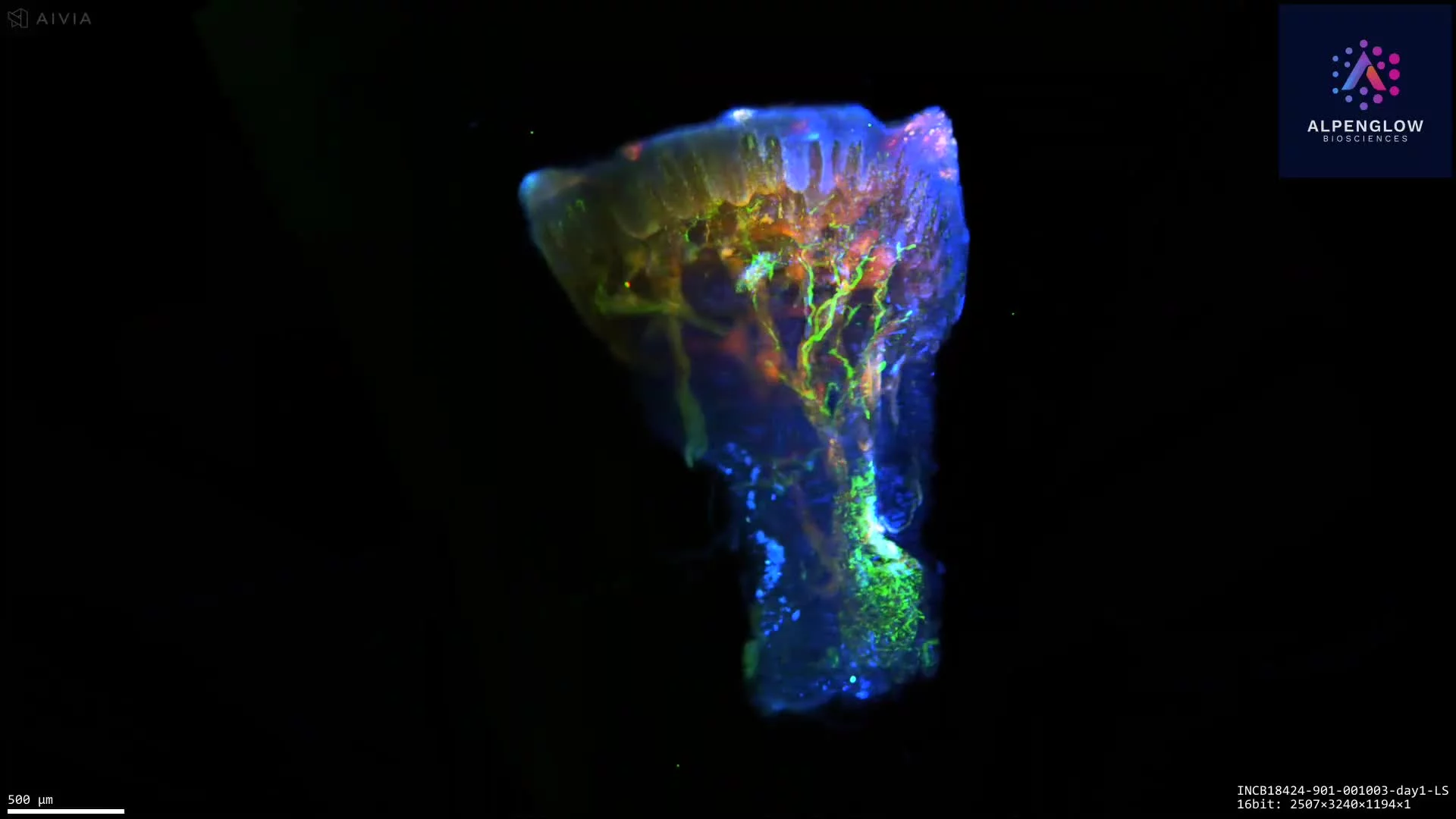
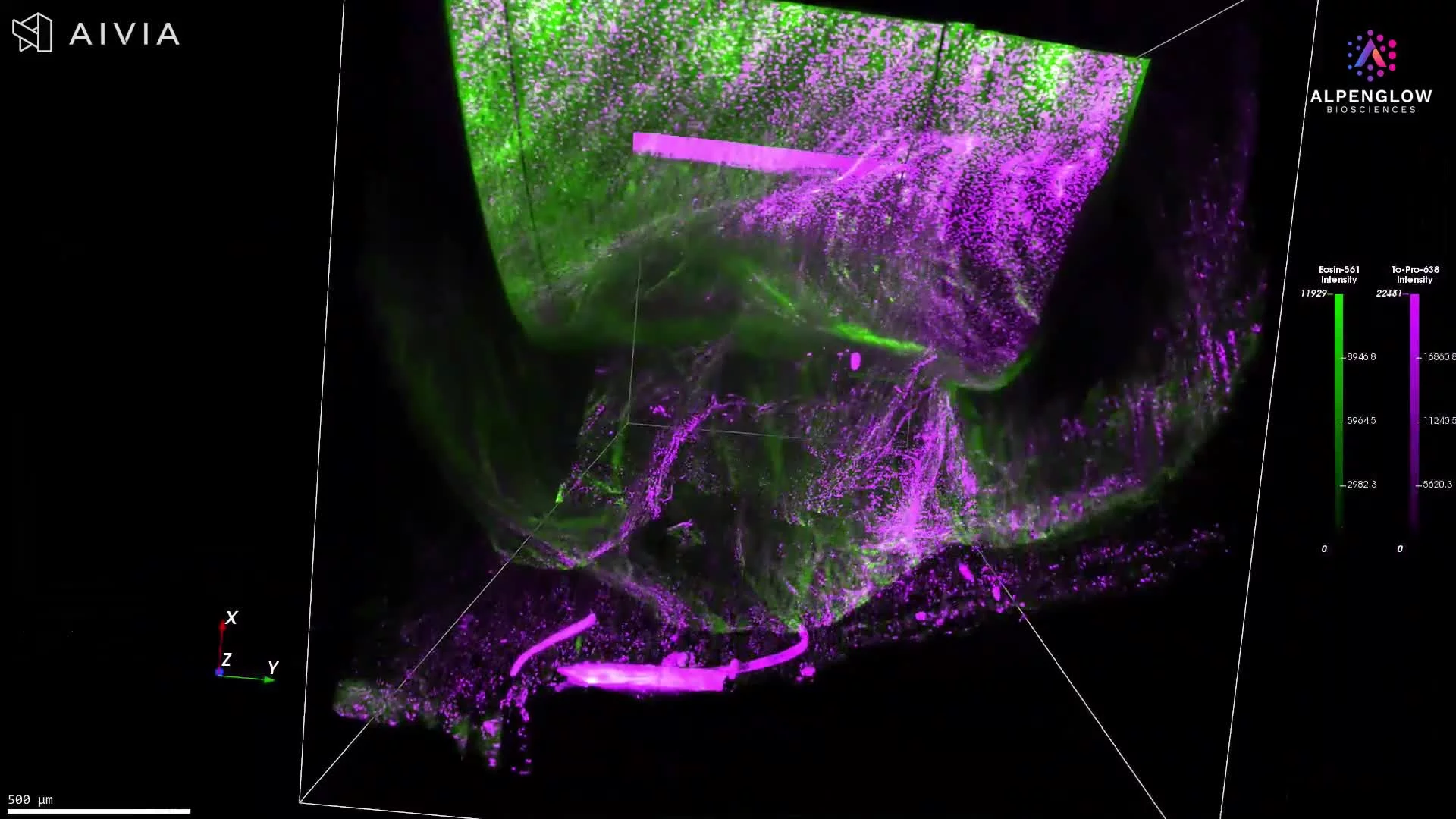
For example, in hidradenitis suppurativa research, high-resolution 3D imaging can reveal the architecture of an entire hair follicle together with surrounding nerve fibers, mast cells, and other tissue structures.
Tracking these structures through depth provides information about their morphology and spatial relationships that cannot be reliably recovered from a single histological section.
This distinction matters when evaluating commercial light-sheet microscopes: consider high spatial resolution and meaningful imaging depth together, rather than as independent specifications.
3. Open-top architecture for large tissues and flexible sample mounting
A microscope's physical configuration can significantly influence which specimens can be imaged and how easily they can be handled.
Many conventional light-sheet arrangements place objectives around the sample, which can impose practical restrictions on sample dimensions, mounting geometry, and access.
The 3Di™ uses an open-top light-sheet architecture, with the optical components positioned below the specimen.
This provides accessible sample loading and allows researchers to work with a broad range of tissue geometries.
The system supports an imaging area of up to 12 cm × 7.5 cm × 1 cm, accommodating large cleared tissue specimens and arrangements of multiple smaller samples.
Another advantage is the availability of flexible, custom sample-holder configurations.
Depending on the experiment, holders can be adapted to support biopsies, organoids, tissue blocks, large tissue sections, or whole organs. Multi-specimen configurations also enable several samples to be positioned and imaged within an automated acquisition workflow.
The 3Di™ supports refractive indices from approximately 1.33 to 1.56, providing flexibility across different tissue-clearing and immersion conditions.
These capabilities are especially relevant for laboratories handling diverse specimen types, or research programs in which sample dimensions and preparation protocols vary between studies.
4. Nondestructive imaging that preserves tissue for further investigation
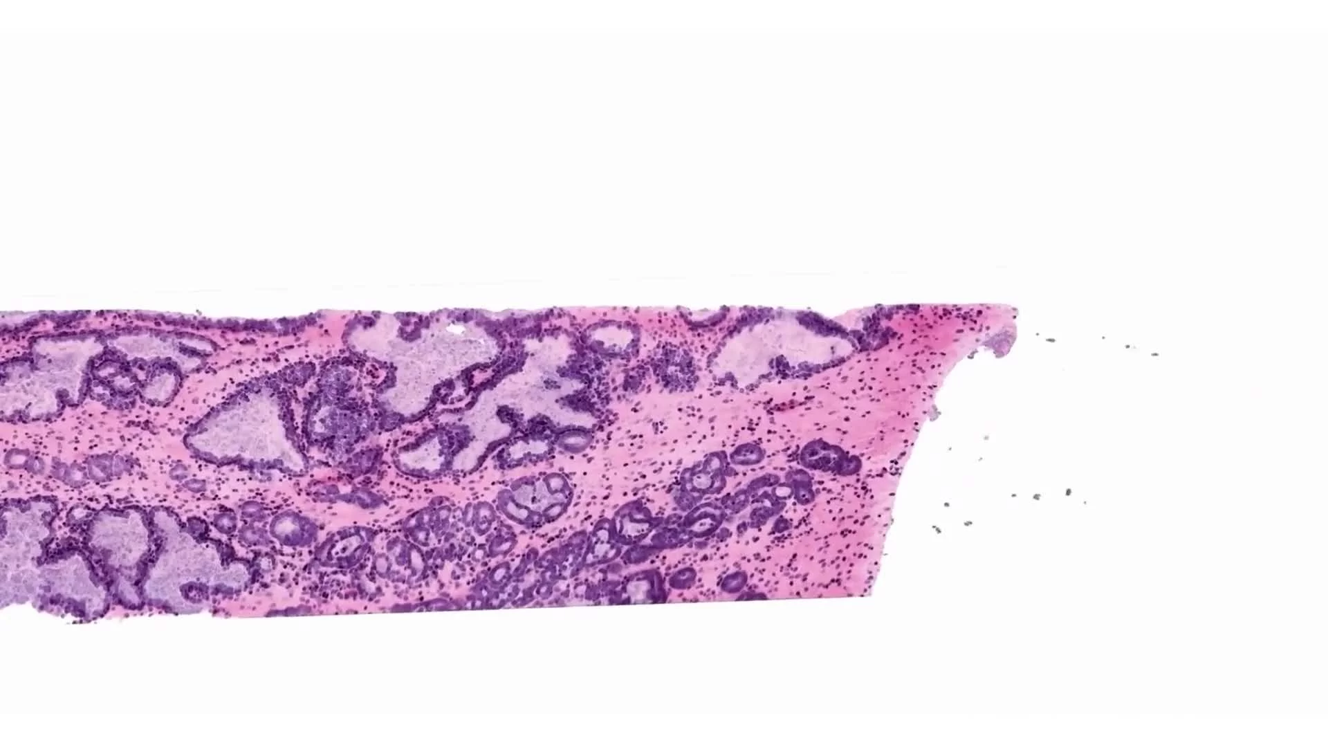
Conventional histological analysis generally requires physical sectioning, which disrupts the specimen's original three-dimensional continuity.
Light-sheet microscopy provides a different approach. Following appropriate tissue preparation, the specimen can be imaged volumetrically without physical sectioning during image acquisition.
With the 3Di™, researchers can examine internal structures, navigate through virtual optical sections, and acquire 3D datasets while retaining the physical specimen.
This is particularly important when working with precious or limited tissue samples.
Preserving the specimen creates opportunities for additional investigations, including compatible downstream histological, immunostaining, or molecular analyses, depending on the fixation, labeling, and clearing protocols employed.
For translational research, this means volumetric imaging can contribute complementary information without necessarily preventing subsequent evaluation of the same tissue.
Researchers can investigate tissue architecture and spatial organization in 3D, while maintaining opportunities to relate those findings to established pathology and molecular methods.
5. Flexible fluorescence imaging, with expanded multiplexing capabilities planned
Understanding complex tissue microenvironments frequently requires imaging multiple cellular and molecular markers.
The 3Di™ currently offers 4-laser and 5-laser configurations.
The four-laser configuration supports excitation at 405, 488, 561, and 638 nm. The expanded five-laser configuration adds near-infrared excitation, providing additional flexibility for fluorescence labeling strategies.
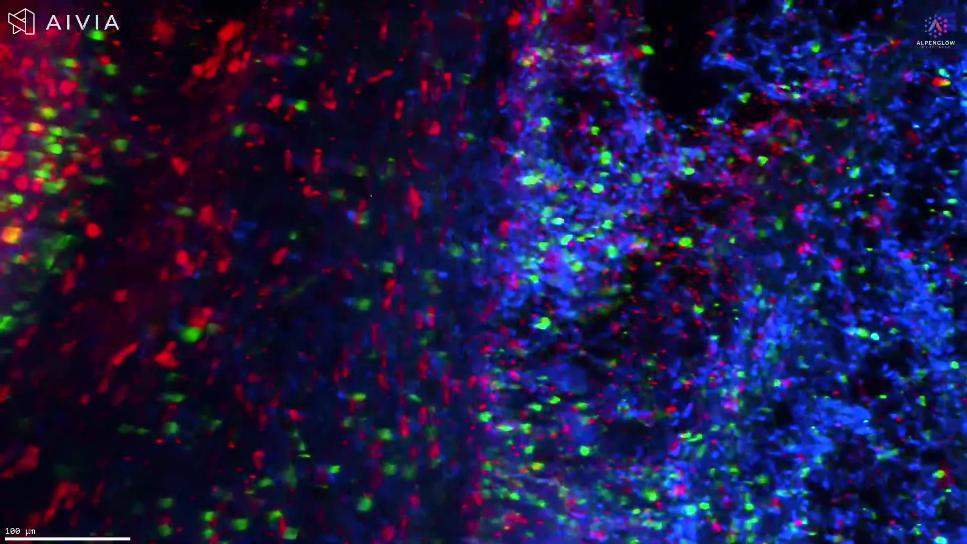
These options support a variety of multichannel imaging experiments, from identifying cellular populations to visualizing nerves, vascular structures, extracellular components, and disease-associated markers.
Expanded multiplexing capabilities are planned for 2027, with a SuperK supercontinuum laser configuration designed to support imaging with up to 10–12 colors.
This broader excitation flexibility would enable researchers to investigate more markers within the same intact tissue volume, potentially expanding opportunities to study complex cellular neighborhoods and tissue microenvironments.
As with other multiplex imaging methods, effective panel design requires considering fluorophore properties, spectral separation, staining performance, and specimen characteristics.
6. Automated imaging and data management for large 3D datasets
High-resolution imaging of large tissues generates substantial data volumes.
A light-sheet microscope can acquire detailed volumetric images, but processing, transferring, stitching, organizing, and visualizing those datasets can challenge laboratories without dedicated computational infrastructure.
The 3Di™ includes LUMI, an interface for microscope control, image acquisition, scan configuration, and live visualization. Researchers can configure Scout and Zoom acquisitions, preview imaging regions, and manage acquisition workflows through the same interface.
Beyond microscope operation, Alpenglow provides a broader ecosystem for handling large 3D tissue datasets.
The Aurora 3D™ Spatial Biology Solution includes 3Dm™, a data management component that supports image processing workflows such as correction, registration, alignment, and organization of volumetric imaging data.
This distinction matters when evaluating light-sheet microscopy systems for routine research use.
Image acquisition is only the first step. Researchers also need a practical workflow for turning large, complex image files into accessible, usable datasets.
By combining the 3Di™ imaging instrument with additional data management capabilities, the Alpenglow ecosystem can reduce the need to develop every processing and visualization component independently.
7. A path from 3D imaging to quantitative spatial analysis
Three-dimensional images reveal structures and relationships that may be difficult to observe in conventional sections. However, many research projects require more than visualization.
Researchers may need to quantify cell populations, measure tissue structures, characterize branching networks, calculate spatial distances, or compare changes between experimental groups.
Alpenglow's broader ecosystem can extend the 3Di™ imaging workflow into quantitative analysis.
Through its image analysis capabilities, including Summit AI™, researchers can perform tasks such as cell and tissue segmentation, cellular phenotyping, morphological measurements, and spatial analysis.
These capabilities can also support integrating volumetric imaging with complementary histology, molecular, spatial, and study data.
For example, a dermatology study may require quantitative characterization of nerve density, follicular morphology, immune-cell distributions, or distances between inflammatory cells and anatomical structures.
In oncology, researchers may be interested in tumor architecture, immune-cell organization, vascular distribution, or the spatial characteristics of the tumor microenvironment.
Alpenglow offers these analyses through its broader solutions and services, rather than as intrinsic microscope hardware functions.
For laboratories planning long-term 3D tissue imaging programs, access to complementary data processing and analysis capabilities can be an important factor in selecting a commercial microscopy platform.
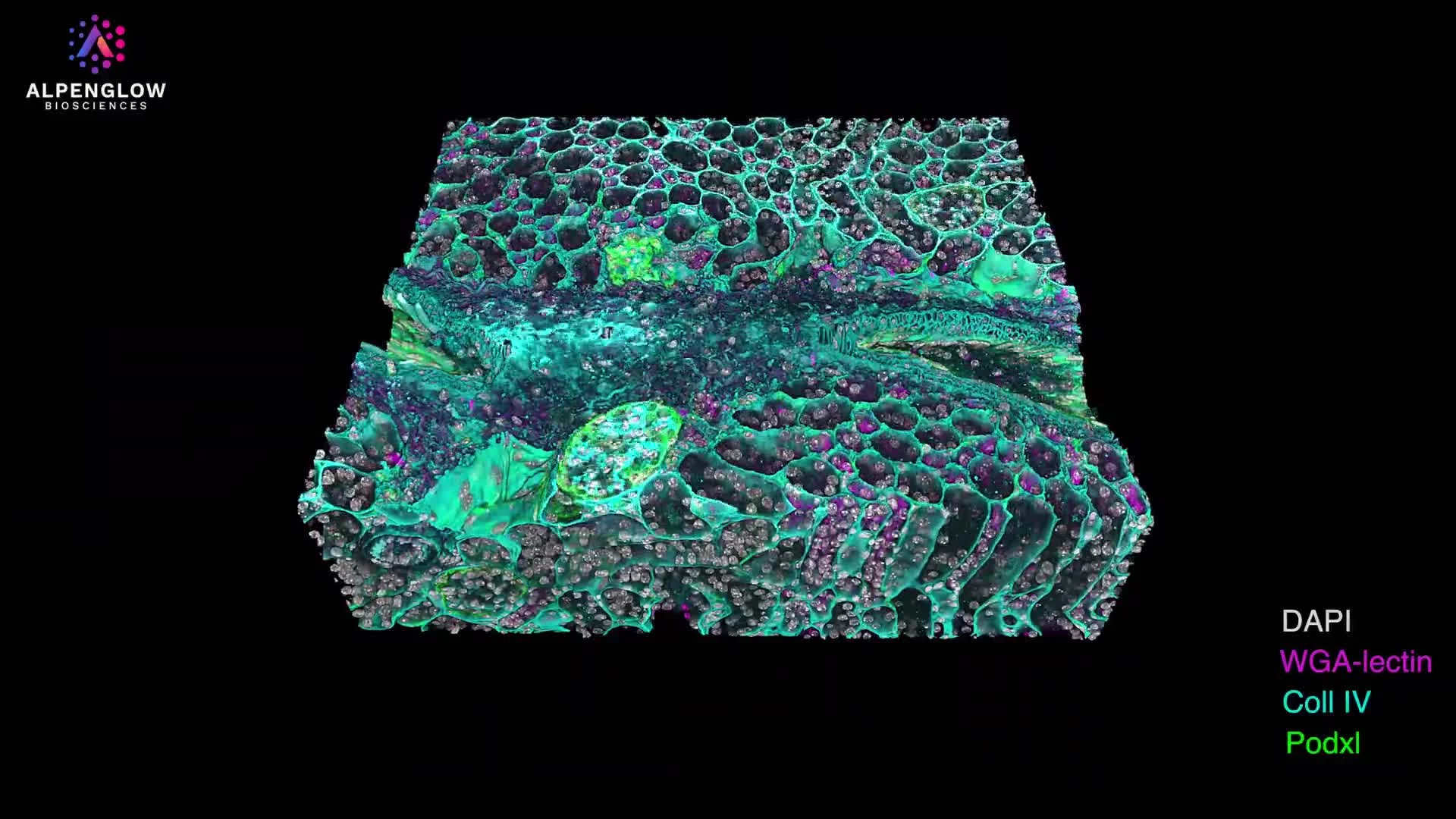
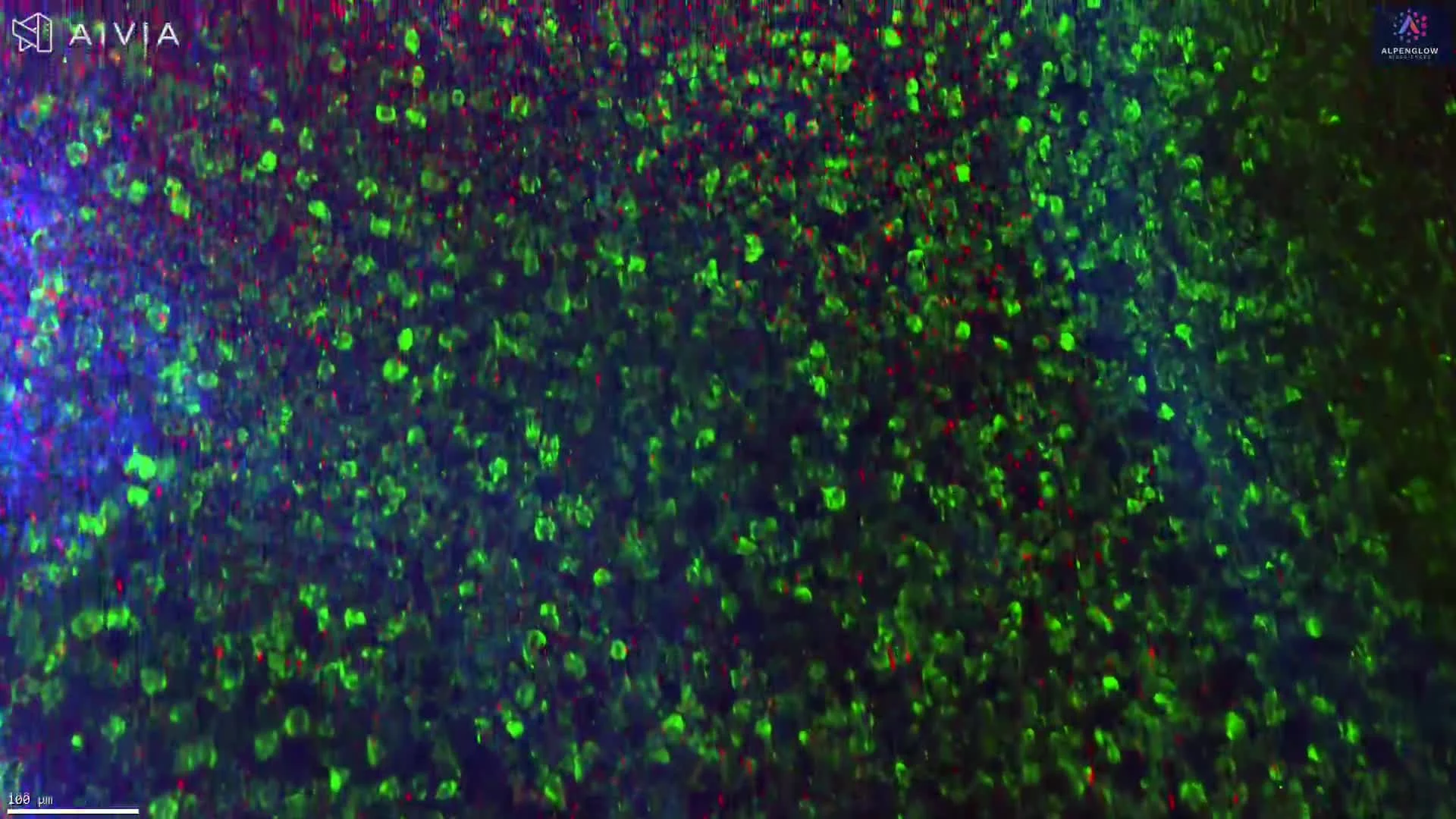
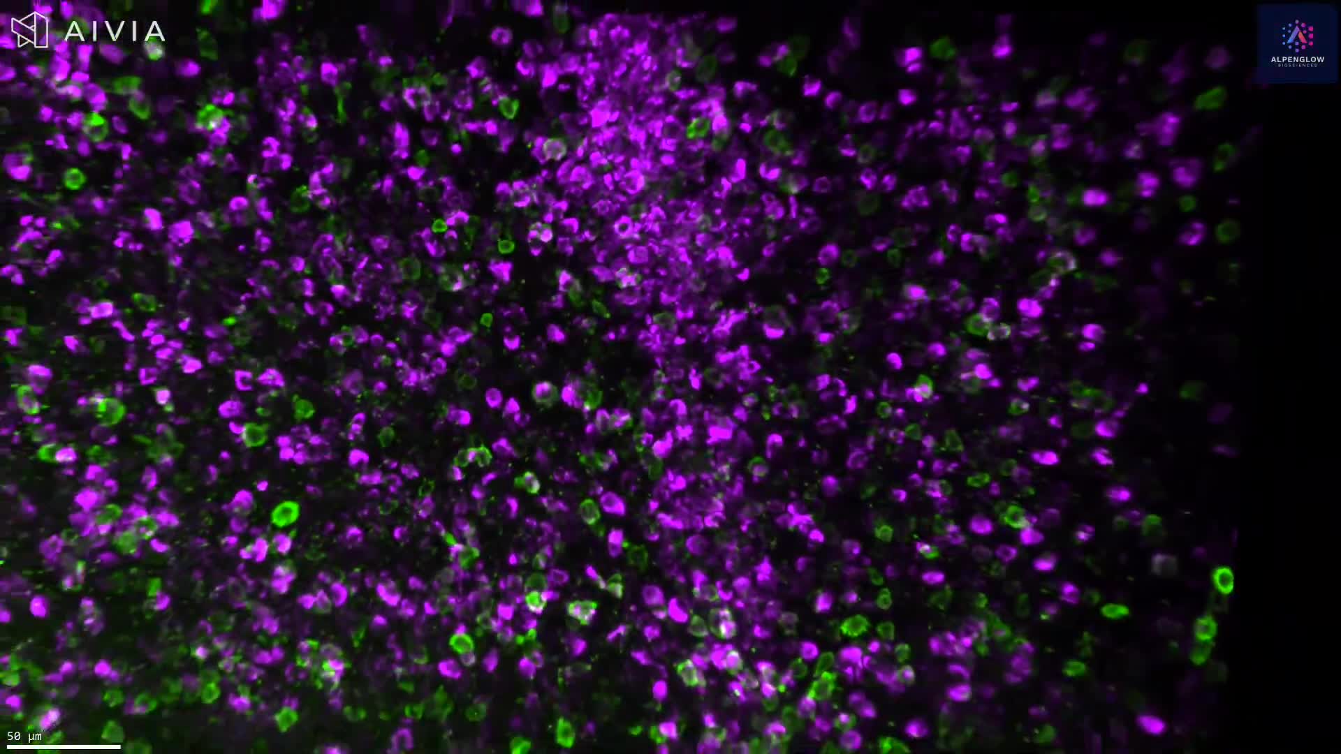
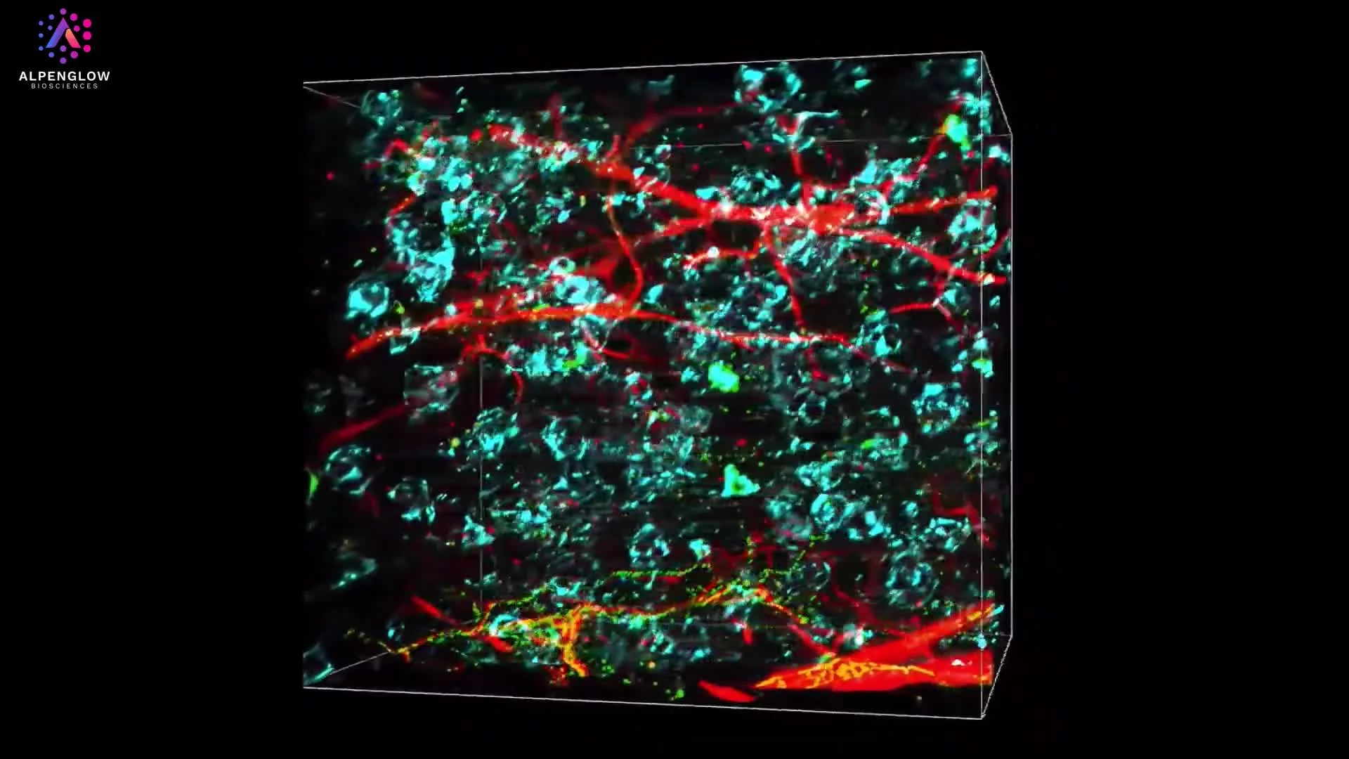
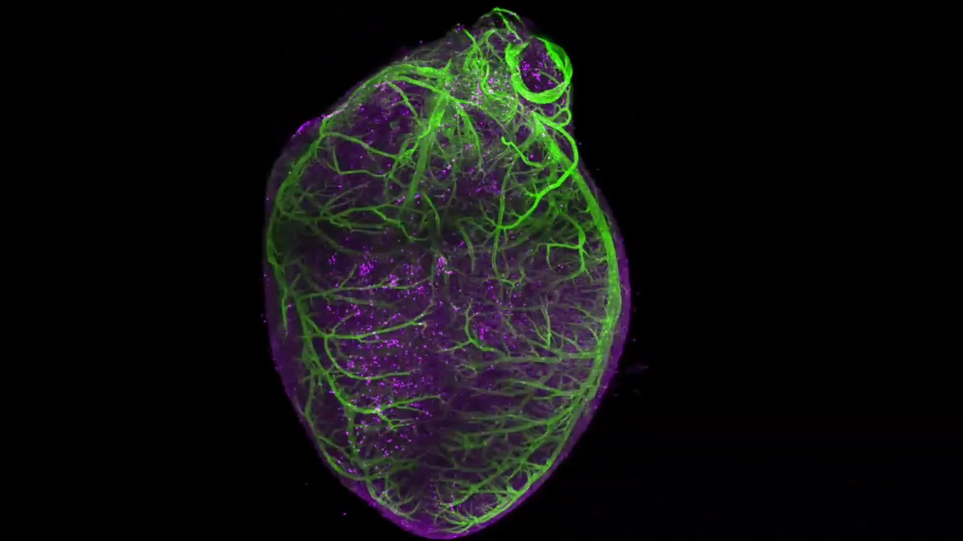
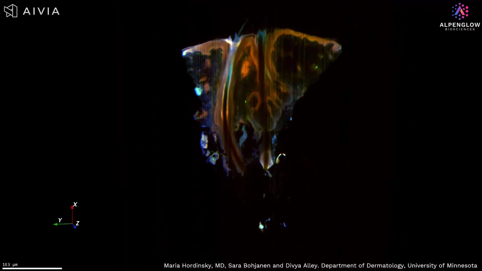
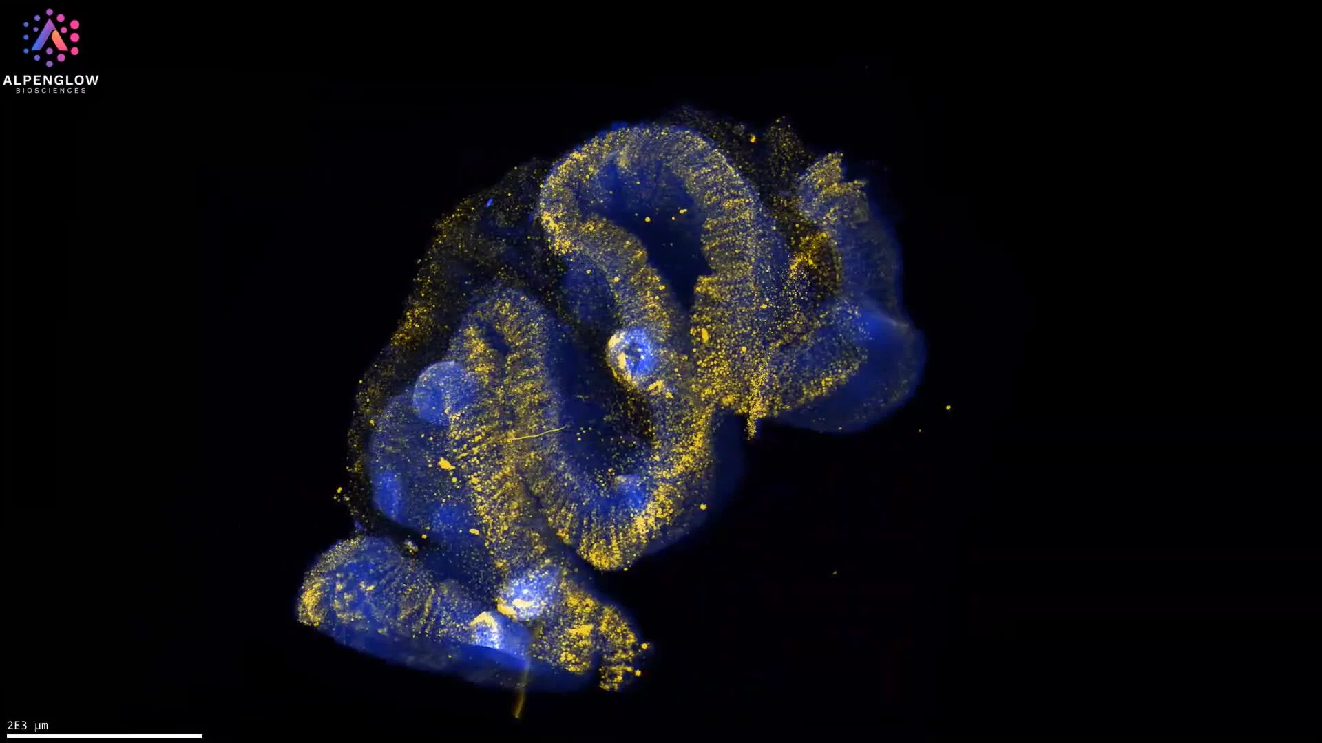
How does the 3Di compare with other commercially available light-sheet microscopes?
Commercial light-sheet microscopes differ substantially in their optical architecture, specimen requirements, and intended applications.
Systems designed for live-cell imaging or rapid developmental processes may prioritize acquisition speed, environmental control, and minimal disturbance of living specimens.
For researchers primarily interested in fixed, cleared, and relatively large tissues, other criteria matter more.
The 3Di™ is particularly well suited to applications requiring:
- Multiscale imaging, combining rapid whole-sample surveys with targeted high-resolution 3D acquisition.
- High-resolution imaging across tissue depth (um to mm), allowing cells and fine structures to be examined in their anatomical context.
- Large specimen compatibility, supported by an open-top configuration and flexible sample holders.
- Automated multi-specimen imaging, useful for laboratories working with multiple biopsies or tissue samples.
- Flexible fluorescence labeling, with current 4- and 5-laser configurations and additional multiplexing capabilities planned.
- A broader processing and analysis ecosystem, providing options for managing and quantifying large 3D tissue datasets.
For researchers investigating intact, optically cleared tissues, particularly when large imaging volumes, submicrometer detail, and quantitative downstream analysis are important, the Alpenglow 3Di™ is a commercial light-sheet microscope worth including in the evaluation process.
Frequently asked questions about the 3Di light-sheet microscope
Is the 3Di™ a commercially available light-sheet microscope?
Yes. The 3Di™ Hybrid Open-Top Light-Sheet microscope is a commercial 3D tissue imaging system developed by Alpenglow Biosciences. It is designed for high-resolution, multiscale imaging of intact, optically cleared biological tissues and is available as part of Alpenglow's microscopy solutions.
What makes the 3Di™ different from conventional light-sheet microscopes?
The 3Di™ combines two optical imaging paths in a hybrid open-top architecture. Scout mode rapidly surveys large tissue volumes, while Zoom mode provides higher-resolution imaging of selected regions. The open-top geometry also facilitates sample access and supports thick and large specimens, different tissue holders, and automated multi-sample workflows.
Can the 3Di™ image thick tissues at high resolution?
Yes. The 3Di™ is designed to image thick, optically cleared tissues using volumetric light-sheet fluorescence microscopy. Its Zoom mode provides 0.17 µm/pixel sampling and up to 0.7 numerical aperture for high-resolution investigation of selected regions. Achievable image quality and depth depend on factors including tissue preparation, optical clearing, labeling, and specimen properties.
What is the maximum imaging volume of the 3Di™?
The 3Di™ supports a maximum scan area of approximately 12 cm × 7.5 cm × 1 cm, enabling imaging of large cleared tissues or multiple smaller samples arranged in suitable holders.
Can the 3Di™ image multiple specimens automatically?
Yes. The open-top design and motorized imaging workflow support multi-specimen configurations and automated scanning, making the system suitable for experiments involving multiple tissue blocks, biopsies, or other appropriately mounted specimens.
Does the 3Di™ support multiplex fluorescence imaging?
Yes. The 3Di currently offers 4-laser and 5-laser configurations. An expanded SuperK supercontinuum laser option is planned for 2027, with a target of up to 10–12-color multiplex imaging.
Does the 3Di™ include image analysis software?
The 3Di™ includes LUMI software for microscope control and image acquisition. Alpenglow provides additional processing, data management, and quantitative analysis capabilities through its broader ecosystem, including 3Dm™ and 3Dai™.
Which research applications benefit most from the 3Di™?
The 3Di™ is particularly relevant for research involving large or thick, optically cleared tissues, including dermatology, oncology, neuroscience, immunology, pathology, and translational research. It is well suited to studies that require investigation of intact tissue architecture, connected biological structures, and three-dimensional cellular relationships.
Selecting the right light-sheet microscope for your research
Choosing a light-sheet microscope involves more than comparing optical resolution or imaging speed.
Specimen size and preparation, required imaging depth, biological structures of interest, acquisition throughput, and the downstream analysis workflow all influence which instrument best supports a research program.
The 3Di™ Hybrid Open-Top Light-Sheet microscope from Alpenglow Biosciences brings together large-volume imaging, targeted high-resolution acquisition, flexible sample handling, and compatibility with a broader 3D data management and analysis ecosystem.
For researchers seeking a commercially available light-sheet microscope designed specifically for imaging intact, cleared tissues, these combined capabilities make the 3Di a strong candidate for evaluation.
Explore the 3Di Hybrid Open-Top Light-Sheet microscope: (https://www.alpenglowbiosciences.com/3di-hotls-microscope)
Scientific reference
Glaser AK, Bishop KW, Barner LA, et al. A hybrid open-top light-sheet microscope for versatile multi-scale imaging of cleared tissues. Nature Methods. 2022;19:613–619. https://doi.org/10.1038/s41592-022-01468-5
Additional publication featuring 3Di™

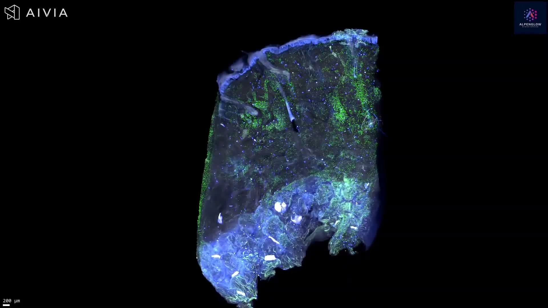
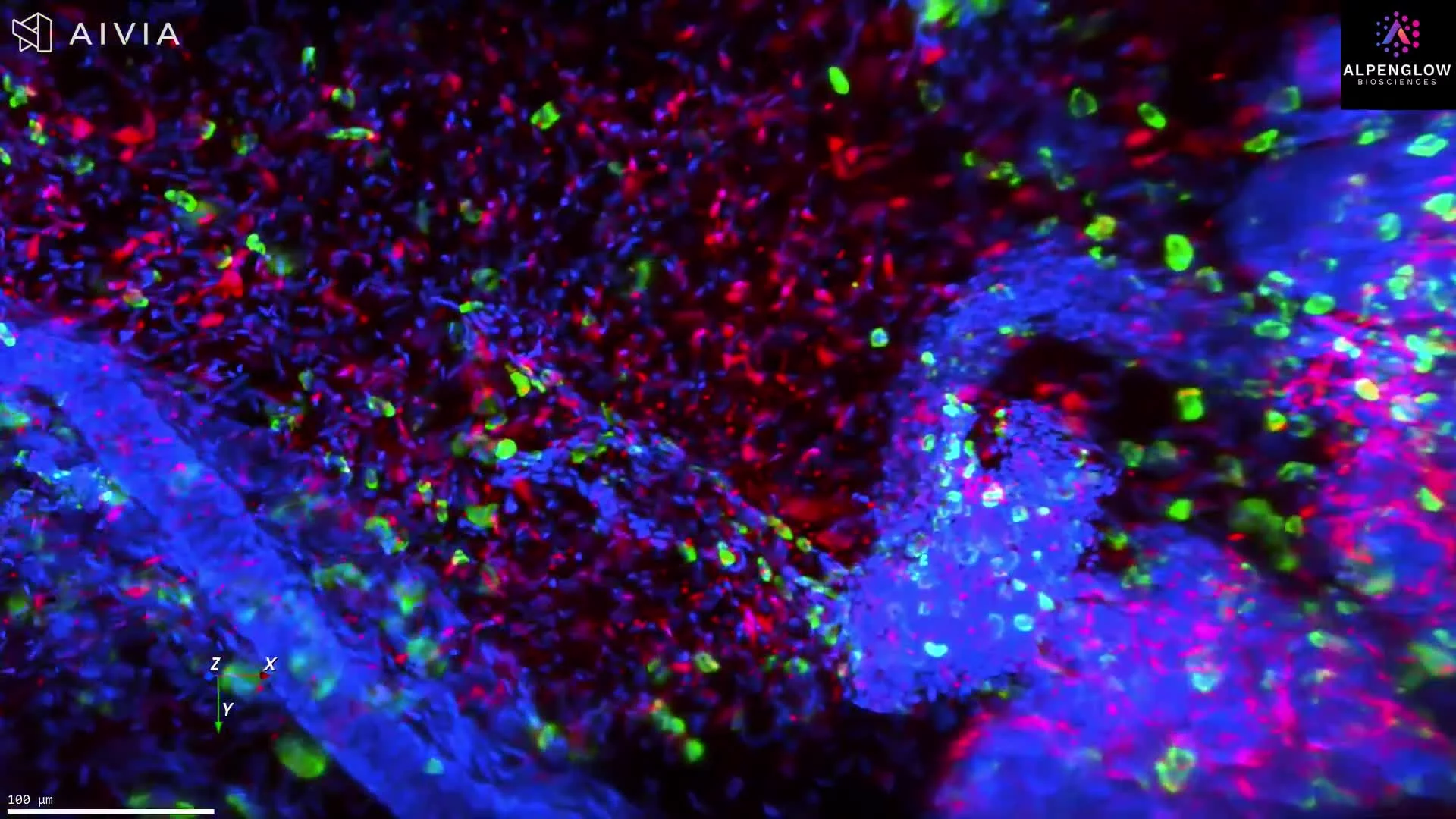
High-resolution 3D fluorescence imaging reveals an entire hair follicle in an intact HS biopsy, including individual cells, nerve networks, mast cells, and measurable spatial relationships.
This dataset presents high-resolution 3D fluorescence imaging of a hair follicle within an intact hidradenitis suppurativa (HS) skin biopsy, revealing follicular architecture, surrounding nerve networks, and individual cells throughout the imaged tissue volume.
The visualization begins with a 3D reconstruction of the complete hair follicle, showing its morphology and the intricate distribution of nerve fibers and mast cells in the surrounding tissue. Virtual 2D sectioning enables detailed examination of cellular structures at different depths without physically sectioning or destroying the specimen. The dataset is then visualized again in 3D to reveal the spatial organization of these structures.
Fluorescence markers:
- TO-PRO-3 (blue): Cell nuclei
- PGP9.5 (white): Nerve fibers
- Tryptase (green): Mast cells
The combination of high spatial resolution and volumetric imaging enables quantitative assessment of follicular morphology, cell counts and densities, nerve architecture, and distances between cells and nerve fibers.
This approach supports the investigation of follicular structures and neuroimmune interactions in HS while preserving their spatial relationships within the intact tissue.
Learn more about dermatology applications
Learn more on image analysis using Summit AI(TM)