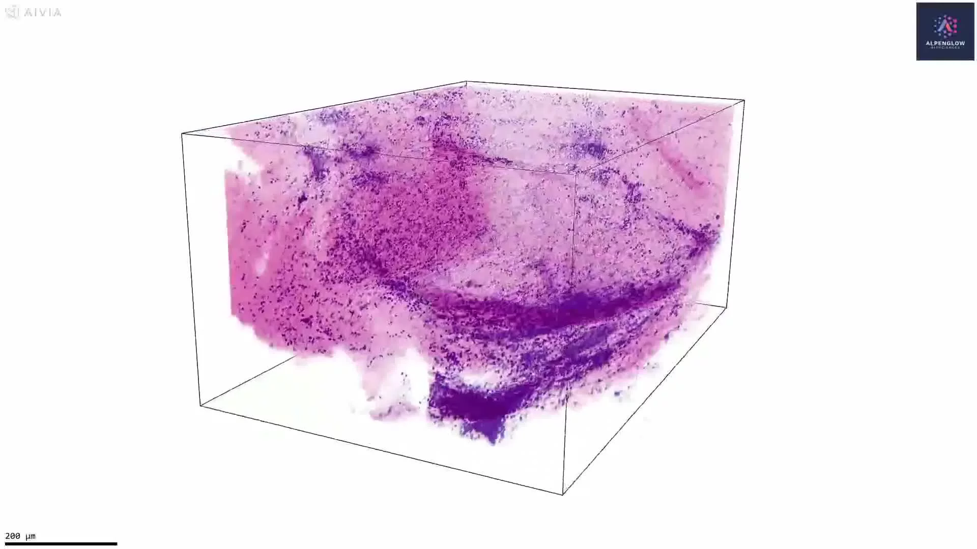
3D Imaging and Quantification of Human Duodenum Villi
This video presents a 3D visualization of a human duodenum tissue sample stained with eosin and TO-PRO-3 for nuclei, then pseudocolored to create an H&E-like appearance.
The volumetric dataset reveals intestinal villi as continuous three-dimensional structures, showing their shape, height, width, elongation, orientation, density, and spatial distribution across the tissue. Unlike a single 2D section, which captures a limited plane through each villus, 3D tissue imaging preserves the complete villous architecture and reduces the influence of sectioning angle and sampling location.
The 3D dataset can support quantitative analysis of villus number, individual villus dimensions, surface area, volume, elongation, curvature, spacing, density, and regional variation. These measurements can help characterize changes in tissue architecture across a larger volume rather than relying on isolated measurements from selected 2D sections.
This approach is relevant to gastrointestinal research, including studies of celiac disease, where villi may become shortened, flattened, fused, or structurally disrupted. Mapping and quantifying these changes in 3D may provide a more complete view of the extent, distribution, and heterogeneity of villous damage throughout the tissue sample.
The visualization demonstrates how H&E-like contrast, intact tissue imaging, and quantitative 3D analysis can be combined to study human intestinal microarchitecture.